
1. Introduction
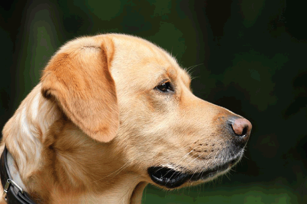
Eyelids are a vital part of the canine visual apparatus, acting as protective shutters, spreading the tear film, and facilitating the removal of debris. When the eyelid deviates from its normal anatomy or function, a cascade of ocular problems can ensue—ranging from mild irritation to irreversible vision loss. Abnormal eyelid conditions are among the most common ophthalmic complaints seen in veterinary practice, especially in brachy‑ and dolichocephalic breeds that are predisposed by genetics or conformation.
This guide pulls together the latest peer‑reviewed literature (up to 2024), clinical experience, and evidence‑based recommendations to give veterinarians, veterinary technicians, breeders, and dog owners a one‑stop reference for recognizing, diagnosing, treating, and preventing eyelid abnormalities in dogs.
2. Anatomy & Physiology of the Canine Eyelid
Understanding normal eyelid structure is the cornerstone for detecting pathology.
| Structure | Function | Key Features |
|---|---|---|
| Skin | Barrier, protection | Thin on the palpebral (inner) surface, thicker on the external surface. |
| Orbicularis oculi muscle | Contraction leads to blinking and closure | Circular muscle; innervated by the facial nerve (CN VII). |
| Tarsal plate (tarsus) | Provides rigidity; maintains shape | Dense connective tissue with meibomian glands embedded. |
| Meibomian (sebaceous) glands | Secrete lipid layer of tear film | Prevents tear evaporation. |
| Conjunctiva (palpebral) | Moistens lid, immune defense | Contains goblet cells creating mucin. |
| Nerves & blood supply | Sensation, reflexes, nutrition | Sensory from trigeminal nerve (CN V); vascularized by facial and infra‑orbital arteries. |
| Lacrimal & infra‑orbital glands | Tear production | Contribute to aqueous component of tear film. |
A healthy eyelid maintains a delicate balance of blink frequency (≈ 15–20 blinks/min), tear distribution, and protective tone. Disruption of any of these components can cause exposure, inflammation, or mechanical trauma to the cornea and conjunctiva.
3. Classification of Abnormal Eyelid Conditions
Below is the most commonly encountered spectrum of eyelid abnormalities in dogs. Each entity may coexist with others (e.g., ectropion + entropion) and often requires a multifactorial treatment plan.
3.1 Ectropion
Definition: Outward turning of the eyelid margin, exposing the palpebral conjunctiva.
Typical Breeds: Bloodhound, Basset Hound, Saint Bernard, Neapolitan Mastiff.
Pathophysiology: Laxity of the tarsal plate or excessive skin (often congenital).
3.2 Entropion
Definition: Inward rolling of the eyelid margin, causing the eyelashes and skin to rub against the cornea.
Typical Breeds: Shar‑Pei, Pug, Bulldog, Chow Chow, Cocker Spaniel.
Pathophysiology: Over‑tightening of the palpebral structures, muscular imbalance, or conformation‑related.
3.3 Ptosis (Blepharoptosis)
Definition: Drooping of the upper eyelid, leading to exposure keratitis.
Typical Breeds: Labrador Retriever, Golden Retriever, Beagle (often acquired).
Etiology: Neuromuscular disease (myasthenia gravis), trauma, or congenital muscle weakness.
3.4 Lagophthalmos
Definition: Incomplete closure of the eyelids, typically due to facial nerve paralysis or severe ectropion.
Clinical Significance: Corneal drying and ulceration.
3.5 Trichiasis & Distichiasis
Trichiasis: Abnormal growth of hairs (often from eyelash follicles) on the palpebral conjunctiva.
Distichiasis: An extra row of eyelash‑like hairs emerging from meibomian glands.
Common Breeds: Poodles, Shih Tzu, Lhasa Apso.
3.6 Dermoid & Cystic Lesions
Dermoid: Congenital inclusion of skin adnexa within the eyelid; appears as a firm, sub‑cutaneous nodule.
Cysts: Sebaceous or apocrine gland cysts, often secondary to chronic inflammation.
3.7 Neoplasia & Masses
Benign: Papillomas, adenomas.
Malignant: Squamous cell carcinoma, melanomas, sebaceous gland tumors.
Important Note: Malignant lesions may mimic chronic inflammation; biopsy is essential.
3.8 Inflammatory & Infectious Eyelid Disorders
Conjunctivitis with lid involvement: Bacterial, viral (canine distemper), fungal (cryptococcosis), allergic.
Blepharitis: Inflammation of the lid margin often secondary to meibomian gland dysfunction or parasites (e.g., Cheyletiella).
4. Causes & Risk Factors
| Category | Specific Causes | Explanation |
|---|---|---|
| Genetic / Congenital | Breed‑predisposed conformation, hereditary collagen disorders | Mutations affecting connective tissue (e.g., COL5A1 in Shar‑Pei) cause laxity or tightening. |
| Traumatic | Lacerations, blunt force, foreign bodies | May lead to scar contracture (entropion) or loss of tissue (ectropion). |
| Inflammatory / Allergic | Atopic dermatitis, flea allergy, contact irritants | Chronic lid margin inflammation promotes edema and laxity. |
| Infectious | Bacterial (Staph, Pseudomonas), fungal (Aspergillus), parasitic (Demodex) | Direct invasion or secondary inflammation can distort lid anatomy. |
| Neoplastic | Tumors (SCC, melanoma) | Mass effect and tissue destruction alter eyelid contour. |
| Systemic Neuromuscular | Myasthenia gravis, facial nerve paralysis (idiopathic or due to otitis media) | Causes ptosis or lagophthalmos. |
| Age‑related Degeneration | Decreased collagen elasticity, chronic blepharitis | Older dogs often develop ectropion secondary to skin laxity. |
| Iatrogenic | Improper suturing, over‑resection during cosmetic procedures | May create postoperative entropion/ectropion. |
| Nutritional Deficiencies | Vitamin A, essential fatty acid deficits | Poor tear film quality predisposes to inflammation and lid margin changes. |
Key Take‑away: While many conditions are breed‑specific, any dog can develop secondary eyelid pathology from trauma, infection, or systemic disease. Early recognition of predisposing factors helps in prevention and timely intervention.
5. Signs & Symptoms
| Clinical Sign | Potential Underlying Abnormality |
|---|---|
| Redness & Swelling of the Lid Margin | Blepharitis, allergic dermatitis, early ectropion. |
| Rubbing or Scratching of the Face | Entropion, trichiasis, foreign bodies. |
| Excessive Tearing (Epiphora) | Lacrimal duct obstruction secondary to lid malposition, ectropion. |
| Corneal Ulceration / Scarring | Chronic entropion, lagophthalmos, exposure keratitis. |
| Visible Skin Redundancy / “Floppy” Lid | Ectropion, especially in senior dogs. |
| Drooping Upper Eyelid | Ptosis, facial nerve paralysis. |
| Hair Growth on Conjunctival Surface | Trichiasis or distichiasis. |
| Mass or Nodule on Lid | Dermoid cyst, neoplasm, granuloma. |
| Blepharospasm (continuous blinking) | Painful corneal irritation, entropion, ulcer. |
| Discharge (serous, mucopurulent) | Conjunctivitis, bacterial infection, tear film instability. |
Red‑flag signs requiring immediate veterinary attention include: sudden onset of corneal ulceration, intense pain, severe swelling, or any visible mass with rapid growth.
6. Diagnostic Work‑up
A systematic approach ensures accurate diagnosis and optimal treatment planning.
6.1 Clinical Examination
- Full Physical Exam – Assess overall health, breed‑specific predispositions, and systemic signs.
- Ophthalmic Assessment – Use the “Moorfields” protocol (menace response, palpebral reflex, pupillary light reflex, intra‑ocular pressure).
- Eyelid Palpation – Evaluate skin tension, tarsal plate rigidity, and any palpable masses.
6.2 Ophthalmic Tests
| Test | Purpose | Interpretation |
|---|---|---|
| Schirmer Tear Test (STT‑1) | Quantify aqueous tear production | < 15 mm/min = dry eye (keratoconjunctivitis sicca). |
| Fluorescein Staining | Detect corneal epithelial defects | Positive uptake = ulcer or abrasion. |
| Lissamine Green / Rose Bengal | Identify devitalized cells, mucin deficiency | Staining of conjunctiva = dry eye or chronic irritation. |
| Tonometry (Tonopen/Rebound) | Measure intra‑ocular pressure (IOP) | High IOP may indicate secondary glaucoma from chronic keratitis. |
| Slit‑lamp Biomicroscopy | Detailed view of lid margin, meibomian glands, cornea | Helpful for diagnosing marginal blepharitis, meibomian gland dysfunction. |
| Imaging (Ultrasound, CT, MRI) | Evaluate deep masses, bony involvement, orbital structures | CT preferred for bony involvement; MRI for soft‑tissue extension. |
| Cytology/Histopathology | Differentiate inflammatory vs. neoplastic lesions | Fine‑needle aspirate (FNA) for cytology; excisional biopsy for definitive diagnosis. |
| Allergy Testing (Serum IgE, Intradermal) | Identify environmental allergens causing chronic blepharitis | Useful in atopic dogs with recurrent lid inflammation. |
6.3 Laboratory Work‑up
- CBC & Chemistry Panel – Detect systemic disease (e.g., autoimmune, endocrine).
- Serology (e.g., Toxoplasma, Leishmania) – In endemic regions, these can cause ocular inflammation.
- PCR/Culture – Identify bacterial or fungal pathogens when infection suspected.
7. Treatment Options
Management is individualized based on the type, severity, age, and overall health of the dog.
7.1 Medical Management
| Condition | Therapeutic Goal | Medications |
|---|---|---|
| Blepharitis / Conjunctivitis | Reduce inflammation & infection | Topical ophthalmic antibiotics (e.g., ofloxacin, gentamicin), NSAID eye drops (diclofenac), artificial tears. |
| Allergic Dermatitis | Control pruritus, barrier repair | Antihistamines (e.g., cetirizine), corticosteroids (short‑term), omega‑3 supplements, hypoallergenic shampoos. |
| Dry Eye (Keratoconjunctivitis Sicca) | Stimulate tear production | Cyclosporine A ophthalmic (0.2 %), Tacrolimus (0.03 %). |
| Corneal Ulceration secondary to entropion/ectropion | Promote healing, prevent infection | Topical antibiotics, Atropine (mydriasis), mucolytics (e.g., cautery or tarsorrhaphy if needed). |
| Ptosis (neuromuscular) | Improve lid elevation | Anticholinesterase drugs (e.g., pyridostigmine) for myasthenia gravis; treat underlying cause. |
| Trichiasis/Distichiasis | Remove offending hairs | Electro‑cautery, cryotherapy, laser ablation, or cryosurgical removal. |
| Dermoid Cysts | Reduce size/inflammation | Sclerotherapy (e.g., OK‑432) is experimental; surgical excision preferred. |
| Neoplasia | Control tumor growth | Surgical excision + wide margins, adjuvant radiotherapy, chemotherapy (e.g., carboplatin for SCC). |
NOTE: Systemic antibiotics are reserved for deep tissue infection or pre‑/post‑operative prophylaxis. Topical therapy remains the mainstay for superficial lid disease.
7.2 Surgical Corrections
| Procedure | Indications | Key Steps & Technical Pearls |
|---|---|---|
| Entropion Repair (Hotz‑Celsus, Lateral Canthoplasty) | Inward rolling lids causing corneal trauma | Lateral canthotomy to release tension, sutures placed through the sub‑orbicularis fascia, placement of a permanent suture to evert the lid margin. |
| Ectropion Correction (Miller’s/Portmann–Boehm technique) | Excess skin/eyelid laxity leading to exposure | Excisional skin with muscle plication, sub‑cutaneous sutures to tighten tarsal plate, removal of redundant skin. |
| Ptosis Repair (Levator Muscle Resection, Muller’s Muscle Tightening) | Droopy upper lid impairing visual axis | Exposure of levator palpebrae superioris, muscle shortening (5‑10 mm), adjustable sutures for fine‑tuning. |
| Lagophthalmos Surgical Tarsorrhaphy | Incomplete lid closure from facial nerve paralysis or severe ectropion | Partial or temporary tarsorrhaphy using non‑absorbable sutures, silicone sponges or punctal plugs for temporary support. |
| Blepharoplasty (Cosmetic/Functional) | Excessive folds, aesthetic concerns, or functional obstruction | Reductive skin removal, resection of abnormal folds, careful preservation of lacrimal apparatus. |
| Excision of Dermoid/Cystic Lesions | Sub‑cutaneously located masses | En‑bloc removal with margins of healthy tissue; histopathology mandatory. |
| Tumor Resection + Reconstructive Flaps | Malignant eyelid tumors | Wide local excision (≥ 5 mm margin), advancement or rotational flaps to restore lid integrity; cryotherapy of margins if needed. |
| Laser/ Cryo‑ablation of Trichiasis | Aberrant hairs causing irritation | CO₂ laser for precise ablation, cryotherapy (liquid nitrogen) for hair follicle destruction. |
Post‑operative care includes analgesia (opioids, NSAIDs), antibiotic prophylaxis, topical lubricants, and protective Elizabethan collars to prevent self‑trauma. Re‑evaluation at 7‑10 days and again at 4‑6 weeks is standard to assess wound healing and functional outcome.
7.3 Adjunctive Therapies
- Topical Cyclosporine for chronic dry eye after eyelid surgery.
- Autologous Serum Eye Drops for persistent corneal epithelial defects.
- Low‑Level Laser Therapy (LLLT) to accelerate wound healing.
- Nutraceuticals (see Section 10) for long‑term ocular health.
8. Prognosis, Complications & Long‑Term Outlook
| Condition | Prognosis (with appropriate treatment) | Common Complications |
|---|---|---|
| Entropion | Excellent; most dogs regain normal vision. | Over‑correction (ectropion), suture dehiscence, recurrence (especially in growing puppies). |
| Ectropion | Good; cosmetic and functional improvement. | Persistent skin laxity, wound infection, recurrence if not fully excised. |
| Ptosis | Variable; depends on underlying cause. | Continued drooping if neuromuscular disease persists; need for repeated surgery. |
| Lagophthalmos | Guarded; may require permanent tarsorrhaphy. | Chronic keratitis, corneal ulceration, secondary infection. |
| Trichiasis/Distichiasis | Good with proper hair removal. | Regrowth of hairs, scarring. |
| Dermoid Cyst | Excellent after complete excision. | Recurrence if cyst wall left behind. |
| Neoplasia | Depends on tumor type & stage. | Local recurrence, metastasis, eyelid contracture after radical excision. |
| Blepharitis/Conjunctivitis | Often resolves with medical therapy. | Chronic dry eye, corneal scarring. |
Key points for owners: Early intervention dramatically reduces the risk of irreversible corneal damage. Regular veterinary ophthalmic examinations (at least annually for predisposed breeds) are essential for monitoring and early detection.
9. Prevention Strategies
- Selective Breeding – Avoid mating dogs with known hereditary eyelid abnormalities. Genetic screening (where available) for collagen disorders or facial nerve anomalies reduces incidence.
- Puppy Screening – Examine newborns for ectropion/entropion; early surgical correction in puppies (< 6 months) yields better cosmetic outcomes and reduces corneal injury.
- Environmental Management –
- UV Protection: Use canine‑safe sunglasses or hats for breeds with prominent eyes (e.g., Pugs, Bulldogs).
- Allergen Control: Frequent bathing, hypoallergenic bedding, and regular flea/parasite prophylaxis.
- Nutrition – Providing a diet rich in omega‑3 fatty acids, antioxidants (vitamins A, C, E), and adequate protein supports healthy skin and tear film.
- Routine Ophthalmic Check‑ups – Early detection of lid laxity or hair misdirection can be addressed surgically before corneal involvement.
- Owner Education – Teach owners to recognize early signs (excess tearing, rubbing, visible skin folds) and seek prompt veterinary care.
10. Diet, Nutrition & Supplements for Ocular Health
| Nutrient | Physiological Role | Food Sources / Supplements | Recommended Daily Intake (approx.) |
|---|---|---|---|
| Vitamin A (Retinol) | Maintains conjunctival epithelium; essential for tear production | Liver, carrots, sweet potatoes, egg yolk; Retinol supplements (e.g., 5000 IU/kg diet) | 500–700 IU/kg (AAFCO) |
| Omega‑3 Fatty Acids (EPA/DHA) | Anti‑inflammatory; stabilizes tear film | Fish oil, krill oil, algae‑derived supplements | 100–300 mg EPA/DHA per kg body weight |
| Vitamin C | Antioxidant; protects corneal cells from oxidative stress | Citrus fruits (dog‑safe), berries, ascorbic acid supplements | 10–20 mg/kg |
| Vitamin E (α‑tocopherol) | Protects membrane lipids; aids wound healing | Wheat germ oil, sunflower seeds, tocopherol supplements | 5–10 IU/kg |
| Taurine | Supports retinal function; deficiency linked to retinal degeneration | Meat, fish, taurine‑fortified kibble | 0.1 g/kg (minimum) |
| Zinc | Cofactor for antioxidant enzymes; aids epithelial repair | Beef, pork, zinc gluconate | 30–50 mg/kg |
| L‑Carnitine | Improves oxidative metabolism in ocular muscles | Beef, chicken, carnitine supplements | 50–100 mg/kg |
| Beta‑Carotene (pro‑vitamin A) | Antioxidant, skin health | Carrots, pumpkin, beta‑carotene tablets | 1–3 mg/kg |
Practical Feeding Guidelines
- Balanced Commercial Diets that meet AAFCO nutrient profiles generally cover most requirements.
- For high‑risk breeds (e.g., Shar‑Pei, Bulldog), consider adding fish‑oil capsules (0.5 ml per 10 kg body weight) and vitamin E (25 IU per 10 kg).
- Hydration is critical; ensure constant access to fresh water and consider wet food for dogs with dry‑eye predisposition.
- Avoid excessive vitamin A supplementation, as hypervitaminosis can cause bone abnormalities and ocular toxicity.
11. Owner Education & When to Seek Veterinary Care
| Situation | Action |
|---|---|
| Mild redness or occasional rubbing | Monitor for 24‑48 h; apply a warm compress; schedule a routine check‑up if persists. |
| Constant tearing, obvious eyelid fold or droop | Book an appointment within 48 h; possible ectropion/entropion requiring surgical evaluation. |
| Sudden corneal opacity, ulcer, or loss of vision | Emergency visit – urgent ophthalmic treatment needed. |
| Visible mass or cyst | Prompt evaluation – rule out neoplasia. |
| Signs of systemic disease (fever, lethargy) with ocular changes | Full physical exam and blood work, as ocular signs may be secondary to systemic illness. |
Key messages for owners
- Early detection saves sight. Even subtle changes can lead to serious complications.
- Do not attempt home removal of hairs (trichiasis) without veterinary guidance; improper removal can scar the lid.
- Never use human eye drops unless explicitly prescribed by a veterinarian; canine ocular pharmacology differs.
- Maintain vaccination schedule (especially distemper) to prevent viral ocular disease.
12. Summary & Key Take‑aways
- Abnormal eyelid conditions (ectropion, entropion, ptosis, lagophthalmos, trichiasis, dermoid, neoplasia) are common, breed‑predisposed, and potentially vision‑threatening.
- Comprehensive examination (clinical, Schirmer, fluorescein, imaging, cytology) is essential to differentiate primary lid disease from secondary ocular pathology.
- Medical therapy (antibiotics, anti‑inflammatories, tear substitutes) controls infection and inflammation but surgical correction remains the definitive treatment for structural abnormalities.
- Prognosis is generally excellent when intervention occurs early; however, delayed treatment can lead to chronic corneal scarring, glaucoma, or permanent blindness.
- Prevention hinges on responsible breeding, regular eye examinations, environmental management, and a diet rich in ocular‑supportive nutrients.
- Owner vigilance is critical—recognizing early signs and seeking prompt veterinary attention dramatically improves outcomes.
By integrating clinical expertise, evidence‑based therapeutics, and preventive nutrition, veterinarians can safeguard canine ocular health and preserve the precious gift of sight for our four‑legged companions.
#CanineEyeHealth, #DogEyelidCare, #VeterinaryOphthalmology, #EntropionFix, #EctropionTreatment, #DogPtosis, #Lagophthalmos, #Trichiasis, #Distichiasis, #DogDermoid, #EyeHealthTips, #PetNutrition, #Omega3ForDogs, #VitaminAForPets, #DogEyeCheck, #HealthyEyesHappyDogs, #PetWellness, #DogOwnersGuide, #VetAdvice, #DogEyeSurgery, #PreventBlindness, #CanineVision, #DogHealth, #PetCare, #DogLovers, #DogMedicalInfo, #PetSafety, #DogLifestyle, #DogBreeds, #DogHealthTips.


Add comment