
Adenoviruses are non‑enveloped, double‑stranded DNA viruses that infect a wide variety of vertebrate hosts, including birds, mammals, reptiles, and fish. In avian species, they are responsible for a spectrum of clinical entities ranging from sub‑clinical infections to severe systemic disease. Among waterfowl, Duck Adenovirus (commonly abbreviated as DAdV) is a notable pathogen that can cause high morbidity, occasional mortality, and considerable economic loss in both commercial duck production and backyard flocks.
While adenovirus infections have been extensively studied in chickens (e.g., Fowl adenovirus causing Inclusion Body Hepatitis) and turkeys (e.g., Turkey adenovirus), the literature on ducks is comparatively sparse, leading to gaps in disease recognition, management, and preventive strategies. This guide consolidates current scientific knowledge, field observations, and practical recommendations into a single, exhaustive resource for veterinarians, duck‑keepers, researchers, and policy‑makers.
Key take‑away: Adenovirus in ducks is a multifactorial disease whose expression depends on viral strain, host genetics, age, concurrent stressors, and management practices. Understanding each component is essential for successful control.
2. Etiology – Causes
2.1. Virus Taxonomy
| Taxonomic Level | Designation |
|---|---|
| Order | Herpesvirales (historically mis‑assigned) – Correct: Family Adenoviridae |
| Family | Adenoviridae |
| Sub‑family | Aviadenovirinae (bird‑specific adenoviruses) |
| Genus | Mastadenovirus (most duck isolates) |
| Species | Anatid adenovirus 1 (AnHV‑1) – the classic “Duck Adenovirus” |
| Additional species | Anatid adenovirus 2–4 (emerging strains, often with distinct tissue tropism) |
The prototype Anatid adenovirus 1 (AnHV‑1) was first isolated from domestic ducks in the United Kingdom in the 1970s. Molecular phylogeny places it close to Fowl adenovirus serotypes but with distinct capsid proteins that confer host specificity.
2.2. Virus Structure & Replication
- Capsid: Icosahedral, ~70‑90 nm diameter, composed of hexon, penton, and fiber proteins. The hexon contains hypervariable regions that determine serotype and antigenicity.
- Genome: Linear, ~30‑35 kb double‑stranded DNA; encodes ~30–35 open‑reading frames including DNA polymerase, penton base, and viral protease.
- Replication Cycle:
- Attachment – fiber protein binds to a sialic‑acid‑containing receptor on duck epithelial or endothelial cells.
- Entry & Uncoating – endocytosis followed by release of viral DNA into the nucleus.
- Early Gene Expression – DNA polymerase and regulatory proteins are produced, hijacking host transcription.
- DNA Synthesis – viral genome replication via a rolling‑circle mechanism.
- Late Gene Expression – structural proteins (hexon, penton, fiber) are synthesized.
- Assembly & Release – capsids assemble in the nucleus; virions are released by cell lysis, often after forming characteristic intranuclear inclusion bodies.
2.3. Transmission Routes
| Route | Description | Relative Importance |
|---|---|---|
| Fecal‑oral | Infected duck excreta contaminates water, feed, or litter; susceptible birds ingest virus. | Primary |
| Aerosol/Respiratory | Droplet spread during coughing, sneezing, or high‑density housing. | Moderate |
| Vertical (Egg‑to‑Embryo) | Transovarial passage of low‑level virus; hatchlings may be infected at birth. | Low‑to‑moderate (strain‑dependent) |
| Fomites | Contaminated equipment, boots, nets, or vehicles. | Variable, heavily influenced by biosecurity |
| Wild Waterfowl Reservoirs | Migratory ducks, geese, and swans can harbor related adenoviruses; spill‑over to domestic flocks. | Significant in extensive/free‑range systems |
2.4. Predisposing Factors
- High stocking density – amplifies fecal contamination and aerosol load.
- Poor water quality – stagnant or chlorinated water can facilitate viral persistence.
- Nutritional deficiencies – especially Vitamin A, E, and selenium, which modulate mucosal immunity.
- Concurrent infections – Mycoplasma, Salmonella, Escherichia coli or Duck virus hepatitis can compromise the immune system, allowing adenovirus to proliferate.
- Stressors – transport, abrupt temperature changes, handling, and crowding elevate corticosterone, suppressing antiviral defenses.
3. Clinical Manifestations – Signs & Symptoms
Adenovirus infection in ducks can be sub‑clinical, acute, or chronic, depending on the virulence of the strain, age of the bird, and environmental conditions. The following table outlines the most common presentations:
| Clinical Form | Age Group | Primary Organs Affected | Typical Signs |
|---|---|---|---|
| Inclusion Body Hepatitis (IBH) | 1‑4 weeks (young ducklings) | Liver, spleen, pancreas | Lethargy, anorexia, yellowish discoloration of skin (jaundice), abdominal distension, sudden death. |
| Enteric Adenovirosis | 2‑8 weeks | Intestine, bursa of Fabricius | Watery or mucoid diarrhea, drooping wings, dehydration, weight loss. |
| Respiratory Adenovirosis | 4 weeks‑adult | Trachea, lungs | Nasal discharge, coughing, sneezing, dyspnea, conjunctivitis, occasional hemorrhagic tracheitis. |
| Systemic/Multisystemic | Any age (more severe in immunocompromised) | Liver, kidney, heart, pancreas, brain | Depression, fever (38‑41 °C), ataxia, seizures, petechial hemorrhages, mortality up to 30 % in severe outbreaks. |
| Chronic Carrier State | Adult | Low‑grade shedding from gut and respiratory tract | No outward signs, but intermittent virus shedding for months. |
3.1. Pathognomonic Lesions
- Intranuclear basophilic inclusion bodies – large, eosinophilic to basophilic “Smudge cells” within hepatocytes, pancreatic acinar cells, and endothelial cells.
- Hepatomegaly with pale, friable liver – often accompanied by petechial hemorrhages.
- Intestinal ulceration and necrosis – may appear as hemorrhagic spots on the mucosal surface.
- Respiratory mucosal hyperplasia – thickened tracheal epithelium with focal necrosis.
3.2. Differential Diagnosis
| Condition | Overlapping Signs | Distinguishing Features |
|---|---|---|
| Duck viral hepatitis (DVH) – Duck hepatitis B virus | Jaundice, sudden death | Absence of inclusion bodies; liver necrosis without intranuclear inclusions; serology positive for DVH. |
| Mycoplasma anatis infection | Respiratory distress, conjunctivitis | Mycoplasma cultured from trachea; no hepatic lesions. |
| Salmonella septicemia | Diarrhea, fever, mortality | Gram‑negative rods on culture; endotoxemia signs. |
| Riemerella anatipestifer (RA) | Septicemia, edema, mortality | Gram‑negative coccobacilli; characteristic “RA lesions” in bone and joints. |
| Avian Influenza (low pathogenic) | Respiratory signs | Hemagglutination assay positive; high mortality only in HPAI. |
4. Duck Breeds at Risk
Although adenovirus can infect any duck species, certain domestic breeds show heightened susceptibility due to genetic, physiological, and management factors.
4.1. High‑Risk Domestic Breeds
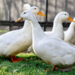
- Pekin (American Pekin) – The most widely farmed meat duck worldwide. Its rapid growth rate creates metabolic stress, compromising hepatic function and rendering it more vulnerable to hepatic inclusion body hepatitis.
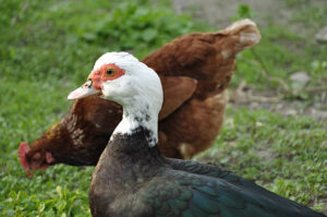
- Muscovy (Cairina moschata) – Muscovy ducklings have a slower immune maturation compared with mallards and are prone to severe enteric disease when exposed to adenovirus at 2‑3 weeks of age.
- Rouen – A larger, heritage breed often kept in free‑range or semi‑intensive systems where contact with wild waterfowl is frequent; increased exposure leads to higher infection rates.
- Khaki Campbell – Highly prolific egg layers; the reproductive stress and potential vertical transmission make them a noteworthy group for sub‑clinical carriage.
4.2. Why These Breeds Are More Susceptible
- Genetic Bottlenecks: Commercial lines have undergone intensive selection for production traits (growth, egg yield) at the expense of immune diversity. This reduced heterozygosity can diminish the ability to mount robust antiviral responses.
- Metabolic Demands: Rapidly growing meat ducks (e.g., Pekin) demand high protein and energy, which can cause transient hepatic overload, creating a permissive environment for viral replication.
- Management Practices: High‑density indoor rearing, use of communal waterers, and limited outdoor access increase fecal‑oral transmission.
- Maternal Antibody Transfer: In egg‑laying breeds, the quantity and specificity of maternal antibodies transferred to the yolk vary. Inadequate passive immunity leads to early‑life susceptibility.
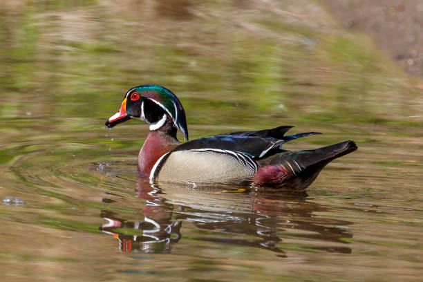
4.3. Wild and Semi‑Domesticated Ducks
- Mallard (Anas platyrhynchos) – Natural reservoirs for many avian adenoviruses; generally asymptomatic carriers.
- Northern Pintail (Anas acuta) – Frequently mingle with domestic flocks in wetland areas, facilitating cross‑species spill‑over.
5. Affected Life‑Stages
| Life‑Stage | Typical Age (days) | Predominant Clinical Form | Reason for Vulnerability |
|---|---|---|---|
| Embryo | 0‑21 (incubation) | Vertical transmission, embryonic death | Transovarial infection; virus can cross the chorioallantoic membrane. |
| Hatchling | 1‑7 | Sub‑clinical or mild enteric disease | Immature gut barrier; maternal antibodies waning. |
| Duckling | 8‑28 | Inclusion Body Hepatitis, severe enteric disease | Rapid tissue growth; immune system still developing. |
| Juvenile | 29‑90 | Respiratory disease, systemic infection | Exposure to environment and stressors (e.g., weaning). |
| Adult | >90 | Carrier state, occasional respiratory flare‑ups | Fully developed immunity, but stress can reactivate shedding. |
Key point: The first 4 weeks of life are the most critical window for clinical disease, especially hepatic and intestinal forms. Proper early‑life management dramatically reduces disease incidence.
6. Diagnosis
Accurate diagnosis relies on a combination of clinical observation, pathological examination, and laboratory testing.
6.1. Field Diagnosis
- History – Sudden onset of mortality in ducklings, recent introduction of new birds, or recent water source change.
- Clinical exam – Jaundice, diarrhea, respiratory discharge, and depressed behavior.
- Post‑mortem observations – Enlarged, pale liver with hemorrhagic foci; inclusion bodies visible on gross exam in severe cases.
While field diagnosis can be suggestive, definitive confirmation requires laboratory work.
6.2. Laboratory Methods
| Test | Sample Type | Turn‑around Time | Sensitivity/Specificity | Comments |
|---|---|---|---|---|
| Histopathology | Liver, pancreas, intestine, trachea (fixed in 10 % neutral buffered formalin) | 24‑48 h | High (presence of characteristic intranuclear inclusions) | Gold standard for lesion identification. |
| Immunohistochemistry (IHC) | Same tissues as histopathology | 48‑72 h | High (virus‑specific monoclonal antibodies) | Confirms adenovirus antigen within inclusions. |
| Polymerase Chain Reaction (PCR) – conventional or real‑time | Cloacal swab, tracheal swab, tissue homogenate, blood | 6‑12 h (real‑time) | Very high (detects <10 copies) | Allows strain typing via sequencing. |
| Virus Isolation | Embryonated duck eggs (10‑day old) or primary duck embryo fibroblast (DEF) cultures | 5‑7 days | Moderate (requires viable virus) | Useful for vaccine development. |
| Serology (ELISA/HAI) | Serum | 2‑3 days | Moderate (detects antibodies; not useful for acute infection) | Helpful for herd immunity surveys. |
| Electron Microscopy (EM) | Tissue homogenate | 24‑48 h | High (visualization of icosahedral particles) | Rarely needed; confirmatory. |
6.2.1. Recommended Diagnostic Algorithm
- Collect samples from at least three dead ducklings (liver, pancreas, intestine, trachea) and fresh cloacal swabs.
- Perform rapid PCR on swabs for preliminary confirmation (real‑time assay targeting hexon gene).
- Send tissue for histopathology and IHC – confirm inclusion bodies and viral antigen.
- If PCR is negative but suspicion high, repeat sampling and consider virus isolation in embryonated duck eggs.
6.3. Differential Diagnosis – Laboratory Distinction
- Fowl adenovirus – PCR primers specific to FAdV hexon differ from AnHV‑1; sequencing confirms species.
- Duck hepatitis B virus – Serology (HBsAg) and PCR for DHBV polymerase gene.
- Bacterial cultures – Salmonella and Riemerella grown on selective media; not present in viral lesions.
7. Treatment
Currently, no antiviral drug is licensed specifically for avian adenoviruses. Management therefore focuses on supportive therapy, secondary bacterial infection control, and optimizing the host’s immune response.
7.1. Supportive Care
| Intervention | Dosage/Regimen | Rationale |
|---|---|---|
| Fluid therapy (electrolyte‑balanced isotonic solution) | 5–10 mL kg⁻¹ h⁻¹ subcutaneously or via oral drippers | Counteract dehydration from diarrhea and restore plasma volume. |
| Vitamin A (retinyl acetate) | 50 000 IU kg⁻¹ intramuscularly, once daily for 3 days | Supports mucosal integrity and immune function. |
| Vitamin E + Selenium (e.g., Se‑Vit) | 0.5 mL L⁻¹ water for 5 days | Antioxidant protection of hepatic cells. |
| Probiotics (Lactobacillus spp., Bifidobacterium) | 1 mL L⁻¹ drinking water, daily | Restores gut flora after viral‑induced dysbiosis. |
| N-Acetylcysteine (NAC) | 100 mg kg⁻¹ PO, once daily | Hepatoprotective thiol donor, reduces oxidative stress. |
7.2. Antibacterial Therapy
Secondary bacterial infections are common, especially E. coli septicemia arising from gut barrier breakdown. Empirical choice should be guided by culture and sensitivity when possible, but common regimens include:
- Enrofloxacin – 10 mg kg⁻¹ PO, once daily for 5 days (broad‑spectrum gram‑negative coverage).
- Gentamicin – 5 mg kg⁻¹ IM, once daily for 3 days (if renal function acceptable).
Important: Avoid routine use of broad‑spectrum antibiotics without bacterial confirmation; overuse contributes to resistance and may suppress beneficial microbiota.
7.3. Immunostimulants
- Poly I:C (synthetic dsRNA) – 2 mg kg⁻¹ IM, single dose; stimulates innate interferon response.
- Levamisole – 2 mg kg⁻¹ PO, daily for 3 days; enhances cell‑mediated immunity.
Evidence for efficacy is limited; these agents should be used as adjuncts rather than primary therapy.
7.4. Prognostic Indicators for Treatment Decision
| Positive Prognostic Sign | Negative Prognostic Sign |
|---|---|
| Age < 14 days, mild lesions, normal blood chemistry | Age > 21 days with severe hepatic necrosis |
| Rapid response to fluid therapy (improved activity within 12 h) | Persistent hypoglycemia despite glucose supplementation |
| No concurrent bacterial sepsis | High bacterial load on culture (>10⁶ CFU mL⁻¹) |
| Low viral load on qPCR (Ct > 30) | High viral load (Ct < 20) |
8. Prognosis & Complications
8.1. Expected Outcomes
| Scenario | Mortality Rate | Recovery Time | Long‑Term Consequences |
|---|---|---|---|
| Mild enteric disease (ducklings ≤ 14 days) | < 5 % | 5‑7 days | Full recovery, possible carrier state. |
| Inclusion Body Hepatitis (acute, high viral load) | 15‑30 % (up to 50 % in high‑density farms) | 7‑14 days for survivors | Hepatic fibrosis, reduced growth performance. |
| Respiratory adenovirosis (mixed infections) | 5‑10 % | 10‑14 days | Chronic sinusitis, decreased egg production in layers. |
| Chronic carrier (adult ducks) | < 1 % | N/A | Intermittent shedding, risk of outbreak in naïve flocks. |
8.2. Complications
- Secondary Bacterial Sepsis – E. coli, Salmonella, or Riemerella may invade damaged liver and intestine, leading to endotoxemia and rapid death.
- Hepatic Fibrosis – Persistent inflammation can cause collagen deposition, decreasing liver functional reserve and affecting feed conversion.
- Immune Suppression – Adenovirus infection depletes lymphoid tissues (bursa, thymus), predisposing birds to other viral (e.g., avian influenza) or parasitic infections.
- Reproductive Impact – In laying ducks, adenovirus can cause decreased egg quality, shell defects, and reduced hatchability.
- Economic Losses – Mortality, reduced weight gain, and increased medication costs can translate into significant financial setbacks for producers.
9. Prevention
Because treatment options are limited, prevention is the cornerstone of control. A multi‑layered strategy (“biosecurity‑vaccination‑management”) is essential.
9.1. Biosecurity Measures
| Measure | Practical Implementation |
|---|---|
| All‑in‑all‑out (AIAO) | Empty and disinfect barns between production cycles; avoid mixing ages. |
| Controlled entry | Footbaths (10 % bleach), dedicated clothing, and vehicle disinfection at the farm gate. |
| Water management | Use fresh, clean water daily; filter or UV‑treat communal waterers. |
| Litter hygiene | Replace litter at least every 4 weeks; avoid excessive moisture that promotes viral persistence. |
| Rodent and wild bird control | Install netting, fences, and deterrents; limit access of wild waterfowl to feed and water. |
| Egg handling | Disinfect eggs with a mild chlorine solution before incubation; avoid cross‑contamination between hatcheries. |
| Quarantine | Isolate new stock for a minimum of 21 days; test for adenovirus by PCR before integration. |
9.2. Vaccination
To date, no commercially licensed vaccine exists specifically for duck adenovirus in most countries. However, experimental and autogenous vaccines have shown promise.
| Vaccine Type | Production Method | Efficacy | Availability |
|---|---|---|---|
| Live attenuated (passage‑reduced) AnHV‑1 | Serial passage in duck embryo fibroblasts; attenuation confirmed by reduced pathogenicity. | 70‑85 % protection against hepatic disease when administered at 3 days of age. | Limited to research institutions; not widely commercialized. |
| Inactivated (killed) vaccine | Virus purified, inactivated with binary ethyleneimine (BEI), adjuvanted with oil‑in‑water (e.g., Montanide). | 60‑70 % reduction in mortality; stimulates humoral antibodies detectable by ELISA. | Autogenous formulations possible under veterinary oversight. |
| Recombinant subunit (hexon protein) | Hexon gene cloned in baculovirus expression system; purified protein + adjuvant. | Early trials show strong neutralizing antibody titers; field efficacy pending. | Experimental; not yet market‑ready. |
Practical recommendation: For high‑value breeding operations, an autogenous inactivated vaccine prepared from locally circulating strains can be administered to breeders (2 ml IM at 6 weeks of age) and a booster at 12 weeks. Ensure compliance with local regulatory frameworks.
9.3. Nutritional & Management Strategies
- Balanced ration – 15–18 % crude protein for ducklings, 12–14 % for adults; include high‑quality vegetable oil for essential fatty acids.
- Vitamin‑mineral premix – Provide at least 10,000 IU kg⁻¹ vitamin A, 200 mg kg⁻¹ vitamin E, and 0.3 mg kg⁻¹ selenium.
- Probiotic supplementation – Incorporate Lactobacillus‑based products (10⁸ CFU g⁻¹) in starter feeds.
- Environmental enrichment – Provide floating platforms and dry areas to reduce crowding and stress.
- Temperature control – Maintain 30 °C for the first week, then gradually reduce to 22 °C; avoid abrupt drops that elevate corticosterone.
10. Diet and Nutrition
10.1. Energy and Protein
- Starter (0‑3 weeks) – 3000–3200 kcal kg⁻¹ metabolizable energy; 22–24 % crude protein.
- Grower (4‑8 weeks) – 2800 kcal kg⁻¹; 18–20 % protein.
- Finisher (≥ 9 weeks) – 2600 kcal kg⁻¹; 16–18 % protein.
High‑quality soybean meal, fish meal, or cottonseed meal should be the primary protein sources.
10.2. Micronutrients Critical for Antiviral Immunity
| Micronutrient | Function | Recommended Level | Sources |
|---|---|---|---|
| Vitamin A | Maintains mucosal epithelium, promotes IgA production | 5000–10 000 IU kg⁻¹ feed | Carrots, alfalfa, pre‑formed retinyl acetate. |
| Vitamin E | Antioxidant; protects cell membranes from lipid peroxidation | 200–300 mg kg⁻¹ feed | Wheat germ oil, sunflower oil. |
| Selenium | Component of glutathione peroxidase; counters oxidative stress | 0.2–0.3 mg kg⁻¹ feed | Sodium selenite, selenomethionine. |
| Zinc | Supports thymic development, lymphocyte proliferation | 80–120 mg kg⁻¹ feed | Zinc oxide, zinc sulfate. |
| Copper | Enzyme cofactor for superoxide dismutase | 15–25 mg kg⁻¹ feed | Copper sulfate, copper proteinate. |
| Vitamin D₃ | Modulates innate immunity; important for calcium metabolism (egg shell quality) | 4000–6000 IU kg⁻¹ feed | Sun‑exposed feed, vitamin D₃ premix. |
10.3. Hydration and Water Quality
- Provide clean, fresh water at all times; replace daily.
- pH: 6.5–7.5.
- Chlorine residual: < 0.5 ppm (higher levels can irritate mucosa).
10.4. Feed Additives with Antiviral Potential
- Herbal extracts (e.g., oregano oil, thymol) – demonstrated in vitro inhibition of adenovirus replication at 0.1 % inclusion.
- Beta‑glucans – immunomodulatory polysaccharides; 0.05 % of diet may enhance macrophage activity.
Note: Always test additive tolerance in a small pilot group before full‑scale implementation.
11. Zoonotic Risk
Adenoviruses are species‑specific in most cases. Anatid adenovirus 1 does not infect humans, and no documented zoonotic transmission has been reported. However, certain considerations remain:
- Occupational Exposure – Farm workers handling sick birds or contaminated litter may experience irritant reactions (conjunctivitis, dermatitis) due to viral particles or secondary bacterial contaminants, not true infection.
- Cross‑Species Transmission – While human infection is improbable, inter‑species transmission within avian species (e.g., from wild waterfowl to domestic ducks) is well documented and can affect biosecurity.
- Laboratory Safety – Researchers working with live adenovirus should follow BSL‑2 containment practices (protective eyewear, gloves, and biosafety cabinets).
Bottom line: Adenovirus infection in ducks poses no direct public health threat, but standard hygiene (hand washing, protective clothing) remains essential to prevent secondary bacterial infections and maintain overall farm biosafety.
12. Integrated Management Plan – Putting It All Together
| Phase | Action Items | Timeline |
|---|---|---|
| Pre‑stocking | • Quarantine and PCR‑screen new birds. • Prepare autogenous vaccine (if used). • Clean and disinfect facility (10 % bleach). |
3‑4 weeks before placement |
| Placement (Day 0) | • Administer vaccine (if live attenuated, at day 3). • Provide high‑quality starter feed with vitamin A/E/Se. • Install footbaths and water filters. |
Day 0‑1 |
| First 2 Weeks | • Monitor for diarrhea, jaundice, respiratory signs. • Provide oral electrolytes and vitamin A if any signs appear. • Collect cloacal swabs for PCR if suspicion arises. |
Days 1‑14 |
| Weeks 3‑4 | • Booster vaccination (if inactivated vaccine schedule). • Rotate litter; add fresh bedding. • Begin probiotic water additive. |
Days 15‑28 |
| Weeks 5‑8 | • Continue balanced grower diet. • Perform weekly health checks and weight gains. • Conduct serology to assess immunity (optional). |
Days 29‑56 |
| Beyond 8 Weeks | • Maintain biosecurity, especially limiting wild bird contact. • Offer adult diet enriched with vitamin D₃ and calcium for layers. • Periodic PCR surveillance of a random 5 % of flock. |
Ongoing |
| Outbreak Response | • Immediate isolation of affected birds. • Initiate supportive therapy (fluids, vitamins). • Collect samples for rapid PCR. • Intensify disinfection (quaternary ammonium + UV). |
Within 24 h of suspicion |
13. Key Take‑Home Messages
| Point | Practical Implication |
|---|---|
| Adenovirus is primarily fecal‑oral | Emphasize water hygiene and litter management. |
| Young ducklings (≤ 4 weeks) are most vulnerable | Prioritize monitoring and early vaccination. |
| Inclusion bodies are diagnostic hallmarks | Use histopathology + IHC to confirm. |
| No specific antivirals exist | Focus on supportive care and secondary bacterial control. |
| Vaccination (autogenous) can reduce mortality | Consider for high‑value or intensive operations. |
| Zoonotic risk is negligible | Standard farm hygiene suffices for worker safety. |
| Nutrition underpins immunity | Provide adequate vitamins A/E/Se and antioxidants. |
| Biosecurity is the cornerstone | Apply AIAO, quarantine, and wild‑bird exclusion. |
#DuckAdenovirus, #WaterfowlHealth, #PekinDuck, #MuscovyDuck, #DuckDiseaseGuide, #AvianVeterinary, #FarmBiosecurity, #DuckHepatitis, #InclusionBodyHepatitis, #DuckNutrition, #VeterinaryMedicine, #PoultryScience, #DuckFarming, #WildBirdRisks, #DuckVaccines, #AnimalHealth, #VeterinaryTips, #DuckCare, #BirdHealth, #ZoonoticSafety, #EggProduction, #DuckLiverDisease, #DuckRespiratory, #FarmManagement, #DucksOfInstagram, #DuckLovers, #LiveBirds, #PoultryHealth, #DuckResearch, #VeterinaryPractice, #AnimalWelfare, #DucksInTheWild, #DuckBreeds, #FeedFormulation, #WaterQuality, #FarmHygiene, #BiosecurityFirst, #DuckSupportiveCare, #VeterinaryDiagnostics, #DuckImmunology, #DucksDaily, #PetDuckCare, #BackyardDucks, #CommercialDucks, #DuckFarmSafety, #VeterinaryEducation, #AnimalScience, #DuckVirus, #OIEGuidelines, #USDA, #FAO, #GlobalAvianHealth, #DucksAndHumans, #NoZoonosis, #HealthyDucks, #DuckWorld, #DuckFacts, #BirdMedicine, #DuckCommunity, #FarmLife, #SustainableFarming, #DucksAndHealth, #Ducks2026, #DucksOfTheWorld, #DuckExperts, #DuckLab, #DucksOnline, #AvianDisease, #ViralInfections, #DuckPrevention, #DucksInfo, #DuckCareTips, #DuckHealthCheck, #DuckLoversClub.
Add comment