
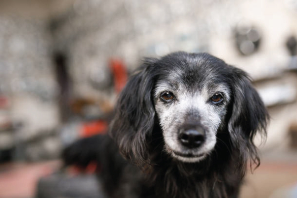
Aqueocentesis (also known as anterior chamber paracentesis) in dogs is a procedure involving the collection of a small sample of aqueous humor from the anterior chamber of the eye. This fluid is then submitted for various diagnostic analyses. It is a specialized ophthalmic procedure, typically performed by a veterinary ophthalmologist or a veterinarian experienced in advanced ocular techniques.
What is Aqueous Humor?
Aqueous humor is a clear, watery fluid that fills the anterior and posterior chambers of the eye. It is produced by the ciliary body and serves several vital functions:
Maintains intraocular pressure (IOP).
Nourishes the avascular cornea and lens.
Removes metabolic waste products.
Purpose/Indications for Aqueocentesis
Aqueocentesis is performed primarily for diagnostic purposes when there is suspicion of intraocular disease that can be identified by analyzing the aqueous humor. Common indications include:
Uveitis: To differentiate between infectious, immune-mediated, or neoplastic causes of intraocular inflammation.
Intraocular Neoplasia (Cancer): To identify neoplastic cells that may be shedding into the anterior chamber.
Intraocular Infection: To identify bacterial, fungal, or protozoal organisms causing endophthalmitis or panophthalmitis.
Glaucoma: In some cases, to rule out secondary causes of glaucoma (e.g., inflammatory cells blocking the drainage angle).
Crystallinopathy/Cholesterolosis: To identify cholesterol or other crystals within the aqueous humor.
Therapeutic: Less commonly, it may be performed to rapidly reduce dangerously high intraocular pressure, or to inject therapeutic agents directly into the anterior chamber, though this is not strictly “aqueocentesis for sampling.”
Contraindications
Perforated Globe: Pre-existing perforation of the cornea or sclera.
Severe Corneal Ulceration: Risk of iatrogenic perforation.
Uncontrolled Coagulopathy: Risk of intraocular hemorrhage (hyphema).
Highly Uncooperative Patient: Without adequate sedation or general anesthesia.
Absence of a Clear Diagnostic Goal: Due to the inherent risks, it should not be performed without a specific reason.
Preparation
Patient Preparation:
General Anesthesia or Heavy Sedation: Essential to ensure the patient remains still and to minimize stress and pain. A local anesthetic (e.g., proparacaine topical drops) is also used.
Periocular Clipping and Sterile Prep: The fur around the eye is clipped, and the area is prepped with a dilute povidone-iodine solution or similar antiseptic.
Eyelid Speculum: To hold the eyelids open and prevent blinking.
Equipment:
Operating Microscope: Highly recommended for magnification and illumination, ensuring precision and minimizing complications.
Sterile Gloves and Drapes: To maintain asepsis.
Fine-Gauge Needle: Typically a 25-gauge to 30-gauge needle. A 27-gauge needle is often preferred for optimal sample collection.
Small Syringe: A 1 mL tuberculin or insulin syringe for precise aspiration of a very small volume (0.05-0.1 mL) of aqueous humor.
Microcollection Tubes: For appropriate sample handling (e.g., EDTA for cytology, plain for culture/biochemistry).
Indirect Ophthalmoscope: For pre- and post-procedure examination of the posterior segment.
Procedure Steps (Simplified Overview)
Anesthesia and Positioning: The dog is placed under general anesthesia, and the head is positioned appropriately for access to the eye.
Aseptic Preparation: The periocular area is prepped and draped.
Corneal Anesthesia: Topical anesthetic drops are applied to the cornea.
Entry Site Selection: The needle is typically inserted through the dorsotemporal limbus (the junction between the cornea and sclera), which is the thickest part of the cornea/sclera junction and minimizes risk to the lens and iris.
Needle Insertion: The needle is inserted slowly and tangentially, parallel to the plane of the iris, into the anterior chamber. A small dimpling of the cornea is often seen before entry.
Aspiration: Once the tip is clearly within the anterior chamber, a very small volume (0.05-0.1 mL) of aqueous humor is slowly aspirated. Rapid aspiration can cause iris prolapse or sudden hypotony.
Withdrawal: The needle is carefully withdrawn in the same trajectory it entered.
Post-Procedure Care: Topical broad-spectrum antibiotic drops are applied, and intraocular pressure is monitored. Anti-inflammatory drops may also be prescribed.
Sample Handling and Analysis
The collected aqueous humor sample is very small and must be handled with care to ensure accurate results.
Gross Examination: Observe the color, clarity, presence of fibrin, and any visible cells.
Cytology: The most common and valuable test. The sample is used to prepare a direct smear, which is then stained and examined microscopically for cell types (neutrophils, lymphocytes, macrophages, neoplastic cells, red blood cells), microorganisms, and other cellular debris.
Protein Concentration: Elevated protein suggests inflammation.
Glucose Concentration: Can be compared to blood glucose; lower levels in aqueous humor may suggest active inflammation or infection.
Culture and Sensitivity: If an infection is suspected, part of the sample is submitted for bacterial and/or fungal culture to identify the causative agent and determine appropriate antibiotic/antifungal therapy.
PCR: Polymerase Chain Reaction can be used to detect specific viral or bacterial DNA/RNA, though less commonly performed on aqueous humor in dogs.
Cholesterol/Lipid Analysis: If crystallinopathy is suspected.
Interpretation of Results
Increased Inflammatory Cells (neutrophils, lymphocytes, macrophages) and High Protein: Consistent with uveitis. Further differentiation (e.g., predominance of specific cell types) can help narrow down the cause (e.g., infectious vs. immune-mediated).
Atypical or Neoplastic Cells: Suggestive of an intraocular tumor.
Presence of Bacteria or Fungi: Confirms an infectious etiology.
Cholesterol Crystals: Indicative of cholesterolosis, often associated with systemic hyperlipidemia or degenerative processes.
Potential Complications
While generally safe, aqueocentesis carries potential risks:
Hyphema: Bleeding into the anterior chamber, usually transient.
Lens Injury: Puncture of the lens can lead to cataract formation or lens luxation.
Corneal Edema: Localized swelling at the puncture site.
Iatrogenic Infection: Introduction of bacteria or fungi from the skin surface.
Acute Hypotony: Temporary marked decrease in intraocular pressure.
Iris Prolapse: If the entry site is too large or the needle is withdrawn improperly.
Retinal Detachment: A rare, but severe complication.
Pain/Discomfort: Post-procedure ocular discomfort requiring pain management.
Prognosis and Follow-up
The prognosis following aqueocentesis depends heavily on the underlying condition diagnosed. The procedure itself usually has a good short-term outcome with proper technique. Follow-up care involves monitoring for complications and initiating appropriate treatment based on the diagnostic findings.
Aqueocentesis is a valuable diagnostic tool in selected cases of canine intraocular disease, providing critical information that can guide specific and effective therapeutic interventions.
=================================
aqueocentesis, canineaqueocentesis, dogeyehealth, veterinaryophthalmology, canineeyecare, dogeyedisease, dogglaucoma, canineuveitis, vetmed, veterinarycare, animalhospital, pethealth, dogsofinstagram, petparents, doglovers, ocularhealth, eyework, vetprocedure, anmialeye, dogdiagnosis, caninediagnosis, eyedrainage, aqueoushumor, dogtreatment, canineglaucoma, canineeyedisease, veterianarian, vetstudent, vetnurse, dogcare


Add comment