
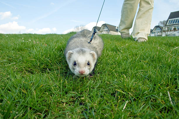
Botulism, a paralytic disease caused by the potent neurotoxins of Clostridium botulinum, is most often associated with livestock, poultry, and human food‑borne outbreaks. Nevertheless, it is an under‑recognized threat to domestic ferrets (Mustela putorius furo), especially those housed in environments where raw‑food diets, scavenging, or exposure to contaminated carcasses occur.
Ferrets are obligate carnivores with a high metabolic rate and a short gastrointestinal transit time, conditions that can favor the germination of C. botulinum spores if they ingest a suitable substrate. Because ferrets are small (typically 0.7–2 kg) and have limited physiological reserves, the onset of botulism can progress rapidly from subtle weakness to fatal respiratory failure.
Understanding the epidemiology, clinical presentation, and evidence‑based management of botulism in ferrets is vital for:
- Prompt recognition and early therapeutic intervention, which dramatically improve survival.
- Designing feeding and husbandry protocols that eliminate exposure.
- Protecting the health of humans who handle ferrets, given the zoonotic potential of the toxin.
2. Etiology & Causes – The Bacterium, Toxin Types, and Sources of Exposure
2.1 Clostridium botulinum – An Overview
Clostridium botulinum is an anaerobic, spore‑forming, gram‑positive bacillus found ubiquitously in soil, marine sediments, and the gastrointestinal tracts of many animals. The organism produces seven serologically distinct neurotoxins (A–G); for mammals, types A, B, E, and rarely F are clinically relevant.
- Type A – Most potent; associated with severe, rapid‑onset disease.
- Type B – Slightly less potent; common in wildlife and domestic animal cases.
- Type E – Linked to fish and marine environments; less common in ferrets.
- Type F – Rare, occasionally seen in exotic pet outbreaks.
2.2 Routes of Exposure in Ferrets
| Source | Typical Scenario | Likelihood in Ferrets |
|---|---|---|
| Contaminated raw meat/fish | Feeding raw chicken, turkey necks, or fish entrails | High when owners practice “raw‑food” or “whole prey” diets |
| Spoiled commercial pet food | Canned or kibble stored > 6 months, opened containers left at ambient temperature | Moderate; toxins can survive cooking |
| Scavenged carrion | Outdoor ferrets that hunt or chew on dead rodents, birds, or reptile carcasses | High in free‑range or semi‑wild settings |
| Environmental spores | Soil dust inhalation, contaminated bedding, or water | Low to moderate; requires anaerobic conditions for germination |
| Iatrogenic contamination | Use of contaminated syringes, feeding tubes, or improperly sterilized equipment | Rare but possible in veterinary clinics or breeding facilities |
2.3 Predisposing Factors
- High‑protein, low‑carbohydrate diets that are under‑cooked or left unrefrigerated.
- Gut dysbiosis (e.g., after broad‑spectrum antibiotics) that reduces competing flora.
- Immunosuppression (e.g., ferrets receiving corticosteroids).
- Environments with limited ventilation (e.g., sealed crates or hutch boxes) that favor anaerobic spore germination.
3. Pathophysiology – How Botulinum Toxin Disrupts Neuromuscular Transmission
The botulinum neurotoxin (BoNT) is a protein complex comprised of a heavy chain (binding & translocation) and a light chain (zinc‑dependent endopeptidase). After ingestion, the toxin traverses the intestinal epithelium, enters the bloodstream, and binds with high affinity to presynaptic cholinergic nerve terminals at the neuromuscular junction (NMJ).
- Binding: The heavy chain attaches to synaptic vesicle protein 2 (SV2) receptors on motor neurons.
- Internalization: The toxin-receptor complex undergoes clathrin‑mediated endocytosis.
- Translocation: Acidic vesicular pH triggers conformational changes, allowing the light chain to cross into the cytosol.
- Proteolysis: The light chain cleaves specific SNARE proteins (e.g., SNAP‑25, synaptobrevin, or syntaxin) depending on the toxin type. This halts vesicle fusion, preventing acetylcholine (ACh) release.
The result is flaccid paralysis because the muscle fibers receive no excitatory signal. The effect is dose‑dependent and irreversible at the affected synapse; recovery requires the growth of new nerve terminals, a process that can take weeks.
In ferrets, the short neuromuscular junction distance and rapid respiration make even a modest toxin load clinically significant, especially in the diaphragm and intercostal muscles.
4. Clinical Signs & Symptoms – Recognizing Botulism in Ferrets
Botulism in ferrets follows a characteristic, progressive timeline that can be divided into three phases: prodromal, paralytic, and terminal. Early detection during the prodromal stage dramatically improves therapeutic success.
| Phase | Time Frame (post‑exposure) | Typical Signs |
|---|---|---|
| Prodromal | 12 – 48 h | Lethargy, decreased appetite, mild weakness, reluctance to climb or jump. |
| Early Paralytic | 24 – 72 h | Diffuse flaccid weakness, drooping ears, reduced tail tone, staggering gait, inability to maintain balance. |
| Advanced Paralytic | 48 – 96 h | Head bobbing, ptosis, facial muscle laxity, loss of gag reflex, dysphagia, urinary retention, constipation. |
| Respiratory Failure | 72 – 120 h | Labored breathing, shallow thoracic excursions, cyanosis, apnea. |
| Terminal | > 120 h | Complete paralysis, absent reflexes, death if not ventilated. |
4.1 Key Physical Findings
- Neurological: Absence of proprioceptive reflexes (e.g., righting reflex), diminished menace response, normal cranial nerve function initially (except for facial weakness later).
- Muscular: Flaccid tone without pain; no rigidity or spasticity.
- Cardiovascular: Normal heart rate but possible bradycardia secondary to hypoxia.
- Gastrointestinal: Constipation, abdominal distension due to reduced motility.
4.2 Differential Diagnosis
- Myasthenia gravis (autoimmune) – Similar neuromuscular weakness but often has a chronic course and can be differentiated by edrophonium testing.
- Tick paralysis – Seasonal, rapid onset; tick removal resolves symptoms.
- Toxoplasmosis, rabies, or distemper – Accompanied by fever, CNS signs, and other systemic signs.
- Hematologic or metabolic disorders (e.g., hypoglycemia) – Usually present with different laboratory abnormalities.
5. Diagnostic Approach – From History to Laboratory Confirmation
A systematic diagnostic protocol maximizes the chance of early, accurate identification of botulism.
5.1 History & Exposure Assessment
- Dietary review: Raw or undercooked meat, fish, or “prey” items; recent changes in food source; duration of storage.
- Environmental audit: Presence of dead wildlife, contaminated bedding, or recent excavation work exposing soil spores.
- Medication record: Recent antibiotics, anti‑inflammatories, or immunosuppressants.
5.2 Physical Examination
- Document neurological deficits (graded on a 0‑5 scale for each limb).
- Observe respiratory mechanics (rate, effort, thoracic excursion).
- Evaluate gastrointestinal motility (palpation for gas, fecal output).
5.3 Laboratory Tests
| Test | Purpose | Typical Findings in Botulism |
|---|---|---|
| Complete Blood Count (CBC) | Detect secondary infection or anemia | Usually normal; mild leukocytosis may appear late. |
| Serum Chemistry | Assess organ function | May show mild azotemia secondary to dehydration. |
| Coagulation Profile | Rule out DIC | Typically normal. |
| Botulinum Toxin Assay (Mouse Bioassay or Endopep‑MS) | Gold‑standard confirmation of toxin in serum, feces, or gastric contents | Positive for type A/B/E (most common). |
| Clostridium spp. Culture (Anaerobic) | Identify bacterial presence | Growth of C. botulinum from GI contents confirms source but may be hampered by prior antibiotics. |
| Electromyography (EMG) | Detect reduced motor unit potentials | Decreased amplitude and recruitment, consistent with presynaptic block. |
| Edrophonium (Tensilon) Test | Differentiate from myasthenia gravis | No improvement in botulism; transient improvement in MG. |
Note: The mouse bioassay, while highly sensitive, requires a specialized laboratory and can take 24–48 h. In practice, treatment is often initiated based on clinical suspicion alone, especially if the animal is deteriorating.
5.4 Imaging
- Thoracic radiographs – Rule out pulmonary infiltrates; may show a normal lung field despite severe hypoxia.
- Abdominal ultrasound – Occasionally reveals gas‑producing organisms in the intestines, but not specific.
6. Therapeutic Management – Antitoxin, Supportive Care, and Specific Interventions
Effective therapy for botulism in ferrets is multimodal, targeting toxin neutralization, symptomatic support, and prevention of secondary complications.
6.1 Antitoxin Administration
- Equine‑derived Botulism Antitoxin (BAT) – The only toxin‑neutralizing product currently licensed for veterinary use (off‑label in ferrets).
- Dose: 0.5–1 IU/kg IV; repeat dosing may be required if clinical signs progress.
- Timing: Ideally within 12 h of onset; efficacy diminishes as toxin binds irreversibly to nerve terminals.
- Adverse Effects: Serum sickness, hypersensitivity; pre‑medicate with antihistamines (e.g., diphenhydramine 1 mg/kg SC) and corticosteroids (e.g., dexamethasone 0.2 mg/kg IV) if signs of anaphylaxis appear.
- Human Botulism Antitoxin (HBAT) – Occasionally used under compassionate‑use protocols when equine BAT is unavailable. Dose adjustments are extrapolated from adult human dosing (0.5 IU/kg).
6.2 Supportive Care
| Component | Details |
|---|---|
| Fluid Therapy | Crystalloid bolus 20 ml/kg IV over 15 min, followed by maintenance (40–60 ml/kg/day) to correct dehydration and maintain perfusion. |
| Nutritional Support | Early enteral feeding via a gastrostomy tube or, if the esophagus is functional, a nasogastric tube. Use highly digestible, low‑fat formulas (e.g., commercial ferret critical care diet). |
| Respiratory Support | Oxygen supplementation (100 % FiO₂ via mask). For progressive respiratory compromise, mechanical ventilation (volume‑controlled) with low tidal volumes (6–8 ml/kg) and PEEP 3–5 cm H₂O. Intubation using a 2.0–2.5 mm cuffed endotracheal tube. |
| Analgesia & Sedation | Minimal sedation to avoid further respiratory depression. Low‑dose butorphanol (0.1–0.2 mg/kg IM) for comfort; midazolam (0.1 mg/kg IV) for anxiety while preserving respiratory drive. |
| Antibiotic Therapy | Broad‑spectrum coverage against secondary bacterial infection (e.g., ampicillin‑sulbactam 30 mg/kg IV q8h). Avoid agents that affect gut flora dramatically unless warranted. |
| Antiemetics | Maropitant (1 mg/kg SC) to reduce the risk of aspiration. |
| Prokinetics | Metoclopramide (0.2 mg/kg SC q8h) if gastric motility is reduced. |
| Physical Therapy | Passive range‑of‑motion exercises to prevent joint contracture during prolonged recumbency. |
6.3 Monitoring & Re‑assessment
- Neurological scoring every 4 h to gauge improvement or progression.
- Arterial blood gases (if ventilated) to maintain PaO₂ > 80 mmHg and PaCO₂ < 45 mmHg.
- Serum electrolytes (especially potassium) every 12 h; replace as needed.
- Urine output > 1 ml/kg/h indicates adequate perfusion.
6.4 Duration of Care
Most ferrets that survive the acute phase begin neuromuscular recovery within 5–10 days, with full return to baseline after 2–4 weeks. Some may retain residual weakness or dysphagia for up to 6 weeks; continued physiotherapy and dietary modification are recommended.
7. Prognosis & Potential Complications – Expected Outcomes and Long‑Term Concerns
7.1 Survival Statistics
- Untreated cases – Mortality > 90 % (rapid progression to respiratory failure).
- Treated with antitoxin + intensive care – Survival rates rise to 45 %–70 %, depending on time to treatment and severity at presentation.
7.2 Factors Influencing Prognosis
| Positive Prognostic Indicator | Negative Prognostic Indicator |
|---|---|
| Early antitoxin administration (< 12 h) | Advanced respiratory paralysis requiring prolonged ventilation |
| Mild to moderate weakness (neurological score ≥ 3/5) | Metabolic acidosis, severe hypoxia, or multi‑organ dysfunction |
| No concurrent systemic disease | Chronic gastrointestinal stasis or severe secondary infection |
| Young age (< 2 years) | Advanced age (> 5 years) or immunocompromised status |
7.3 Common Complications
- Ventilator‑Associated Pneumonia (VAP) – Requires culture‑directed antibiotics.
- Pressure Ulcers – Prevented by frequent repositioning and soft bedding.
- Aspiration Pneumonitis – Minimized by proper tube placement and anti‑emetics.
- Secondary GI Dysmotility – May necessitate prokinetic drugs and fiber‑enriched feeds.
- Neuromuscular Residual Deficits – Persistent weakness of the tail, hindlimbs, or facial muscles; physiotherapy can improve outcomes.
8. Prevention Strategies – Practical Steps to Keep Ferrets Safe
8.1 Dietary Management
- Avoid Raw/Undercooked Meat – If a raw diet is chosen, freeze at –20 °C for ≥ 3 weeks to inactivate spores, then cook to an internal temperature of 74 °C before feeding.
- Use Commercially Processed Foods – Choose reputable brands with proven shelf‑life and storage guidelines.
- Discard Leftovers Promptly – Remove uneaten food within 30 min to prevent bacterial overgrowth.
- Separate Feeding Areas – Prevent cross‑contamination from other pets or wildlife.
8.2 Environmental Hygiene
- Clean, dry bedding – Replace weekly; use low‑dust, absorbent materials (e.g., paper‑based bedding).
- Ventilation – Ensure cages have adequate airflow; avoid sealed environments where anaerobic conditions can develop.
- Rodent Control – Employ traps or safe poisons to limit ferret access to dead rodents.
8.3 Biosecurity in Breeding & Veterinary Settings
- Sterilize feeding equipment (bowls, syringes) using autoclave or high‑temperature dishwasher cycles.
- Hand hygiene – Wash hands with soap and water before handling each ferret.
- Quarantine new animals for at least 14 days, monitoring for neurological signs.
8.4 Vaccination & Prophylaxis
- No commercial botulism vaccine is approved for ferrets. In high‑risk breeding facilities, a custom‑made toxoid vaccine (off‑label use) may be considered under veterinary supervision, but efficacy data are limited.
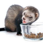
9. Diet & Nutrition – Feeding Practices That Reduce Risk and Aid Recovery
9.1 Baseline Nutritional Requirements
Ferrets require high‑protein (≥ 35 % of calories), high‑fat (≥ 20 % of calories), and low‑carbohydrate diets to mimic their obligate carnivore physiology. Adequate taurine (≥ 250 mg/kg) is essential for cardiac health.
9.2 Safe Food Options
| Food Category | Recommended Items | Reasoning |
|---|---|---|
| Commercial kibble | Premium ferret-specific kibble (e.g., FerretOne, Mazuri). | Formulated with balanced nutrients; low risk of C. botulinum contamination. |
| Cooked meat | Boiled chicken breast, turkey thigh, lean beef – cooked to 74 °C. | Eliminates spores; easy to digest. |
| Eggs | Hard‑boiled, scrambled (no added butter). | High‑quality protein and choline; low risk of toxin. |
| Limited treats | Freeze‑dried insects (e.g., crickets) sourced from reputable suppliers. | Low bacterial load; provide enrichment. |
9.3 Nutritional Support During Recovery
- Enteral formulas – High‑calorie, semi‑liquid diets such as Esbilac or Purina Pro Plan Veterinary Diets (ferret critical care).
- Supplemental electrolytes – Add 0.5 % sodium chloride solution to fluids if hyponatremia develops.
- Probiotics – Enterococcus faecium or Lactobacillus spp. cultures (e.g., Proviable‑DF) can help restore gut flora after antibiotic therapy.
9.4 Feeding Frequency
- Initial phase (first 48 h): Offer small, frequent meals (every 2–3 h) to prevent gastric overload and aid motility.
- Rehabilitation phase: Gradually increase portion size and return to 2–3 meals per day as strength improves.
10. Zoonotic Risk – Human Health Considerations and Occupational Safety
Botulinum toxin is highly potent in humans; however, ferret‑to‑human transmission is rare because the disease is primarily toxin‑mediated rather than bacterial. The principal zoonotic concerns are:
- Handling contaminated material – Contact with spores or toxin in raw meat, feces, or vomitus may lead to accidental ingestion or mucosal exposure.
- Laboratory exposure – Personnel performing mouse bioassays or culturing C. botulinum must follow biosafety level 2 (BSL‑2) practices, wearing gloves, lab coats, and eye protection.
- Veterinary staff – Use of antitoxin can provoke hypersensitivity reactions; staff should be trained to manage anaphylaxis.
Preventive measures for humans:
- Wear disposable gloves when cleaning cages, handling food, or disposing of waste.
- Wash hands thoroughly with soap and water after any ferret contact.
- Avoid eating or drinking while handling raw meat or cleaning equipment.
- Educate owners about the risks of feeding raw diets without proper preparation.
If a human develops botulism symptoms after exposure to a ferret (rare), immediate medical evaluation is required. Human antitoxin (HBAT) is the treatment of choice, administered under hospital supervision.
11. Key Take‑aways – Quick Reference for Clinicians
| Topic | Essential Point |
|---|---|
| Etiology | C. botulinum spores → toxin (A, B, E) → flaccid paralysis. |
| Risk Factors | Raw or improperly stored meat, scavenging, gut dysbiosis, anaerobic environment. |
| Clinical Signs | Progressive weakness → cranial nerve deficits → respiratory failure. |
| Diagnosis | History + neuro exam; confirm with mouse bioassay or Endopep‑MS; EMG supportive. |
| Treatment | Prompt antitoxin (0.5–1 IU/kg IV) + intensive supportive care (fluids, ventilation, nutrition). |
| Prognosis | Survival 45–70 % with early antitoxin; mortality > 90 % untreated. |
| Prevention | Cooked diets, proper storage, clean bedding, avoid scavenging, hygiene. |
| Zoonosis | Low but possible via toxin exposure; practice strict hand hygiene and PPE. |
| Recovery | Neuromuscular function returns in 2–4 weeks; monitor for residual deficits. |
#FerretBotulism, #BotulismInFerrets, #FerretHealth, #ExoticPetCare, #VeterinaryMedicine, #PetSafety, #FerretNutrition, #RawFoodRisks, #AnimalToxin, #VetEducation, #ZoonosisAwareness, #BotulinumToxin, #FerretRescue, #PetFirstAid, #FerretWellness, #VeterinaryScience


Add comment