
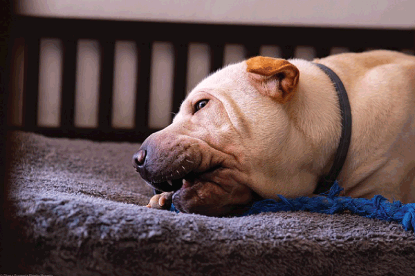
The eyes are windows to the soul, and for our canine companions, they are vital tools for navigating their world, communicating, and expressing affection. When a dog’s eye health is compromised, it can be distressing for both the animal and its guardian. One particularly alarming condition that can affect a dog’s eye is buphthalmos – the irreversible enlargement of the eyeball. Far from being a mere cosmetic concern, buphthalmos is almost always a serious sign of underlying, often painful, and vision-threatening disease, most commonly advanced glaucoma.
This comprehensive guide will delve deep into buphthalmos in dogs, providing an elaborate understanding of its causes, the signs and symptoms to watch for, the diagnostic process, available treatments, the prognosis and potential complications, preventative measures, and even considerations for diet and nutrition. Our aim is to equip dog owners with the knowledge necessary for early detection, proactive management, and ultimately, ensuring the best possible quality of life for their beloved pets.
Understanding the Canine Eye: A Brief Anatomical Overview
Before we dive into buphthalmos, a brief review of the canine eye’s structure and function is helpful. The eye is a complex organ designed to detect light and transmit visual information to the brain. Its shape and internal pressure (intraocular pressure or IOP) are maintained by a delicate balance of aqueous humor production and drainage.
The aqueous humor is a clear, watery fluid produced by the ciliary body (a ring of tissue behind the iris). It fills the anterior and posterior chambers of the eye, nourishing the lens and cornea. This fluid continuously flows through the pupil and drains out of the eye primarily through a meshwork of tissues located in the iridocorneal angle (often referred to as the drainage angle), into the bloodstream. This constant turnover of aqueous humor is vital for maintaining a healthy IOP, which is typically between 10-25 mmHg in dogs.
What is Buphthalmos? The Pathophysiology of an Enlarged Eye
Buphthalmos is defined as the pathological, irreversible enlargement of the entire eyeball, resulting from chronically elevated intraocular pressure. It is crucial to distinguish buphthalmos from exophthalmos, which is the protrusion of a normal-sized eyeball from the socket, typically caused by a mass or inflammation behind the eye pushing it forward. In buphthalmos, the eye itself is structurally altered and physically larger.
The mechanism behind buphthalmos involves the stretching and thinning of the ocular tunics – specifically the sclera (the fibrous, white outer layer of the eye) and the cornea (the transparent front part that covers the iris and pupil). When IOP remains abnormally high for an extended period, the constant internal pressure acts on these external layers. In younger dogs or those with more elastic ocular tissues, the sclera and cornea gradually distend and stretch, leading to a visibly larger globe. As the tissues stretch, they also thin, becoming more fragile.
This enlargement is not merely a cosmetic change; it signifies profound and often irreversible damage to the internal structures of the eye, most notably the optic nerve. The optic nerve, responsible for transmitting visual information from the retina to the brain, is highly sensitive to pressure. Chronic compression from elevated IOP leads to its progressive degeneration and atrophy, resulting in irreversible blindness. Moreover, the stretching of the globe can disrupt other internal structures such as the ciliary body (affecting aqueous production), the zonular fibers (which hold the lens in place), and the retina itself.
Once an eye becomes buphthalmic, the structural changes are permanent. Even if the IOP is surgically or medically brought under control, the eye will remain enlarged, and the damage to the optic nerve and retina will persist. Therefore, buphthalmos itself is a clinical sign of severe, chronic, end-stage ocular disease, almost exclusively chronic glaucoma. Its presence indicates that significant time has passed with uncontrolled high IOP, making a visual prognosis for that eye extremely poor.
Why Are Dogs Prone to Glaucoma and, Consequently, Buphthalmos?
Dogs are particularly susceptible to glaucoma compared to many other species, largely due to a genetic predisposition in numerous breeds. The anatomy of the canine iridocorneal angle, specifically the development of the pectinate ligament, can make them more vulnerable to drainage abnormalities. This unique susceptibility means that glaucoma is a relatively common and devastating eye disease in dogs, with buphthalmos being its visible, chronic manifestation.
Causes of Buphthalmos: Unraveling the Root of the Problem
The vast majority of cases of buphthalmos are a direct consequence of long-standing, uncontrolled glaucoma. Glaucoma is not a single disease but rather a group of eye conditions characterized by progressive damage to the optic nerve due to abnormally high intraocular pressure. This elevated pressure occurs when there’s an imbalance between the production and drainage of aqueous humor, with drainage being the most common issue.
Glaucoma can be broadly categorized into two main types: primary and secondary.
1. Primary Glaucoma
Primary glaucoma arises from inherited anatomical or functional abnormalities in the eye’s drainage system (the iridocorneal angle), without any other identifiable underlying eye disease. This means the eye itself is otherwise healthy, but its genetic predisposition leads to a clogged, malformed, or dysfunctional drainage pathway. Primary glaucoma is almost always bilateral, meaning it affects both eyes, though often not simultaneously. One eye may develop symptoms months or even years before the other, making prophylactic treatment of the “normal” eye crucial.
- Primary Open-Angle Glaucoma (POAG): In this form, the drainage angle appears structurally normal upon examination (gonioscopy), but there’s a microscopic dysfunction in the trabecular meshwork (the network of tissues within the angle that filters aqueous humor). This leads to a gradual, insidious increase in IOP. It tends to progress slowly, often with subtle signs initially, making buphthalmos a later, more dramatic presentation. Beagles, Basset Hounds, and Norwegian Elkhounds are commonly affected.
- Primary Angle-Closure Glaucoma (PACG): This is the most common and aggressive form of primary glaucoma in dogs. It involves a physically narrowed or closed drainage angle, which can suddenly block aqueous humor outflow, leading to acute, painful, and severe spikes in IOP. This is often due to an inherited condition called goniodysgenesis, where the structures of the drainage angle are abnormally developed, predisposed to collapsing or having broad, imperforate sheets of tissue (pectinate ligament dysplasia) that impede drainage. This form often leads to rapid vision loss and is the type most commonly associated with buphthalmos due to chronic, recurrent angle closure.
Breeds Predisposed to Primary Glaucoma: Many breeds have a strong genetic predisposition to primary glaucoma, highlighting the importance of genetic screening and early vigilance in these dogs. Some common breeds include:
- American Cocker Spaniels and English Cocker Spaniels: Highly predisposed to PACG.
- Basset Hounds: Known for POAG and PACG.
- Beagles: Often affected by POAG.
- Siberian Huskies: Susceptible to PACG.
- Samoyeds: Prone to PACG.
- Norwegian Elkhounds: Known for POAG.
- Chow Chows: High incidence of PACG.
- Shih Tzus: Often affected by PACG.
- Boston Terriers: Predisposed to PACG.
- Great Danes: Can develop PACG.
- Dachshunds: Susceptible to PACG.
- Border Collies: Some lines are predisposed.
- Flat-Coated Retrievers: Prone to PACG.
- Poodles (Standard and Miniature): Can also be affected.
The insidious nature of POAG and the acute, recurrent nature of PACG both contribute to chronic high IOP that, if left untreated or poorly managed, will inevitably lead to buphthalmos in susceptible individuals.
2. Secondary Glaucoma
Secondary glaucoma develops as a consequence of another pre-existing eye disease, injury, or systemic condition that obstructs or damages the drainage angle. The underlying condition causes inflammation, physical blockage, or structural changes that impede aqueous humor outflow. Left untreated, these conditions can lead to chronic elevation of IOP and eventually, buphthalmos.
Common causes of secondary glaucoma include:
- Uveitis (Inflammation of the Uvea): This is one of the most common causes of secondary glaucoma. The uvea consists of the iris, ciliary body, and choroid. Inflammation inside the eye can cause inflammatory cells, fibrin, and protein to block the drainage angle, dramatically reducing outflow. Additionally, chronic uveitis can lead to the formation of adhesions (synechiae) between the iris and the lens (posterior synechiae) or the iris and the cornea (anterior synechiae), physically closing the angle. Uveitis can be caused by various factors, including infections (bacterial, viral, fungal, parasitic), immune-mediated diseases (e.g., Vogt-Koyanagi-Harada-like syndrome), trauma, or certain systemic illnesses (e.g., tumors elsewhere in the body, ehrlichiosis). Chronic, recurrent uveitis is a significant risk factor for buphthalmos.
- Lens Luxation or Subluxation: This occurs when the lens of the eye shifts from its normal position due to weakened or broken zonular fibers (the tiny ligaments that hold the lens in place).
- Anterior Luxation: If the lens moves forward into the anterior chamber, it can physically block the pupil and the drainage angle, causing a sudden and severe increase in IOP, rapidly leading to glaucoma.
- Posterior Luxation: If the lens moves backward into the vitreous, it can still cause inflammation (uveitis) leading to secondary glaucoma, or its presence can disrupt vitreous dynamics. Lens luxation can be hereditary (e.g., in Terriers like Jack Russell, Miniature Bull Terriers, and Border Collies) or secondary to trauma or advanced glaucoma itself (where the stretching of the globe damages zonules).
- Intraocular Tumors: Masses or growths within the eye (e.g., melanoma, adenocarcinoma, lymphosarcoma) can obstruct the drainage angle, infiltrate the ciliary body (interfering with aqueous humor production and drainage), or cause inflammation, all leading to glaucoma. As these tumors grow, they exert pressure and disrupt normal ocular function.
- Intraocular Hemorrhage (Hyphema): Bleeding inside the eye (e.g., due to trauma, systemic bleeding disorders, hypertension, severe inflammation) can lead to red blood cells, fibrin, and inflammatory debris clogging the delicate drainage pathways of the trabecular meshwork, causing a physical blockage and increased IOP.
- Trauma: Direct blunt or penetrating injury to the eye can cause immediate inflammation, bleeding, or direct structural damage to the drainage angle, leading to acute or chronic glaucoma. Even seemingly minor trauma can initiate a cascade of events.
- Retinal Detachment: While less common, severe or chronic retinal detachment can sometimes lead to secondary uveitis and subsequent glaucoma due to the release of inflammatory mediators.
- Advanced Intraocular Cysts or Granulomas: Large cysts or granulomas (e.g., secondary to fungal infections) within the eye can physically block drainage structures or cause chronic inflammation, leading to elevated IOP.
3. Other Rare Causes (Non-Glaucomatous)
While glaucoma is overwhelmingly the primary driver of buphthalmos, it’s worth noting that very rarely, other conditions might contribute to eye enlargement, though they often present differently or are less common:
- Congenital Hydrophthalmos: This is a rare congenital anomaly where the eye is abnormally large at birth or shortly after due to developmental defects in the aqueous outflow system; it is essentially a form of congenital primary glaucoma. The enlarged eye is present from a very young age.
- Orbital Masses/Cellulitis (Indirect Pressure): While typically causing exophthalmos (protrusion of a normal-sized eye), in extremely chronic and severe cases, a massive orbital tumor or diffuse, severe orbital cellulitis (infection/inflammation behind the eye) could theoretically exert enough sustained anterior pressure to contribute to some globe distension, particularly if it also interferes with venous drainage from the eye, indirectly increasing IOP. However, this is an atypical and very complex scenario, not a direct cause of buphthalmos in the same way glaucoma is.
Understanding the underlying cause is crucial because it dictates the specific diagnostic and treatment strategies. However, by the time buphthalmos is evident, the damage is often extensive, irreversible, and the visual prognosis for that eye is poor, irrespective of the initial cause.
Signs and Symptoms: Recognizing the Alarm Bells
Early detection of glaucoma is paramount for preserving vision, as buphthalmos represents a chronic, advanced, and often end-stage manifestation. Dog owners must be vigilant for subtle changes in their dog’s eyes and behavior, as waiting until the eye is visibly enlarged often means irreversible blindness has already occurred. The signs can vary in intensity depending on the chronicity and severity of the elevated IOP.
1. Early Signs of Glaucoma (Before Buphthalmos is Apparent):
These signs indicate an active glaucoma crisis or chronic elevation of IOP and warrant immediate veterinary attention, even if the eye hasn’t visibly enlarged yet. This is the critical window for potential vision preservation.
- Ocular Pain: This is often the first and most prominent sign of acutely elevated IOP. Dogs are incredibly stoic, but pain in the eye can be severe and manifest as:
- Blepharospasm: Squinting or holding the affected eye partially or completely closed due to discomfort.
- Rubbing the Eye/Face: Pawing at the eye, rubbing the face on furniture, the floor, or with paws.
- Head Pressing: Pressing their head against objects or into corners, a non-specific sign of headache or generalized pain.
- Lethargy or Depression: A general decrease in activity, enthusiasm, and interaction.
- Loss of Appetite: Reluctance to eat due to discomfort or systemic malaise from pain.
- Avoidance of Touch: Shying away or reacting negatively when touched near the head or eye.
- Unusual Aggression or Withdrawal: Behavioral changes due to chronic discomfort.
- Redness of the Eye:
- Conjunctival Hyperemia: Redness and congestion of the conjunctiva, the pink membranes lining the eyelids and covering the white part of the eye.
- Episcleral Injection: Deep, engorged, purple-red blood vessels visible in the sclera, often radiating away from the limbus (the junction of the cornea and sclera). Unlike superficial conjunctival vessels, these vessels do not blanch (whiten) with topical phenylephrine, making them a strong indicator of increased IOP.
- Corneal Edema (Cloudiness): The normally clear, transparent cornea may develop a bluish haze, cloudy, or “steamy” appearance. This occurs when the high pressure interferes with the normal function of the corneal endothelial pump (which actively removes fluid from the cornea), allowing fluid to accumulate within the corneal layers, leading to opacification. This can range from a subtle haziness to a dense, opaque blue eye.
- Pupil Dilation (Mydriasis): The pupil of the affected eye may appear noticeably larger (more dilated) than the unaffected eye and may be sluggish or completely unresponsive to light. This is due to pressure damage to the optic nerve (affecting the afferent visual pathway) and/or direct compression of the nerves supplying the iris muscles.
- Vision Loss: Owners might notice:
- Bumping into objects, especially in dim light or unfamiliar surroundings.
- Hesitation to jump onto furniture or go down stairs.
- Difficulty locating toys, treats, or food bowls.
- Changes in gait, confidence, or an increased reliance on scent and sound. Vision loss can be rapid and complete in acute glaucoma (within hours to days).
- Epiphora (Excessive Tearing) or Ocular Discharge: The eye may water excessively as a pain response, and sometimes a clear to mucoid discharge may be present.
- Prominence of the Third Eyelid (Nictitating Membrane): The third eyelid may be more visible, potentially indicating ocular pain and retraction of the globe.
2. Advanced Signs (When Buphthalmos is Present):
By this stage, the disease is chronic, and the eye has undergone significant, irreversible remodeling due to sustained high pressure. Vision is typically already lost.
- Visible Enlargement of the Eyeball: This is the defining characteristic of buphthalmos. The affected eye will appear noticeably and often dramatically larger or more prominent than the unaffected eye (or the other eye if both are affected). Owners might notice the eyelids struggling to close fully over the enlarged globe. This can be subtle at first but becomes progressively obvious as the condition advances.
- Prominence of the Third Eyelid (Nictitating Membrane): The enlarged globe often pushes the third eyelid forward, making it permanently visible and sometimes inflamed.
- Scleral Thinning and Pigmentation: The white sclera may appear thinner and more fragile, sometimes allowing the underlying darker uveal tissue to show through, giving it a bluish or greyish tint. In some cases, the chronically inflamed and distended sclera may develop pigmentary changes (melanosis).
- Corneal Striae (Stretch Marks) or Haab’s Striae: Fine, white, linear lines may be visible within the cornea, particularly in the deeper layers. These are indicative of stretching, tearing, or damage to the corneal collagen fibers and Descemet’s membrane due to chronic distension.
- Fixed and Dilated Pupil: The pupil will likely be fully dilated (mydriatic) and completely unresponsive to light, indicating severe or complete optic nerve damage, retinal atrophy, and irreversible blindness.
- Absent Menace Response or Other Visual Reflexes: The dog will not blink in response to a sudden threatening gesture (menace response), and other visual reflexes (e.g., dazzling reflex) will be absent, confirming functional blindness in the affected eye.
- Lens Subluxation/Luxation: The extreme stretching of the globe can weaken and rupture the zonular fibers that hold the lens in place, causing it to shift from its normal position. The lens might move into the anterior chamber (anterior luxation) or posterior chamber (posterior luxation), further exacerbating glaucoma or causing additional inflammation.
- Phthisis Bulbi: Paradoxically, after a prolonged period of severe buphthalmos and chronic inflammation, some eyes may eventually shrink, become soft, and lose their normal shape (“shrunken eye”). This occurs due to complete destruction of the ciliary body and cessation of aqueous humor production. This is an end-stage sign of a dead, non-functional eye that has exhausted its ability to produce aqueous humor, often providing natural (but late) pain relief.
- Chronic Pain and Discomfort: While the acute, severe pain might subside somewhat as the eye becomes blind and nerve endings are irrevocably damaged, a chronically buphthalmic eye is still often uncomfortable due to persistent low-grade inflammation, exposure keratitis (if the eyelid can’t close fully over the enlarged globe), or recurrent corneal ulceration on the stretched, fragile cornea. The dog may become less active, irritable, or withdrawn due to this lingering discomfort.
Any of these signs, especially the combination of redness, cloudiness, pain, and a dilated pupil, warrant an immediate trip to the veterinarian. Waiting can mean the difference between vision and permanent blindness, or prolonged suffering.
Diagnosis: Pinpointing the Problem
Diagnosing buphthalmos and its underlying cause requires a thorough ophthalmic examination, often performed or confirmed by a veterinary ophthalmologist. The diagnostic process aims to confirm elevated IOP, assess the degree of damage, and identify whether the glaucoma is primary or secondary.
- Comprehensive Ophthalmic Examination:
- Visual Assessment: The vet will first observe the dog’s eyes for symmetry, size, any visible enlargement, redness, discharge, and evidence of pain (squinting, rubbing).
- Neuro-ophthalmic Exam:
- Menace Response: Testing whether the dog blinks or withdraws from a threatening gesture. Absent in blind eyes.
- Pupillary Light Reflexes (PLRs): Assessing if the pupils constrict in response to light. Diminished or absent in eyes with significant optic nerve damage.
- Dazzle Reflex: A subcortical reflex where even a blind eye may show a slight pupil constriction or head aversion to very bright light, assessing residual retinal function.
- Direct and Indirect Ophthalmoscopy: Using an ophthalmoscope, the vet examines the back of the eye (fundus), specifically the retina and the optic nerve head. In glaucoma, damage to the optic nerve head, manifesting as “cupping” (a depression in the optic disc), and retinal atrophy are hallmark signs.
- Slit Lamp Biomicroscopy: A specialized microscope (slit lamp) provides a magnified, stereoscopic view of the anterior segment of the eye (cornea, anterior chamber, iris, lens). This allows for detailed assessment of corneal edema, inflammatory cells or fibrin in the anterior chamber, synechiae (adhesions), iris atrophy, and lens position (luxation/subluxation) or opacity (cataract formation).
- Tonometry (Measurement of Intraocular Pressure):
- This is the cornerstone of glaucoma diagnosis. A tonometer is used to accurately measure the IOP. Normal canine IOP is typically between 10-25 mmHg. Readings consistently above this range, especially with other clinical signs, are definitive indicators of glaucoma. Readings can exceed 50-60 mmHg in acute glaucoma.
- Schiotz Tonometer: An older, less commonly used indentation tonometer that estimates IOP by measuring the depth of corneal indentation caused by a known weight.
- Applanation Tonometer (e.g., Tono-Pen): A more precise and widely used electronic device. After applying a topical anesthetic drop, the pen-like device gently touches the cornea multiple times to get average IOP readings. It’s portable and accurate.
- Rebound Tonometer (e.g., TonoVet): Increasingly popular, this device fires a small, lightweight probe at the cornea and measures its rebound characteristics. It is highly accurate, often requires no topical anesthetic (making it very comfortable for the dog), and is excellent for screening. It’s particularly useful for anxious dogs or for initial screening by general practitioners.
- Gonioscopy:
- This specialized procedure is crucial for classifying the type of glaucoma. It uses a special contact lens (goniolens) placed on the cornea, allowing direct visualization of the iridocorneal drainage angle. It helps the vet assess the anatomy of the drainage angle, determining if it is open, narrowed, or completely closed. It can identify congenital abnormalities like goniodysgenesis (malformation of the drainage angle structures, such as pectinate ligament dysplasia), or acquired blockages (e.g., peripheral anterior synechiae, inflammatory debris, or tumor infiltration). Gonioscopy is essential for differentiating between primary (open-angle vs. angle-closure) and secondary glaucoma and is invaluable for prognosis and treatment planning for the unaffected eye.
- Ocular Ultrasonography:
- When the cornea is too opaque (e.g., due to severe edema, advanced pigmentary keratitis) or the internal structures are obscured (e.g., by hyphema, dense cataracts), ultrasound can provide invaluable information about the structures within the posterior segment of the eye. It can help assess the position of the lens (luxation/subluxation), detect masses, identify retinal detachment, evaluate the optic nerve head, and screen for other intraocular pathologies not visible directly.
- Electroretinography (ERG):
- This advanced diagnostic test measures the electrical activity of the retina in response to light stimuli. It helps assess the functional viability of the retina and the photoreceptor cells. It is primarily used to provide a prognosis for vision, especially before contemplating vision-sparing surgeries. If the retina shows severe non-responsiveness (extinguished ERG), the eye is functionally blind, and vision-sparing surgery may not be warranted.
- Blood Work and Imaging (if secondary glaucoma is suspected):
- If secondary glaucoma is suspected, the vet may recommend systemic diagnostic tests. This can include blood tests (complete blood count, biochemistry panel, infectious disease titers) to look for systemic inflammation, infection, immune-mediated diseases, or underlying metabolic disorders.
- Radiographs, CT (Computed Tomography), or MRI (Magnetic Resonance Imaging) of the head may be considered if an orbital mass, foreign body, severe orbital cellulitis, or intracranial disease is suspected of causing secondary glaucoma.
- Referral to a Veterinary Ophthalmologist:
- Given the complexity and urgency of glaucoma, especially once buphthalmos is developing, referral to a board-certified veterinary ophthalmologist is highly recommended. These specialists possess advanced equipment, expertise, and surgical skills for definitive diagnosis, specialized medical management, and complex surgical interventions.
Treatment: Managing Pain and Preserving What’s Left
The treatment goals for buphthalmos are twofold: first, and most importantly, to alleviate pain and discomfort, and second, to preserve any remaining vision, if possible. Unfortunately, by the time buphthalmos is established, vision is often already lost or severely compromised, making pain management the predominant focus.
A. Emergency Treatment (for Acute Glaucoma, before/early buphthalmos):
If a dog presents with acute, severe glaucoma (very high IOP, intense pain, significant corneal edema) before buphthalmos is fully developed, immediate and aggressive intervention is critical to attempt to save vision in the affected eye and to protect the contralateral eye. This is a true ophthalmic emergency.
- Osmotic Diuretics (e.g., IV Mannitol, Glycerin orally): Administered intravenously (Mannitol) or sometimes orally (Glycerin), these agents rapidly draw fluid out of the eye (and brain), dramatically lowering IOP within minutes to hours. Mannitol is the most common choice for acute, severe pressure spikes. It’s a temporary but crucial measure to buy time.
- Topical Glaucoma Medications: These eye drops are started immediately and frequently both to reduce IOP and manage inflammation.
- Prostaglandin Analogues (e.g., Latanoprost, Travoprost, Bimatoprost): These are highly effective for primary open-angle glaucoma in dogs by increasing uveoscleral outflow. They act quickly (within 30-60 minutes). Crucially, they are often contraindicated in secondary glaucoma caused by uveitis or lens luxation, as they can exacerbate inflammation or pain in those specific contexts.
- Carbonic Anhydrase Inhibitors (CAIs) (e.g., Dorzolamide hydrochloride, Brinzolamide): These medications reduce aqueous humor production by inhibiting the enzyme carbonic anhydrase in the ciliary body. They are effective for both primary and secondary glaucoma and can be used safely with uveitis. They are often applied 2-3 times daily.
- Beta-Blockers (e.g., Timolol maleate): These reduce aqueous humor production by blocking beta-adrenergic receptors. They are less potent than prostaglandin analogues or CAIs for IOP reduction in dogs, and must be used with caution in dogs with underlying heart or respiratory conditions (e.g., bradycardia, asthma, congestive heart failure).
- Topical Steroids (e.g., Prednisolone acetate, Dexamethasone): If secondary glaucoma is due to severe uveitis, topical steroids may be used to control inflammation, but this must be done very carefully and only after ruling out corneal ulceration, as steroids can worsen ulcers.
- Systemic Pain Medication: NSAIDs (e.g., Carprofen, Meloxicam) or opioids (e.g., Tramadol, Buprenorphine, Gabapentin) are administered to manage the intense pain associated with acute glaucoma.
B. Chronic Management (for Buphthalmos):
Once buphthalmos has developed, the eye is often already blind, and the focus shifts significantly. Medical management for vision preservation becomes less likely to succeed, and surgical options for pain relief and quality of life are usually considered.
Medical Management for Chronic Glaucoma/Buphthalmos:
If there’s any hope of preserving some residual vision (which is rare with established buphthalmos), or if the eye is only mildly enlarged and IOP can be controlled effectively and painlessly, lifelong topical medications are prescribed. This requires significant owner commitment for daily administration and frequent recheck examinations.
- Topical CAIs and/or Beta-Blockers: These are typically the mainstay for long-term medical management, especially if prostaglandin analogues are contraindicated. They are often used in combination for better IOP control.
- Prophylactic Treatment for the Contralateral Eye: For primary glaucoma, the unaffected eye will invariably develop glaucoma eventually. Aggressive prophylactic treatment with topical CAIs and/or beta-blockers is crucial to delay the onset of glaucoma in this eye, though it rarely prevents it entirely. Owners must be diligent in treating both eyes.
However, medical management often becomes ineffective once buphthalmos is evident. The chronic structural changes within the eye (e.g., extensive scarring of the drainage angle, retinal detachment, optic nerve atrophy) make IOP control extremely difficult to achieve and maintain long-term using only medications. At this stage, surgical intervention becomes the most humane and effective option for pain relief.
Surgical Treatment Options for Buphthalmic Eyes:
When medical management fails, vision is lost, and the eye remains painful, surgical intervention becomes necessary. These surgeries are generally categorized into vision-sparing and vision-sacrificing procedures. By the time buphthalmos is present, vision-sparing options are rarely viable, as vision is already gone. The primary goal of surgery for buphthalmos is pain relief and improved quality of life for the dog.
1. Vision-Sparing Surgeries (Rarely an Option with Established Buphthalmos): These are typically performed in early-stage glaucoma before buphthalmos and blindness occur, but are mentioned for completeness in glaucoma management.
- Laser Cyclophotocoagulation (Transscleral or Endoscopic): This procedure uses a laser to selectively destroy portions of the ciliary body (the tissue that produces aqueous humor), thereby reducing aqueous humor production and consequently IOP. It can be performed transsclerally (laser applied through the sclera) or endoscopically (laser applied internally using a tiny camera). It can be effective but often requires multiple treatments and carries risks of post-operative inflammation or recurrence. Success rates vary.
- Gonioimplantation (Aqueous Shunts/Valves): A small, biocompatible tube and plate device (e.g., Ahmed Glaucoma Valve) is surgically implanted to create an alternative drainage pathway for aqueous humor from the anterior chamber to a subconjunctival reservoir. This surgery is technically demanding and carries risks of complications like fibrous encapsulation, infection, or hypotony (too low IOP).
2. Vision-Sacrificing Surgeries (Common and Recommended for Buphthalmic, Blind, Painful Eyes): These procedures are chosen when the eye is irreversibly blind, chronically painful, and medical management has failed or is no longer sustainable. The primary goal is definitive pain relief and improved quality of life.
- Enucleation (Surgical Removal of the Eyeball):
- Description: This is the most definitive, reliable, and effective treatment for a chronically painful, blind, buphthalmic eye. The entire eyeball is surgically removed, and the eyelids are permanently sutured closed. The orbit fills with tissue, and over time, the area often becomes less conspicuous.
- Pros: Provides immediate and complete pain relief, eliminates the source of chronic suffering, prevents all future complications (e.g., corneal ulcers, infections on a non-functional eye). Dogs typically adapt very quickly to monocular vision, often showing a dramatic improvement in demeanor and activity levels once the chronic pain is gone.
- Cons: Loss of the eye, cosmetic change (though many owners find a closed, pain-free socket less distressing than a bulging, painful eye). Small risk of surgical complications (e.g., bleeding, infection, nerve damage).
- Intraocular Prosthesis / Evisceration with Intrascleral Prosthesis:
- Description: This procedure aims for a better cosmetic outcome. The internal contents of the buphthalmic eye (lens, vitreous, retina, uveal tissue) are surgically removed, but the sclera (the outer shell of the eye) and the extraocular muscles are carefully preserved. A sterile silicone or acrylic prosthetic sphere is then implanted into the scleral shell to maintain the eye’s shape and size, and the sclera is sutured closed. The eyelids remain intact and can close normally over the prosthesis.
- Pros: Excellent cosmetic result, often indistinguishable from a normal eye at a distance, maintains normal eyelid function and movement. Preserves the aesthetic appearance of the pet.
- Cons: Requires a relatively healthy cornea with no active ulcers or severe edema (as the cornea remains the outermost layer). Still carries risks of complications such as post-operative infection, corneal ulceration, prosthesis extrusion (the body rejecting the implant), or residual inflammation. It can technically still generate some intraocular pressure if residual ciliary body tissue remains, though typically much lower and non-painful. There is a risk of gradual reduction in eye size over time. Not suitable for all cases, especially those with severe corneal disease or intraocular infection/tumors.
- Chemical Ciliary Ablation (Intravitreal Gentamicin Injection):
- Description: A destructive chemical (most commonly gentamicin, sometimes combined with a steroid) is injected directly into the vitreous body of the eye. This drug destroys the ciliary body (the tissue that produces aqueous humor), thereby permanently reducing aqueous humor production. The goal is to lower IOP and alleviate pain.
- Pros: Less invasive than surgical removal, good cosmetic outcome if effective (the eye appears shrunken, but without a surgical incision), can be performed under heavy sedation or light anesthesia.
- Cons: Can be highly unpredictable in efficacy (may require repeated injections). Carries risks of severe complications such as intractable uveitis, rapid and severe phthisis bulbi (leading to an unsightly shrunken eye), corneal ulceration, and retinal detachment. Pain relief is not always immediate or complete, and the eye can remain inflamed for several weeks post-injection. Not suitable for eyes with existing inflammation, infection, or a history of specific systemic diseases. It is considered a less reliable and potentially more complicated option than enucleation for definitive pain relief.
The choice of treatment depends on various factors, including the dog’s overall health, the severity of the buphthalmos, the presence of concurrent eye disease (e.g., tumors, severe infection), the owner’s financial capabilities, and their preference for cosmetic outcome versus definitive pain relief. Ultimately, relieving pain and improving the dog’s quality of life are the paramount considerations. For a chronically painful, blind, buphthalmic eye, enucleation is often the most straightforward and effective method for achieving lasting relief.
Prognosis & Complications: The Long-Term Outlook
The prognosis for buphthalmos is generally guarded to poor for the preservation of vision in the affected eye, but good for pain management and overall quality of life with appropriate treatment.
Prognosis:
- Vision: Once buphthalmos is present, the affected eye is highly likely to be permanently blind. The prolonged high intraocular pressure causes irreversible damage to the optic nerve and atrophy of the retina. Even if IOP can be reduced through aggressive treatment, the structural damage is permanent, and visual function rarely returns. The term “buphthalmos” itself implies irreversible blindness.
- Pain: While the initial acute pain of glaucoma can be excruciating, effective surgical treatment (especially enucleation) can lead to complete and lasting pain relief, significantly improving the dog’s comfort, demeanor, and overall well-being. With evisceration or chemical ablation, pain relief is usually good but can have some residual discomfort or complications.
- Contralateral Eye: If the buphthalmos is due to primary glaucoma, the unaffected “fellow” eye has a very high risk (80-100%) of developing glaucoma within 6-24 months, even if it appears normal at the time of diagnosis of the first eye. Aggressive prophylactic treatment with topical medications (e.g., CAIs, beta-blockers) is crucial to delay its onset and potentially preserve vision for as long as possible. However, it rarely prevents it entirely. Owners must be prepared for the possibility of bilateral blindness.
Complications of Buphthalmos (if left untreated or poorly managed):
Leaving a buphthalmic eye untreated or inadequately managed leads to a cascade of severe complications, significantly diminishing the dog’s quality of life:
- Irreversible Blindness: This is almost a certainty for the buphthalmic eye, as the optic nerve and retina are destroyed.
- Chronic and Severe Pain: The dog will live in constant, debilitating discomfort. This chronic pain directly impacts their behavior, leading to lethargy, withdrawal, irritability, aggression, loss of appetite, and a general disinterest in activities they once enjoyed. This significantly diminishes quality of life.
- **Corneal
#Buphthalmos, #DogEyeHealth, #CanineGlaucoma, #DogEyes, #EnlargedEyeball, #VetOphthalmology, #PetHealthAwareness, #DogVisionCare, #EyeConditionsInDogs, #PetHealthMatters, #DogBlindness, #VeterinaryOphthalmology, #DogEyeProblems, #SaveTheirSight, #BlindDogAwareness, #CanineHealth, #PetWellness, #VetCare, #PetsWithSpecialNeeds, #DogLovers


Add comment