
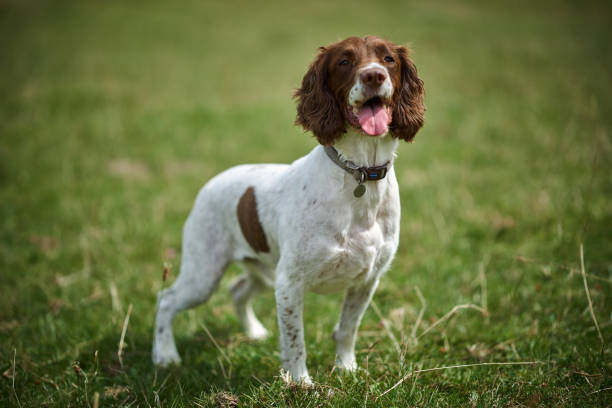
Canine Gallbladder Mucocele (GBM) is a progressively recognized and often life-threatening condition in dogs, characterized by an abnormal accumulation of thick, gelatinous mucus within the gallbladder. This distended, mucus-filled organ can lead to various complications, ranging from chronic inflammation and discomfort to acute gallbladder rupture, bile peritonitis, and even death. Once considered a rare finding, the incidence of GBM has steadily increased, likely due to a combination of improved diagnostic techniques, longer canine lifespans, and a greater understanding of its underlying causes and breed predispositions. This comprehensive guide aims to shed light on every critical aspect of canine gallbladder mucocele, from its obscure origins to its complex diagnosis, intricate treatment, and long-term management strategies.
Understanding the Canine Gallbladder and Mucocele Formation
The gallbladder is a small, hollow organ situated beneath the liver, primarily responsible for storing and concentrating bile produced by the liver. Bile plays a crucial role in the digestion and absorption of fats and fat-soluble vitamins in the small intestine. Under normal circumstances, bile flows freely from the liver through the bile ducts into the gallbladder, where it is concentrated, and then is released into the duodenum (the first part of the small intestine) in response to a meal.
A gallbladder mucocele disrupts this delicate process. It begins with the excessive production of highly viscous mucus by the gallbladder wall’s epithelial cells, coupled with impaired gallbladder motility (contractility). This thick, sticky mucus, often described as having a “kiwi fruit” appearance on ultrasound due to its characteristic stellate or radiating pattern, gradually fills the gallbladder. As the mucocele grows, it can obstruct the cystic duct (the duct connecting the gallbladder to the common bile duct) or even the common bile duct itself, impeding normal bile flow. The accumulation of mucus, bile, and cellular debris leads to progressive distension of the gallbladder, thickening of its wall, and chronic inflammation. In severe cases, the pressure within the distended gallbladder can compromise its blood supply, leading to necrosis (tissue death) and ultimately, rupture.
Causes of Canine Gallbladder Mucocele
The exact pathogenesis of gallbladder mucocele is multifactorial and not fully understood, but it is strongly associated with certain metabolic disorders and breed predispositions. It’s believed to arise from a combination of abnormal mucus production, altered gallbladder motility, and potentially genetic factors.
- Metabolic and Endocrine Disorders: These are frequently implicated as predisposing factors:
- Hyperadrenocorticism (Cushing’s Disease): This condition, characterized by excessive cortisol production, is a significant risk factor. Cortisol can alter bile composition, increase bile viscosity, impair gallbladder motility, and promote mucus secretion. Studies have shown a high co-occurrence of GBM in dogs with Cushing’s.
- Hypothyroidism: Underactive thyroid glands can lead to changes in lipid metabolism, bile flow, and gallbladder function. Hypothyroid dogs often have hyperlipidemia (elevated blood fats) and can develop GBM.
- Diabetes Mellitus: Dogs with diabetes may have altered lipid metabolism and increased susceptibility to inflammation, contributing to mucocele formation. Insulin resistance and hyperglycemia can indirectly affect gallbladder function.
- Hyperlipidemia: Elevated levels of cholesterol and triglycerides in the blood are commonly found in dogs with GBM, even in the absence of other endocrine diseases. While it’s debated whether hyperlipidemia is a cause or an effect of altered bile metabolism, it certainly contributes to the thickened bile and altered gallbladder environment.
- Pancreatitis: Inflammation of the pancreas can lead to local inflammation around the gallbladder, affecting its function and potentially contributing to mucocele formation or exacerbating existing ones.
- Cholesterol and Bile Acid Metabolism Abnormalities: Some dogs may have genetic or acquired defects in how they handle cholesterol and bile acids, leading to more viscous bile and increased mucus production.
- Genetic Predisposition: Certain breeds exhibit a significantly higher incidence, suggesting a strong genetic component. This predisposition might involve specific gene mutations affecting lipid metabolism, bile composition, or gallbladder contractility. For example, some breeds might have genetic variants that make them prone to hyperlipidemia, which then indirectly leads to GBM.
- Dietary Factors: While not a primary cause, diets high in fat or certain types of fats might exacerbate hyperlipidemia or contribute to bile stasis, potentially accelerating mucocele formation in susceptible individuals. However, direct causation has not been definitively established.
- Inflammation and Infection: Chronic inflammation of the gallbladder (cholecystitis) can lead to changes in the gallbladder wall, including hyperplasia of mucus-producing glands, which further contributes to mucocele formation. While bacterial infection (cholangitis/cholecystitis) can occur secondary to bile stasis, it’s generally considered a complication rather than an initial cause of mucocele development.
Signs and Symptoms
The clinical signs of gallbladder mucocele are often non-specific, insidious, and can vary greatly depending on the stage of the disease and whether complications like rupture have occurred. Early-stage mucoceles may be asymptomatic, discovered incidentally during imaging for other conditions.
Non-Specific and Chronic Signs (Often seen in less severe cases or early stages):
- Lethargy: Reduced energy levels and general malaise.
- Anorexia or Hyporexia: Decreased appetite or complete refusal to eat.
- Vomiting: Can be intermittent or persistent, often related to nausea from biliary stasis or pain.
- Diarrhea: Less common but can occur due to digestive upset.
- Abdominal Pain: Often vague and difficult to pinpoint. Dogs may exhibit hunched posture, reluctance to be handled, or general discomfort. This pain is typically more pronounced with inflammation or distension.
- Weight Loss: Chronic illness can lead to gradual weight loss.
Specific and Acute Signs (Often indicative of more severe disease, obstruction, or rupture):
- Jaundice (Icterus): Yellow discoloration of the gums, whites of the eyes (sclera), and skin. This occurs when the mucocele obstructs the common bile duct, preventing bile from flowing into the intestine, leading to a buildup of bilirubin in the bloodstream. This is a critical sign.
- Fever: May indicate inflammation or infection within the gallbladder or secondary to rupture and peritonitis.
- Dehydration: Due to vomiting and reduced water intake.
- Collapse/Weakness: In severe cases, especially if rupture leads to septic shock.
- Severe Abdominal Pain: Intense pain, often localized to the cranial abdomen, if the gallbladder is severely distended, inflamed, or has ruptured. Dogs may cry out, become restless, or resist palpation.
- Peritonitis: If the gallbladder ruptures, bile leaks into the abdominal cavity, causing a severe inflammatory reaction (bile peritonitis), which is a surgical emergency. Signs include extreme pain, distended abdomen, shock, and rapid deterioration.
It’s crucial to note that these symptoms can mimic many other gastrointestinal or liver conditions, making accurate diagnosis challenging without advanced imaging.
Dog Breeds at Risk (with a paragraph explanation)
While any dog can potentially develop a gallbladder mucocele, certain breeds are significantly overrepresented, strongly suggesting a genetic predisposition or a higher incidence of predisposing metabolic conditions within these breeds.
- Shetland Sheepdogs (Shelties): Shelties are one of the most commonly affected breeds, making them a poster child for GBM. Their predisposition is thought to be multifactorial, including a higher incidence of primary hyperlipidemia and potentially a specific genetic mutation affecting bile composition or gallbladder motility. This breed’s genetic background, particularly in lines prone to endocrine disorders, likely contributes to their increased risk, making routine monitoring and early intervention particularly important for Sheltie owners.
- Cocker Spaniels: Both English and American Cocker Spaniels are frequently diagnosed with GBM. Similar to Shelties, Cocker Spaniels often exhibit a propensity for hyperlipidemia, hypothyroidism, and other metabolic syndromes that can influence bile viscosity and gallbladder function. Their genetics likely play a role in their increased susceptibility to these underlying conditions, subsequently raising their risk for developing mucoceles, often requiring careful dietary management and screening.
- Miniature Schnauzers: Miniature Schnauzers are well-known for their genetic predisposition to hyperlipidemia (high triglycerides and cholesterol), a condition that is a significant risk factor for GBM. This breed’s unique lipid metabolism makes them prone to developing excessive amounts of fat in the blood, which can alter bile composition and lead to the formation of thick, sludgy bile and ultimately mucoceles. Regular lipid panel checks are advisable for this breed.
- Pomeranians: Pomeranians also appear on the list of at-risk breeds, though the specific mechanisms are less well-defined compared to Shelties or Schnauzers. Their genetic background might predispose them to subtle metabolic imbalances or altered gallbladder function, which, when combined with other factors, can lead to mucocele development. Owners of Pomeranians should be vigilant for any gastrointestinal signs.
- Chihuahuas: Chihuahuas, despite their small size, are another breed that shows an increased incidence of GBM. The reasons for their susceptibility are still under investigation, but it’s possible they share some genetic predispositions to metabolic disorders or exhibit unique gallbladder physiology that makes them more prone to mucus accumulation. Given their small stature, diagnosis can sometimes be delayed as symptoms might be subtle.
- Beagles: Beagles, often used in research due to their robust genetics, are also found to have a higher risk of developing gallbladder mucoceles. This breed, known for its hearty appetite, may be predisposed to certain metabolic conditions, including hyperlipidemia, which can contribute to the formation of mucoceles. Regular veterinary check-ups and attention to diet are important for Beagles.
- Shetland Sheepdog/Collie Crosses: Given the high incidence in Shelties, it’s not surprising that crosses involving these breeds may also carry an increased genetic risk for GBM. The genetic material from the Sheltie parent can pass on the predisposition to hyperlipidemia or other factors contributing to mucocele formation, highlighting the importance of thorough breed history in mixed-breed dogs.
Other breeds, such as American Eskimo Dogs, Pekingese, and some Terrier breeds, have also been reported with higher frequencies, suggesting that a broader genetic study and understanding of breed-specific metabolic pathways are still needed.
Affects Puppy or Adult or Older Dogs
Gallbladder mucoceles primarily affect middle-aged to older dogs. The vast majority of diagnoses occur in dogs between 6 and 14 years of age, with the average age often cited around 9-10 years. It is rare to see this condition in puppies or very young adult dogs.
The reason for this age predilection is likely linked to the progressive nature of the underlying metabolic conditions that predispose to GBM. Conditions like hyperadrenocorticism, hypothyroidism, and hyperlipidemia are typically diseases of maturity, accumulating deleterious effects on the gallbladder over many years. The abnormal mucus production and impaired gallbladder motility don’t usually develop overnight but rather as a chronic process. Therefore, while the genetic predisposition might be present from birth, the clinical manifestation of a significant mucocele typically requires years to develop to a symptomatic stage.
Diagnosis
Diagnosing a gallbladder mucocele involves a combination of thorough physical examination, blood tests, and advanced imaging, with abdominal ultrasound being the gold standard.
- Clinical Examination:
- A veterinarian will perform a full physical examination, looking for signs such as lethargy, pain on abdominal palpation (especially in the cranial abdomen), jaundice (yellowing of mucous membranes or skin), and signs of dehydration or shock. The abdomen may feel tense or distended.
- Blood Work:
- Complete Blood Count (CBC): May reveal an elevated white blood cell count (leukocytosis), indicating inflammation or infection. Anemia might be present in chronic cases or if there’s significant internal blood loss.
- Serum Biochemistry Panel: This is crucial.
- Liver Enzymes: Significantly elevated Alkaline Phosphatase (ALP) and Gamma-Glutamyl Transferase (GGT) are common, indicating cholestasis (impaired bile flow). Alanine Aminotransferase (ALT) and Aspartate Aminotransferase (AST) may also be elevated, reflecting liver cell damage.
- Bilirubin: Elevated total bilirubin (hyperbilirubinemia) indicates bile duct obstruction, leading to jaundice.
- Cholesterol and Triglycerides: Often elevated (hyperlipidemia), which can be a predisposing factor or an concurrent finding.
- Glucose: May be abnormal if diabetes mellitus is an underlying cause.
- Albumin: May be low in chronic liver disease or inflammation.
- Coagulation Profile: Prothrombin time (PT) and activated partial thromboplastin time (aPTT) should be assessed, especially pre-surgically. Liver disease can impair the production of clotting factors, and severe cholestasis can lead to malabsorption of Vitamin K, essential for several clotting factors. Coagulopathy increases the risk of bleeding during and after surgery.
- Thyroid Testing (T4, TSH): To rule out or confirm hypothyroidism.
- Adrenal Function Testing (ACTH stimulation test, Low Dose Dexamethasone Suppression Test): To rule out or confirm hyperadrenocorticism.
- Urinalysis: May be performed to assess kidney function and rule out other systemic diseases. Bilirubinuria (bilirubin in urine) can be present with hyperbilirubinemia.
- Diagnostic Imaging:
- Abdominal Radiographs (X-rays): Generally not highly diagnostic for GBM itself, as mucoceles are soft tissue densities. However, radiographs may reveal hepatomegaly (enlarged liver), gallstones (choleliths, if mineralized), or signs of peritonitis (loss of serosal detail) if rupture has occurred.
- Abdominal Ultrasound (Gold Standard): This is the definitive diagnostic tool. A highly trained veterinarian or veterinary radiologist can visualize the gallbladder and identify the characteristic features of a mucocele:
- Stellate or Striated Pattern: The highly organized, non-gravid, stellate, or “kiwi fruit” appearance of the mucus within the gallbladder lumen is pathognomonic.
- Gallbladder Distension: The gallbladder is often abnormally large and turgid.
- Thickened Gallbladder Wall: Inflammation often causes the wall to thicken.
- Biliary Sludge/Cholelithiasis: While sludge or gallstones can coexist, the unique pattern of the mucocele differentiates it.
- Dilation of Bile Ducts: If the mucocele is causing obstruction, the common bile duct or intrahepatic bile ducts may appear dilated.
- Pericholecystic Fluid: Fluid around the gallbladder can indicate inflammation or impending rupture.
- Free Abdominal Fluid: Suggests rupture and bile peritonitis.
- Computed Tomography (CT) or Magnetic Resonance Imaging (MRI): These are less commonly used for primary diagnosis of GBM but can be helpful for surgical planning, especially in complex cases or to assess for metastasis if malignancy is suspected.
- Bile Culture and Cytology: If surgery is performed, samples of bile and gallbladder tissue are often collected for bacterial culture and histopathology. This identifies any secondary bacterial infections and confirms the diagnosis of mucocele, often revealing glandular hyperplasia and mucus accumulation.
Treatment
Treatment for gallbladder mucocele depends heavily on the dog’s clinical signs, the size and stage of the mucocele, and the presence of any complications. It can range from medical management to urgent surgical intervention.
1. Medical Management (Non-surgical)
Medical management is typically considered for:
- Asymptomatic or mildly symptomatic mucoceles.
- Dogs with significant anesthetic risks where surgery is not an option.
- Dogs whose owners decline surgery.
- As a temporizing measure while stabilizing a patient for surgery.
However, it’s crucial to understand that medical management does not resolve the mucocele itself; it aims to manage symptoms, improve bile flow, and address underlying conditions. The risk of rupture remains.
- Ursodeoxycholic Acid (UDCA): This synthetic bile acid helps to thin bile, promote bile flow, and has hepatoprotective and anti-inflammatory properties. It is a cornerstone of medical management.
- S-Adenosylmethionine (SAMe) and Silymarin (Milk Thistle): These are hepatoprotective supplements that support liver function and provide antioxidant benefits.
- Antibiotics: If bacterial cholangitis or cholecystitis is suspected or confirmed (e.g., based on fever, neutrophilia, or culture results from bile/tissue, if available). Broad-spectrum antibiotics with good biliary penetration are often used.
- Pain Management: Non-steroidal anti-inflammatory drugs (NSAIDs) or opioid analgesics may be used to control abdominal pain and inflammation, if appropriate for the patient’s liver and kidney status.
- Fluid Therapy: For symptomatic dogs that are dehydrated or unwell.
- Addressing Underlying Conditions: Aggressive management of hyperadrenocorticism, hypothyroidism, or hyperlipidemia is critical. This might involve medications like Trilostane for Cushing’s, levothyroxine for hypothyroidism, or lipid-lowering diets/medications for hyperlipidemia.
- Dietary Modification: A low-fat, highly digestible diet is often recommended to reduce the workload on the gallbladder and pancreas (details below).
Regular follow-up ultrasounds are essential to monitor the mucocele’s size and progression under medical management. If the mucocele continues to grow or the dog becomes symptomatic, surgery often becomes necessary.
2. Surgical Management (Cholecystectomy)
Surgical removal of the diseased gallbladder (cholecystectomy) is the definitive treatment for symptomatic or progressive mucoceles and is highly recommended when:
- The dog is clinically symptomatic (vomiting, anorexia, pain, jaundice).
- The mucocele is large, rapidly growing, or shows signs of impending rupture on ultrasound (e.g., very thin wall, pericholecystic fluid).
- Gallbladder rupture has occurred, leading to bile peritonitis (this is an emergency).
- Medical management has failed, or the mucocele continues to progress.
Pre-operative Stabilization: Before surgery, the patient needs to be carefully stabilized.
- Fluid Therapy: To correct dehydration and electrolyte imbalances.
- Antibiotics: Prophylactic broad-spectrum antibiotics are typically administered due to the risk of bacterial contamination from bile.
- Vitamin K1: If coagulopathy is present (due to liver dysfunction or Vitamin K malabsorption secondary to cholestasis), Vitamin K1 injections are crucial to improve clotting times and minimize surgical bleeding.
- Pain Relief: Analgesics are started even before surgery.
The Surgical Procedure (Cholecystectomy): This is a complex abdominal surgery requiring a skilled veterinary surgeon.
- Exploratory Laparotomy: The abdomen is opened, and the liver and biliary tree are carefully inspected.
- Gallbladder Isolation: The gallbladder is carefully dissected from its attachments to the liver. Great care is taken to avoid rupture, especially if it is distended and fragile.
- Bile Duct Assessment: The patency of the common bile duct is often assessed (e.g., by flushing saline through it) to ensure there are no other obstructions.
- Ligation and Removal: The cystic duct and cystic artery are ligated (tied off), and the gallbladder is removed.
- Peritoneal Lavage: If rupture and bile peritonitis are present, the abdominal cavity is thoroughly flushed with sterile saline to remove bile and inflammatory debris.
- Drain Placement: In cases of peritonitis, an abdominal drain may be placed to allow for continued drainage of fluid.
- Tissue Samples: The removed gallbladder and bile samples are sent for histopathology (to confirm mucocele and assess for other pathology) and bacterial culture/sensitivity.
Post-operative Care:
- Intensive Care: Patients require close monitoring in the immediate post-operative period for complications such as bile leakage, hemorrhage, pancreatitis, and infection.
- Pain Management: Robust analgesia is critical.
- Fluid Therapy: Continued intravenous fluids.
- Antibiotics: Continued for several days to weeks, especially if peritonitis or infection was present.
- Diet: A low-fat, easily digestible diet is gradually reintroduced.
- Monitoring: Blood work (CBC, biochemistry, liver enzymes) is routinely rechecked to monitor recovery.
Prognosis & Complications
The prognosis for dogs with gallbladder mucocele varies significantly depending on the timing of diagnosis, whether rupture has occurred, and the successful management of any underlying conditions.
Prognosis:
- Excellent to Good with Elective Cholecystectomy: If the mucocele is diagnosed and removed surgically before rupture or severe complications, the prognosis is generally good to excellent. Most dogs recover well and have a good quality of life. The liver can adapt to the absence of the gallbladder by releasing bile directly into the duodenum.
- Guarded to Poor with Gallbladder Rupture and Peritonitis: If the gallbladder ruptures, leading to bile peritonitis, the prognosis becomes significantly more guarded, with a higher mortality rate (reported as high as 30-50%). The severe inflammatory response and potential for sepsis make these cases much more challenging to manage, even with aggressive surgical and medical intervention.
- Dependent on Underlying Conditions: The long-term prognosis is also influenced by the successful management of any concurrent diseases (e.g., Cushing’s, hypothyroidism, hyperlipidemia). If these conditions are not controlled, there’s always a risk of other related health issues.
- Medical Management: When surgery is declined or not an option, the prognosis is often unpredictable. While some dogs may live comfortably for a period, the risk of acute rupture or progression to severe symptoms always looms.
Complications:
Pre-operative Complications (related to the mucocele itself):
- Gallbladder Rupture: The most severe complication, leading to bile peritonitis, sepsis, and shock.
- Bile Duct Obstruction: Causes jaundice, severe liver dysfunction, and can predispose to pancreatitis.
- Ascending Infection (Cholangitis/Cholecystitis): Bacterial infection within the biliary tree, leading to inflammation and potentially sepsis.
- Pancreatitis: Inflammation of the pancreas can occur due to inflammation around the common bile duct or severe liver disease.
- Coagulopathy: Impaired blood clotting due to liver dysfunction or Vitamin K malabsorption, increasing surgical risks.
- Disseminated Intravascular Coagulation (DIC): A life-threatening condition of widespread clotting and bleeding, which can be triggered by severe sepsis from peritonitis.
Surgical Complications:
- Hemorrhage: Bleeding during or after surgery, especially from the liver bed.
- Bile Leakage: If the cystic duct ligature slips, or there’s damage to other bile ducts, bile can leak into the abdomen post-operatively, causing peritonitis.
- Damage to Bile Ducts: Accidental ligation or transection of the common bile duct or other hepatic ducts, which can have devastating consequences.
- Anesthetic Complications: Always a risk, especially in older, sick animals.
- Wound Dehiscence/Infection: Breakdown of the surgical incision or infection.
Post-operative Complications:
- Bile Peritonitis (delayed): Can occur if there is a breakdown of the surgical site, leading to continued bile leakage into the abdomen.
- Pancreatitis: Can be initiated or exacerbated by surgical manipulation or post-operative inflammation.
- Infection: Surgical site infection or systemic infection.
- Persistent Liver Dysfunction: If the liver was severely damaged pre-operatively.
- Recurrence (rare after cholecystectomy): While the mucocele itself is removed, the underlying conditions (e.g., hyperlipidemia) persist, and new issues like cholangitis can occasionally arise, although the severe mucocele is gone.
Prevention
Given the strong association with certain breeds and metabolic disorders, prevention focuses on early detection, careful management of predisposing conditions, and appropriate dietary strategies.
- Regular Veterinary Check-ups and Screening for At-Risk Breeds:
- For breeds like Shelties, Cocker Spaniels, and Miniature Schnauzers, annual or bi-annual wellness exams should include thorough abdominal palpation.
- Consider specific blood tests: regular lipid panels (cholesterol, triglycerides) to screen for hyperlipidemia, thyroid function tests, and potentially baseline liver enzyme checks starting in middle age.
- Baseline Abdominal Ultrasound: Some veterinary specialists recommend periodic abdominal ultrasounds (e.g., every 1-2 years starting at age 6-7) for highly predisposed, asymptomatic breeds to detect early signs of mucocele formation or significant biliary sludge before it becomes problematic. This can allow for earlier intervention, potentially averting acute crises.
- Aggressive Management of Predisposing Conditions:
- Hyperadrenocorticism (Cushing’s Disease): If diagnosed, manage diligently with appropriate medication (e.g., Trilostane) to normalize cortisol levels.
- Hypothyroidism: Treat with appropriate thyroid hormone supplementation (levothyroxine).
- Hyperlipidemia: Manage through specific low-fat diets and, in some cases, lipid-lowering medications (e.g., omega-3 fatty acids, fibrates under veterinary guidance).
- Dietary Management:
- Low-Fat, Easily Digestible Diet: Helps reduce the workload on the gallbladder and pancreas. This is especially important for breeds prone to hyperlipidemia.
- Adequate Fiber: May help regulate intestinal motility and lipid absorption.
- Weight Management: Keeping dogs at a healthy body weight reduces the risk of many metabolic disorders.
- Nutritional Supplements (under veterinary guidance):
- Ursodeoxycholic Acid (UDCA): In some asymptomatic at-risk dogs with significant biliary sludge on ultrasound, a veterinarian might prescribe UDCA preventatively to thin bile and promote flow.
- Hepatoprotectants: SAMe and Milk Thistle may support liver health, although their direct preventive role for mucoceles is not definitively proven.
- Genetic Screening: As research progresses, genetic tests might become available for breed-specific predispositions to mucocele formation or related metabolic disorders, allowing breeders to make informed decisions and owners to take proactive steps.
Diet and Nutrition
Diet plays a crucial supportive role in managing dogs with gallbladder mucoceles, both medically and post-surgically, and potentially as a preventive measure in at-risk individuals. The primary goals of dietary intervention are to:
- Reduce the production of viscous bile and cholesterol.
- Support liver health.
- Minimize pancreatic stimulation.
- Manage underlying metabolic conditions.
- Promote overall gastrointestinal health.
Key Dietary Recommendations:
- Low-Fat Diet: This is paramount.
- Why: High-fat meals stimulate strong gallbladder contractions and increase bile acid secretion. For a compromised gallbladder or one filled with thick mucus, this can be problematic. A low-fat diet reduces the workload on the biliary system and can help reduce the severity of hyperlipidemia.
- Specifics: Aim for a diet with a fat content of 10-15% on a dry matter basis (DMB) or less. Many therapeutic gastrointestinal diets are formulated to be low in fat.
- Easily Digestible Carbohydrates and Protein:
- Why: To ensure efficient nutrient absorption and minimize digestive upset, which can be common in dogs with gallbladder issues.
- Specifics: Sources like white rice, sweet potatoes, and lean proteins (chicken breast, turkey, white fish, cottage cheese – low-fat) are good choices.
- Increased Fiber Content (Moderate to High):
- Why: Fiber can help bind bile acids and cholesterol in the gut, promoting their excretion and potentially reducing cholesterol levels. Soluble fiber can also help regulate gut motility and promote a healthy microbiome.
- Specifics: Look for diets with added beet pulp, psyllium, or other fiber sources. You can also add small amounts of cooked vegetables (green beans, pumpkin) or a fiber supplement under veterinary guidance.
- Omega-3 Fatty Acids (EPA and DHA):
- Why: While the diet should be low-fat overall, Omega-3s (from fish oil, krill oil) have anti-inflammatory properties and can help modulate lipid metabolism. They are beneficial for overall systemic health and may reduce inflammation in the biliary tract.
- Specifics: Supplementation should be dosed appropriately by a veterinarian, as excessive amounts can still be problematic.
- Antioxidants and Liver Support Nutrients:
- Why: To combat oxidative stress, support liver cell regeneration, and detoxify.
- Specifics: Vitamin E, C, S-Adenosylmethionine (SAMe), Milk Thistle (silymarin) are often recommended as supplements. These are not typically in foods in therapeutic amounts.
- Small, Frequent Meals:
- Why: Feeding smaller meals more frequently (e.g., 3-4 times a day) can help prevent large surges in bile demand and reduce the overall digestive burden compared to one or two large meals.
Commercial Therapeutic Diets vs. Home-Cooked:
- Commercial Therapeutic Diets: Many prescription veterinary diets are specifically formulated for gastrointestinal issues, pancreatic disease, or liver support, offering controlled fat, protein, and fiber levels. Examples include diets like Royal Canin Gastrointestinal Low Fat, Purina EN Gastroenteric Low Fat, or Hill’s i/d Low Fat. These are generally the safest and most convenient option as they are nutritionally balanced.
- Home-Cooked Diets: Can be an option for dogs with severe allergies or very specific needs, but MUST be formulated by a board-certified veterinary nutritionist to ensure complete and balanced nutrition. Incorrectly balanced home-cooked diets can lead to severe nutritional deficiencies or exacerbate health problems. This is not something to attempt without expert guidance.
Avoid:
- High-fat treats, table scraps, and fatty meats.
- Foods with unknown ingredients or very rich formulations.
- Over-feeding, which contributes to obesity and related metabolic issues.
In all cases, any dietary changes should be made gradually and under the guidance of a veterinarian to ensure they are appropriate for the individual dog’s condition and to monitor for any adverse reactions.
Zoonotic Risk
It is imperative to state clearly: Canine Gallbladder Mucocele is NOT a zoonotic disease.
- What is a zoonotic disease? A zoonotic disease is an infectious disease that can be transmitted from animals to humans.
- Why GBM is not zoonotic: Gallbladder mucocele is a non-infectious, internal physiological disorder of the gallbladder. It involves the abnormal production of mucus and impaired function within the dog’s own body. It is not caused by a virus, bacteria, fungus, parasite, or any other transmissible agent.
- No Risk of Transmission: There is absolutely no risk of a human contracting a gallbladder mucocele or any related illness from a dog suffering from this condition.
While there is no zoonotic risk, pet owners should always practice general good hygiene when caring for any sick animal, such as washing hands after handling, especially if the animal has vomiting or diarrhea. This is a general best practice for pet ownership, not specific to preventing transmission of gallbladder mucocele.
Conclusion
Canine gallbladder mucocele represents a significant and increasingly prevalent health challenge in veterinary medicine. Its complex etiology, often intertwined with metabolic and endocrine disorders, makes early diagnosis and comprehensive management crucial for improving patient outcomes. From non-specific gastrointestinal upset to acute, life-threatening rupture, the clinical presentation of GBM can be varied and insidious, underscoring the importance of vigilance, particularly in at-risk breeds.
While medical management offers a palliative approach for some, surgical cholecystectomy remains the definitive treatment, especially for symptomatic or complicated cases. The prognosis is generally favorable with timely and successful surgery, but it becomes significantly guarded in the face of gallbladder rupture and resultant peritonitis. Prevention strategies, centered on diligent screening, proactive management of predisposing conditions, and targeted nutritional support, offer the best hope for mitigating the impact of this disease.
As our understanding of canine gallbladder mucocele continues to evolve, further research into its genetic underpinnings and refined treatment protocols will undoubtedly emerge. For now, a collaborative approach involving attentive pet owners, precise diagnostic techniques, and skilled veterinary care remains paramount in navigating the complexities of this critical canine health issue, ensuring the best possible quality of life for affected dogs.
#CanineGallbladderMucocele, #DogHealth, #VeterinaryMedicine, #DogGallbladder, #GBMDogs, #DogLiverDisease, #PetHealth, #CushingsDisease, #HypothyroidismDogs, #Hyperlipidemia, #ShetlandSheepdog, #CockerSpaniel, #MiniatureSchnauzer, #DogSurgery, #Cholecystectomy, #DogJaundice, #DogVomiting, #AbdominalPain, #VetCare, #PetWellness, #DogNutrition, #BilePeritonitis, #SeniorDogHealth, #VeterinaryCare, #AskAVet, #PetParents, #DogLover, #HealthyDogs, #PreventativeCare


Add comment