
Table of Contents
- Introduction
- What Is a Cleft Palate? – Anatomy & Types
- Causes of Cleft Palate in Dogs
- 3.1 Genetic Factors
- 3.2 Environmental Influences
- 3.3 Nutritional Deficiencies & Imbalances
- 3.4 Infectious and Teratogenic Agents
- 3.5 Breed Predisposition & Inherited Lines
- Clinical Signs & Symptoms
- 4.1 Neonatal Presentation
- 4.2 Early‑Weaning & Growth Phase
- 4.3 Adult Dogs with Minor Defects
- Diagnostic Work‑up
- 5.1 Physical Examination & Palpation
- 5.2 Imaging Modalities (Radiographs, CT, MRI)
- 5.3 Endoscopy & Fluoroscopy
- 5.4 Genetic Testing & Pedigree Analysis
- 5.5 Differential Diagnosis
- Treatment Options
- 6.1 Medical Management (Supportive Care)
- 6.2 Surgical Repair – Timing, Techniques & Stages
– 6.2.1 Primary Closure (Palatoplasty)
– 6.2.2 Soft‑Palate Reconstruction
– 6.2.3 Use of Autologous Grafts & Alloplastic Materials
– 6.2.4 Staged Repair for Large Defects - 6.3 Post‑Operative Care & Monitoring
- 6.4 Managing Complications (Dehiscence, Infection, Fistula)
- Prognosis & Potential Complications
- 7.1 Short‑Term Outcomes
- 7.2 Long‑Term Quality of Life
- 7.3 Associated Anomalies (Dental, Auditory, Respiratory)
- 7.4 Recurrence & Re‑operation Rates
- Prevention Strategies
- 8.1 Responsible Breeding Practices
- 8.2 Pre‑Breeding Health Screening
- 8.3 Nutritional Supplementation for Pregnant Bitches
- 8.4 Environmental Management (Toxins, Infections)
- Diet & Nutrition for Affected Puppies & Adult Dogs
- 9.1 Feeding Techniques for Neonates (Tube‑feeding, Bottle‑feeding)
- 9.2 Choosing Appropriate Commercial Diets
– 9.2.1 Moist vs. Dry Formulations
– 9.2.2 High‑Protein, Highly Digestible Options - 9.3 Home‑Made & Therapeutic Diets (Recipes, Supplements)
- 9.4 Managing Weight & Growth Rate
- 9.5 Nutritional Support After Surgery
- Frequently Asked Questions (FAQ)
- Conclusion & Take‑Home Messages
1. Introduction
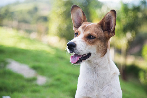
Cleft palate (CP) is one of the most visible congenital craniofacial anomalies in dogs. Though relatively rare, it carries a high mortality risk in neonates because the defect compromises the separation between the oral and nasal cavities, leading to aspiration pneumonia, poor weight gain, and frequent secondary infections. The condition can affect any breed, but it is markedly over‑represented in certain lineages such as the Labrador Retriever, Golden Retriever, German Shepherd, and various miniature breeds (e.g., Pomeranian, Chihuahua).
Understanding cleft palate from a holistic perspective—its embryologic origin, genetic background, clinical presentation, therapeutic options, and preventive measures—empowers veterinarians, breeders, and pet owners to make informed decisions that dramatically improve survival and quality of life. This guide consolidates the most up‑to‑date research, clinical experience, and practical advice into a single, searchable reference.
Key takeaway: Early recognition, prompt supportive care, and appropriately timed surgical repair are the cornerstones of successful management.
2. What Is a Cleft Palate? – Anatomy & Types
2.1 Normal Palatal Anatomy
The canine palate consists of two distinct portions:
| Structure | Composition | Primary Function |
|---|---|---|
| Hard Palate | Bony plate (maxillary and palatine bones) covered by keratinized epithelium | Provides rigid separation, supports teeth, and aids mastication |
| Soft Palate | Muscular tissue (levator veli palatini, tensor veli palatini, palatoglossus) covered by non‑keratinized epithelium | Seals the nasopharynx during swallowing, phonation, and prevents nasal regurgitation |
2.2 Classification of Cleft Palate
| Type | Anatomical Location | Typical Size | Clinical Implications |
|---|---|---|---|
| Complete Cleft of the Hard Palate | Extends from the incisive papilla to the soft palate | Large (>1 cm) | Severe nasal regurgitation, high aspiration risk |
| Partial (Incomplete) Hard Palate Cleft | Limited to anterior or central portion | Small‑moderate | May be tolerated with careful feeding |
| Soft‑Palate Cleft | Isolated defect in the muscular portion | Variable | Often leads to snoring, minor regurgitation |
| Combined Hard & Soft Palate Cleft | Involves both structures | Large | Requires staged surgical approach |
| Midline Cleft (Median) | Central vertical cleft, rarely seen | Small | May coexist with other midline defects (e.g., hydrocephalus) |
The size and location dictate both the urgency of intervention and the complexity of the surgical plan. A “complete” cleft typically means the entire hard palate and a portion of the soft palate are absent, whereas “partial” suggests a segmental defect that can sometimes close spontaneously in very small breeds.
3. Causes of Cleft Palate in Dogs
Cleft palate is a multifactorial disorder, often resulting from an interaction between genetic predisposition and external (environmental, nutritional, infectious) factors. Below is an exhaustive review of each contributing element.
3.1 Genetic Factors
- Polygenic Inheritance – Most breeds display a complex inheritance pattern involving several loci. Genome‑wide association studies (GWAS) in Labrador Retrievers have identified candidate regions on chromosomes CFA14 and CFA20 linked to midline craniofacial defects.
- Autosomal Recessive Mutations – Certain purebred lines harbour recessive alleles that manifest as cleft palate when both parents are carriers. The “PRA‑like” mutation in the IRF6 gene (also implicated in human Van der Woude syndrome) was first described in a family of English Cocker Spaniels.
- Founder Effect & Inbreeding – Small breeding populations amplify harmful alleles. Pedigree analysis often reveals a common ancestor a few generations back.
Practical tip: Conduct pedigree reviews and, when available, use DNA panels targeting known craniofacial mutation markers before breeding.
3.2 Environmental Influences
| Factor | Mechanism | Evidence |
|---|---|---|
| Maternal Stress | Elevated cortisol disrupts neural‑crest cell migration during weeks 3‑5 of gestation. | Experimental models in rodents; anecdotal reports in dogs. |
| Temperature Extremes | Hyperthermia in early gestation impairs palate formation. | Reports of higher CP incidence in litters born during heat waves. |
| Medication Exposure | Teratogenic drugs (e.g., steroids, retinoids) interfere with epithelial fusion. | Case series linking glucocorticoid administration to CP in breeding bitches. |
| Radiation | Ionizing radiation damages DNA of developing palate tissues. | Rare, typically occupational exposure. |
3.3 Nutritional Deficiencies & Imbalances
- Vitamin A (Retinoic Acid) – Both excess and deficiency disturb the signaling pathways that orchestrate palatal shelf growth.
- Folate (Vitamin B9) – Low folate impairs DNA synthesis, crucial during rapid embryonic cell proliferation.
- Zinc & Copper – Essential trace minerals for enzymatic processes in connective‑tissue formation.
Studies in canines fed a diet lacking adequate folic acid showed a 2‑fold rise in CP incidence compared with those on a supplemented regimen.
3.4 Infectious and Teratogenic Agents
| Agent | Mode of Action | Notable Cases |
|---|---|---|
| Canine Parvovirus (CPV) | Directly infects rapidly dividing embryonic cells during early gestation. | Outbreaks in high‑density breeding facilities. |
| Canine Herpesvirus (CHV) | Causes abortion; subclinical infections may affect palate morphogenesis. | Documented in research colonies. |
| Mycotoxins (e.g., Aflatoxin) | Interfere with cellular differentiation. | Rare, but reported after consumption of mold‑contaminated feed. |
| Environmental Teratogens (e.g., pesticides, heavy metals) | Disrupt signaling cascades (SHH, BMP). | Epidemiologic correlation in agricultural regions. |
3.5 Breed Predisposition & Inherited Lines
| Breed | Reported Incidence (per 1,000 litters) | Typical Defect |
|---|---|---|
| Labrador Retriever | 4–6 | Complete hard palate |
| Golden Retriever | 3–5 | Partial hard palate |
| German Shepherd | 2–4 | Combined hard & soft |
| Pomeranian | 1–2 | Small midline cleft |
| French Bulldog | 1–3 | Soft‑palate cleft |
Although numbers vary by geographic region, breeders should treat these breeds as “higher risk” and adopt stricter screening protocols.
4. Clinical Signs & Symptoms
The presentation differs dramatically depending on the age of the puppy, the size of the defect, and whether secondary complications have developed.
4.1 Neonatal Presentation (0‑3 weeks)
| Sign | Description | Why it Happens |
|---|---|---|
| Nasal Regurgitation | Milk or formula dribbles from the nostrils during nursing. | Lack of separation between oral and nasal cavities. |
| Coughing / Gagging | Frequent dry cough after each suckle. | Aspiration of milk into the airway. |
| Failure to Thrive | Weight gain <10 g/day, lethargy. | Inadequate nutrition & chronic pneumonia. |
| Dyspnea & Tachypnea | Labored breathing, especially after feeding. | Aspiration pneumonia, airway obstruction. |
| Poor Body Condition | Runted appearance, visible ribs. | Malabsorption and catabolism from infection. |
Urgent red flag: Any neonate showing signs of respiratory distress should be assessed immediately—aspiration pneumonia can be fatal within 48 hours.
4.2 Early‑Weaning & Growth Phase (3‑12 weeks)
- Continued Nasal Discharge (often serous → purulent).
- Recurrent Upper Respiratory Infections (due to compromised airway clearance).
- Dental Malocclusion (abnormal eruption because of palate anatomy).
- Behavioral Changes (reluctance to eat, fear of water).
4.3 Adult Dogs with Minor Defects
- Snoring or Stertor (soft‑palate insufficiency).
- Intermittent Nosebleeds (trauma from regurgitated material).
- Reduced Exercise Tolerance (due to chronic low‑grade pneumonia).
Even adult dogs with small defects can develop secondary complications if not monitored.
5. Diagnostic Work‑up
A systematic approach ensures an accurate diagnosis, helps identify associated anomalies, and guides surgical planning.
5.1 Physical Examination & Palpation
- Visual Inspection – Use a bright otoscope or a small speculum to expose the palate.
- Palpation – Gently feel the hard palate for bony continuity; note any floating mucosa indicating a soft‑palate cleft.
- Oro‑nasal Endoscopy – Allows assessment of the nasopharynx, especially in combined defects.
5.2 Imaging Modalities
| Modality | Advantages | Limitations |
|---|---|---|
| Plain Radiographs (laterolateral, dorsoventral) | Quick, inexpensive; shows bony defect. | Overlapping structures; poor soft‑tissue detail. |
| CT Scan (Computed Tomography) | High‑resolution 3‑D reconstruction of palate and surrounding sinuses. | Requires anesthesia; higher cost. |
| MRI (Magnetic Resonance Imaging) | Excellent soft‑tissue visualization; useful for soft‑palate assessment. | Longer scan time; limited bone detail. |
| Ultrasound | Bedside evaluation of soft tissue thickness. | Operator dependent; limited for bony assessment. |
Diagnostic tip: A thin‑slice CT scan (≤0.6 mm) with 3‑D rendering is now considered the gold standard before any surgical repair.
5.3 Endoscopy & Fluoroscopy
- Dynamic Fluoroscopy can visualize palate motion during swallowing, helping to decide whether soft‑palate function is adequate.
- Rigid Endoscopy of the nasopharynx identifies any fistulous tracts or concomitant choanal atresia.
5.4 Genetic Testing & Pedigree Analysis
When a breed‑specific mutation is known, a buccal swab can be submitted to a certified laboratory. Even in the absence of a known mutation, whole‑genome sequencing (WGS) of affected puppies and their parents is becoming more accessible and may reveal novel candidate genes.
5.5 Differential Diagnosis
- Submucous cleft palate (appears normal externally but has muscular deficiency).
- Traumatic palate lacerations (rare in neonates).
- Congenital nasal cavity malformations (e.g., choanal atresia).
Distinguishing these entities avoids unnecessary surgeries and directs appropriate management.
6. Treatment Options
6.1 Medical Management (Supportive Care)
| Intervention | Goal | Details |
|---|---|---|
| Tube Feeding (Esophageal or Gavage) | Provide adequate nutrition while bypassing the defect. | Use a 5‑Fr feeding tube; monitor placement with radiographs. |
| Antibiotic Therapy | Prevent/ treat aspiration pneumonia. | Broad‑spectrum (e.g., amoxicillin‑clavulanate) for 7‑10 days; adjust per culture |
#CleftPalateInDogs, #CanineCleftPalate, #DogHealth, #SpecialNeedsDog, #CleftAwareness, #DogSurgery, #VetMed, #VeterinaryMedicine, #PuppyHealth, #DogsofInstagram, #PetHealth, #CleftStrong, #RescueDog, #AdoptDontShop, #CanineHealth, #DogCare, #SpecialNeedsPets, #CleftPalateAwareness, #DogLife, #HealingPaws


Add comment