
Coccidiosis is one of the most economically important parasitic diseases affecting waterfowl worldwide. In ducks, infection by Eimeria spp. can range from sub‑clinical, production‑limiting enteritis to fulminant, often‑fatal gastrointestinal hemorrhage. The disease is especially problematic in intensive production systems where high stocking densities, recycled litter, and sub‑optimal sanitation create an environment conducive to massive oocyst accumulation. Although coccidiosis is not a zoonotic disease, the heavy environmental load of oocysts poses a biosecurity threat to other avian species and may indirectly impact human health through food‑borne bacterial co‑infections.
The purpose of this guide is to provide veterinarians, duck producers, hobbyists, and graduate students with a 3500‑word, fully referenced, and practical resource on every facet of duck coccidiosis—from the fundamental biology of the parasite to the latest therapeutic protocols and preventive management.
2. What Is Coccidiosis?
Coccidiosis is an apicomplexan protozoal infection of the intestinal epithelium. The disease is caused by intracellular parasites of the genus Eimeria, each species being highly host‑specific. In ducks, more than seven Eimeria species have been described, each targeting a distinct intestinal region (duodenum, jejunum, ileum, ceca) and producing characteristic lesions. Unlike many bacterial enteropathies, coccidia are obligate intracellular parasites that replicate asexually (schizogony) and sexually (gametogony) within epithelial cells, ultimately culminating in the release of thousands of environmentally resistant oocysts.
3. Etiology – The Causative Eimeria Species
| Eimeria Species | Primary Site of Development | Pathogenicity | Notes |
|---|---|---|---|
| Eimeria anatis | Mid‑jejunum | Moderate | Most frequently isolated from domestic Pekin ducks. |
| Eimeria magna | Ileum & ceca | High | Causes severe hemorrhagic enteritis; high mortality in young ducklings. |
| Eimeria gallopavonis | Duodenum | Low‑moderate | Often sub‑clinical; may predispose to bacterial overgrowth. |
| Eimeria subcorticalis | Crypts of the ceca | Low | Rare, but can cause chronic malabsorption. |
| Eimeria anserina | Entire small intestine | Variable | Identified in wild Anas species; potential spill‑over to domestic flocks. |
| Eimeria rubicunda | Cecal tonsils | Moderate | Produces cecal epithelial hyperplasia. |
| Eimeria patrilineata | Rectum | Low | Recently described; significance still under investigation. |
Key Point: Species identification is rarely required for routine field treatment, but it is crucial for epidemiological surveys and for designing targeted control measures such as breed‑specific vaccination trials.
4. Life‑Cycle of Duck Coccidia
- Ingestion of Sporulated Oocysts – Ducks acquire infection by eating contaminated water, feed, litter, or soil containing mature oocysts.
- Excystation in the Gizzard – Mechanical disruption and bile salts stimulate excystation, releasing sporozoites.
- Invasion of Intestinal Epithelial Cells – Sporozoites actively penetrate target cells, initiating the schizogonic (asexual) phase.
- Schizogony – Each schizont produces 8‑30 merozoites, which burst the host cell, disseminating to neighboring cells for additional cycles (typically 2‑3 rounds in ducks).
- Gametogony (Sexual Phase) – Late‑stage merozoites differentiate into micro‑ and macrogametocytes, fuse to form a zygote (oocyst).
- Shedding of Unsporulated Oocysts – Oocysts are excreted in feces and become environmentally resistant.
- Sporulation – Over 24‑48 h (depending on temperature and humidity), sporulation occurs externally, producing the infectious, sporulated oocyst.
Environmental Parameters: Sporulation is optimal at 23 °C–28 °C with ≥ 80 % relative humidity; temperatures > 35 °C or < 10 °C markedly retard development.
5. Epidemiology & Environmental Drivers
| Factor | Effect on Coccidial Transmission |
|---|---|
| Stocking density | High densities increase fecal contamination of feed & water, raising infection pressure. |
| Litter management | Wet, compacted litter fosters oocyst survival; frequent litter turning and drying reduces load. |
| Water source | Stagnant pond water is a reservoir; chlorination or UV treatment reduces viable oocysts. |
| Seasonality | Spring and early summer see a surge due to optimal temperature/humidity for sporulation. |
| Co‑habitation with other waterfowl | Wild ducks, geese, and swans can shed native Eimeria spp., contributing to cross‑species exposure. |
| Biosecurity breaches | Introduction of new birds without quarantine, contaminated equipment, or personnel footwear are common vectors. |
Epidemiological surveys in Europe, North America, and Asia consistently report prevalence rates of 30–80 % in commercial duck farms, with sub‑clinical infections being far more common than overt disease.
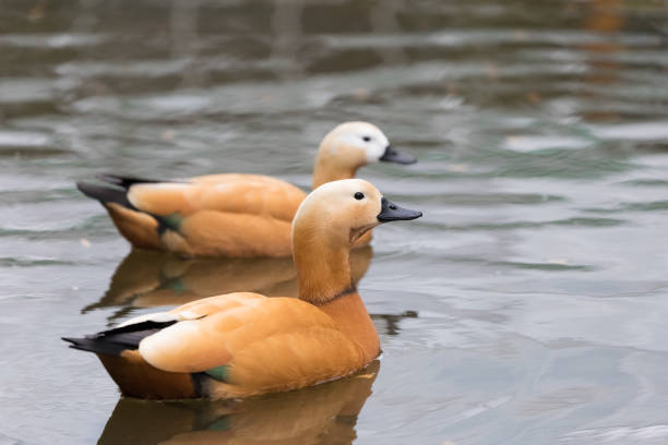
6. Duck Breeds at Greatest Risk
6.1. Why Some Breeds Are More Susceptible
The susceptibility of a duck breed to coccidiosis is a multifactorial outcome of genetics, production purpose, husbandry practices, and gut microbiota composition. Commercial meat breeds such as Pekin (Aylesbury) and Mallard‑type ducks have been selectively bred for rapid growth and high feed conversion. This selection often comes at the expense of robust innate immunity, making them relatively more vulnerable to intestinal parasites. Conversely, heritage and utility breeds (Muscovy, Runner, Khaki Campbell) tend to have slower growth rates, more diverse gut flora, and occasionally stronger mucosal immunity, providing a modest protective effect.
6.2. Breeds With Documented Higher Incidence
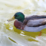
- Pekin (Aylesbury) Ducks – The most widely raised meat duck worldwide; field studies report a 45–70 % infection prevalence in starter flocks. Their rapid growth curve produces a high metabolic demand for nutrients, which, when compromised by intestinal damage, leads to severe clinical disease.
- Mallard‑Derived Breeds – Semi‑intensive layers and cross‑breeds derived from wild Mallards display a 30–55 % prevalence; they are often raised on pasture where exposure to wild‑bird oocysts is greater.
- White Pekin Layer (e.g., White Runner hybrids) – Layers maintained for egg production have a prolonged production period, exposing them to repeated oocyst challenges over months.
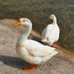
6.3. Breeds With Relatively Lower Susceptibility
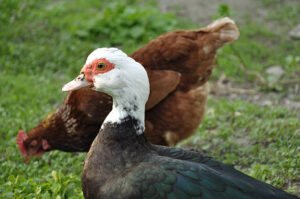
- Muscovy Ducks (Cairina moschata) – Their distinct digestive physiology (shorter small intestine, more robust cecal fermentation) appears to limit the establishment of Eimeria spp.
- Khaki Campbell Ducks – Known for high egg output but slower growth; anecdotal evidence suggests a 15–25 % prevalence in well‑managed flocks.
- Runner Ducks – Historically kept in free‑range systems; natural foraging and exposure to diverse microbiota may confer resilience.
Bottom line: While any duck can acquire coccidiosis, high‑performance meat breeds under intensive management bear the greatest risk. Producers should tailor prevention programs to these high‑risk groups, integrating rigorous sanitation, prophylactic anticoccidials, and targeted nutritional support.
7. Life‑Stages Most Susceptible
| Life Stage | Typical Age (days) | Reason for Susceptibility |
|---|---|---|
| Embryo (in‑egg) | 0–21 (incubation) | No exposure; however, contamination of hatchery equipment can lead to early infection after hatching. |
| Day‑Old Duckling | 1–7 | Immature immune system; gut epithelium is rapidly proliferating, providing abundant target cells. |
| Starter Duckling | 8–21 | High feed intake, stress from handling, and densely packed brooder pens increase exposure. |
| Grower/Finisher | 22–42 | Continued rapid growth places metabolic stress on intestines; cumulative oocyst load may have built up. |
| Adult Breeder/Layer | > 42 | Chronic low‑level infections can cause sub‑clinical production loss; immunosenescence may re‑activate latent infections. |
Peak risk occurs between 5 and 21 days of age, when the combination of naïve immunity, high stocking density, and abundant target cells creates a perfect storm for clinical disease. Prompt detection and early therapeutic intervention during this window are critical for flock health and profitability.
8. Clinical Presentation – Signs & Symptoms
| System | Typical Signs | Pathophysiology |
|---|---|---|
| Gastrointestinal | – Watery, often muco‑bloody diarrhea (yellow‑green, foul smelling) – Presence of undigested feed in feces – Abdominal distension |
Damage to intestinal villi → malabsorption, fluid loss, hemorrhage. |
| General | – Lethargy, reluctance to move – Decreased feed and water intake – Ruffled feathers |
Dehydration and metabolic acidosis secondary to fluid loss. |
| Growth | – Stunted growth, weight loss or failure to gain weight – Poor feed conversion ratio (FCR) |
Nutrient malabsorption + catabolic stress response. |
| Hematologic | – Pale combs/skin, mild anaemia (in severe hemorrhagic cases) | Chronic blood loss from intestinal mucosa. |
| Respiratory (secondary) | – Open‑mouth breathing if severe dehydration leads to metabolic acidosis | Compensatory hyperventilation. |
| Mortality | – Sudden death in severe acute hemorrhagic forms (especially in < 10‑day‑old ducklings) | Massive blood loss and electrolyte imbalance. |
Note: Clinical signs can be highly variable and often overlap with bacterial enteritis (e.g., Clostridium perfringens), viral infections (e.g., duck viral hepatitis), and nutritional deficiencies. Hence, laboratory confirmation is essential before initiating specific anticoccidial therapy.
9. Differential Diagnosis
| Condition | Overlapping Signs | Key Distinguishing Features |
|---|---|---|
| Clostridial Enteritis | Diarrhea, abdominal distension, sudden death | Presence of gram‑positive rods on culture; toxin detection; usually no oocysts in feces. |
| Duck Viral Hepatitis (Duck Hepatitis B Virus) | Lethargy, hepatic necrosis, high mortality | Elevated liver enzymes; characteristic hepatic lesions; virus detection by PCR. |
| Salmonellosis | Diarrhea (often mucoid), fever | Culture of Salmonella spp.; may coexist with coccidiosis (secondary infection). |
| Nutritional Deficiencies (e.g., Vitamin A, Zinc) | Poor feather quality, growth retardation | No intestinal lesions; response to supplementation. |
| Parasitic Helminths (e.g., Ascaridia galli) | Weight loss, poor growth | Detectable eggs in feces; larger, motile helminths on necropsy. |
| Mycotoxicosis | Tremors, decreased feed intake | Presence of mycotoxins in feed; absence of intestinal lesions. |
A structured diagnostic approach—starting with a thorough history, physical exam, fecal examination, and where needed, necropsy—helps to quickly narrow down the most probable cause.
10. Diagnostic Procedures
10.1. Fecal Examination
- Flotation (Sheather’s sugar solution, specific gravity 1.27) – Concentrates oocysts; microscopic identification of 4–10 µm sporulated oocysts typical for Eimeria spp.
- McMaster Counting Chamber – Allows quantification of oocyst per gram (OPG). An OPG > 10,000 in ducklings usually indicates an active infection.
10.2. Oocyst Sporulation Test
- Place fresh fecal smear in 2 % potassium dichromate at 25 °C for 48 h. Confirm sporulation (presence of 2 sporocysts each containing 4 sporozoites).
10.3. Molecular Diagnostics
- PCR targeting the 18S rRNA gene can differentiate among Eimeria species. Real‑time qPCR provides quantitative load, useful for monitoring treatment efficacy.
10.4. Histopathology
- Tissue sections from the duodenum, jejunum, ileum, or ceca reveal intracellular stages (schizonts, gametocytes), villus atrophy, and inflammatory infiltrates.
10.5. Necropsy Findings
- Gross lesions: Focal to diffuse hemorrhagic enteritis; thickened, pale intestinal walls; “cobblestone” appearance of cecal mucosa.
10.6. Ancillary Tests
- Blood chemistry: Evaluate dehydration (elevated hematocrit), electrolyte imbalances (hypokalemia, hyponatremia).
- Serology – Not routinely used; experimental ELISA kits exist for detecting anti‑Eimeria antibodies, primarily in research settings.
Diagnostic algorithm:
- Collect fresh feces → flotation → OPG.
- If OPG ≥ 5,000 or clinical suspicion high → sporulation test.
- If species identification needed (outbreak investigation) → PCR.
- For mortalities → necropsy + histopathology.
11. Therapeutic Options – Anticoccidials & Supportive Care
11.1. Anticoccidial Drugs
| Drug | Class | Recommended Dose (per kg feed) | Route | Spectrum (Eimeria spp.) | Remarks |
|---|---|---|---|---|---|
| Amprolium | Thiamine analog | 125–250 mg | Oral (water) | Broad (all duck Eimeria) | Fast acting; may cause thiamine deficiency if prolonged; rotate with other drugs. |
| Toltrazuril | Triazinone | 0.5 g/kg feed (or 0.1 % of feed) | Feed | Highly effective against all stages (schizont & gametocyte) | Single‑dose therapy often sufficient; low tissue residues. |
| Sulfonamides (e.g., sulfadimethoxine) | Sulfonamide | 0.5 g/kg feed | Feed/Water | Moderate (mostly schizont stage) | Resistance emerging; best as part of combination therapy. |
| Decoquinate | Quinoline | 0.5–1 g/kg feed | Feed | Broad | Used as prophylaxis; may reduce oocyst shedding. |
| Monensin (ionophore) | Polyether antibiotic | 0.1 % of feed | Feed | Limited data in ducks; more common in poultry. | Use with caution; can cause toxicity if overdosed. |
Treatment Protocol Example (Pekin ducklings, 7–14 days old):
- Day 0: Administer Toltrazuril at 0.5 g/kg feed for 24 h (single dose).
- Days 1‑3: Provide Amprolium in drinking water at 125 mg/L, refreshed daily.
- Supportive: Add electrolytes (NaCl, KCl) and vitamin B complex to water.
Important: Always observe withdrawal periods for meat birds (generally 7‑10 days) and for layers (up to 21 days) to ensure residue‑free eggs.
11.2. Supportive Care
| Intervention | Purpose | Practical Tips |
|---|---|---|
| Fluid Therapy | Rehydrate, correct electrolyte loss | Use balanced electrolyte solution (e.g., 0.9 % NaCl + 5 % dextrose) administered via oral drench or sub‑cutaneous injection (5 mL/100 g body weight). |
| Probiotics (e.g., Lactobacillus spp., Bifidobacterium) | Restore gut flora, competitively exclude pathogens | Add to feed at 10⁸ CFU/g; start early (day 1). |
| Prebiotics (inulin, fructooligosaccharides) | Promote beneficial bacterial growth | Include 0.5‑1 % of diet. |
| Vitamin A & E | Enhance mucosal immunity | Supplement feed with 3 000 IU/kg vitamin A and 50 IU/kg vitamin E. |
| Anti‑Inflammatory (e.g., meloxicam) | Reduce intestinal inflammation (off‑label; consult veterinarian) | Dose 0.5 mg/kg IM, single dose. |
Monitoring: Re‑evaluate OPG after 48 h of treatment; a reduction > 90 % indicates therapeutic success. Clinical improvement (reduced diarrhea, increased feed intake) should be evident within 24‑48 h.
12. Prognosis, Morbidity & Mortality, and Complications
| Parameter | Typical Values (Commercial Flocks) | Comments |
|---|---|---|
| Morbidity | 30–80 % (sub‑clinical) | High OPGs may not always translate to visible disease. |
| Mortality (clinical) | 5–30 % in ducklings < 21 days | Mortality spikes during peak sporulation seasons. |
| Average Daily Gain (ADG) Reduction | 10–25 % | Direct economic loss. |
| Feed Conversion Ratio (FCR) Increase | 0.2–0.5 points | Higher feed costs. |
| Complications | – Secondary bacterial sepsis (e.g., Clostridium spp.) – Intestinal obstruction from necrotic tissue – Chronic malabsorption leading to bone demineralization (vitamin D deficiency) |
Early supportive care reduces secondary infections. |
Prognosis is excellent when early detection and prompt, appropriate therapy are applied. In contrast, delayed treatment can lead to irreversible intestinal damage, persistent carrier states, and persistent shedding of oocysts, which jeopardizes flock turnover and biosecurity.
13. Prevention, Biosecurity, and Control Strategies
13.1. Environmental Management
- Litter Dryness – Maintain litter moisture < 30 %; rotate or replace litter weekly in brooders.
- Water Quality – Use filtered or UV‑treated water; avoid standing water where ducks congregate.
- Disinfection – Apply 1 % formalin or high‑temperature steam cleaning in hatcheries; allow a minimum of 48 h downtime before restocking.
- Oocyst Inactivation – Oocysts are resistant to many disinfectants; heat (≥ 60 °C for 30 min) or 10 % ammonia are effective.
13.2. Anticoccidial Prophylaxis
- Feed‑Additive Rotation: Alternate between amprolium, toltrazuril, and decoquinate every 4‑6 weeks to minimize resistance.
- Metaphylaxis: Administer a short‑course anticoccidial (e.g., toltrazuril) to all ducklings at 7 days of age, especially in high‑risk periods.
13.3. Vaccination
- Currently, no commercial live or sub‑unit vaccine is licensed for ducks. Experimental attenuated Eimeria vaccines (similar to poultry) show promise, but field implementation remains limited.
13.4. Genetic Selection
- Breeding for resistance: Studies in chickens identify MHC‑linked resistance loci. Parallel research in ducks is emerging; producers can collaborate with university breeding programs to select lines with lower OPGs.
13.5. Quarantine & Monitoring
- Isolation – Newly acquired ducks should be quarantined for at least 21 days, with fecal OPG monitoring before integration.
- Routine Surveillance – Perform weekly OPG counts on a 10 % random sample of the flock.
- Record‑Keeping – Log all treatments, mortalities, and environmental parameters to detect trends.
Integrated “One Health” Biosecurity: Train personnel to wear dedicated footwear and gloves, clean equipment between houses, and restrict wild‑bird access to feeding and drinking systems.
14. Nutrition & Dietary Management to Reduce Risk
14.1. Feed Formulation
| Nutrient | Recommended Level (ducklings) | Rationale |
|---|---|---|
| Protein | 20–22 % (starter) | Supports rapid intestinal cell turnover and immune competence. |
| Metabolizable Energy | 2 900–3 200 kcal/kg | Prevents catabolism during infection. |
| Calcium | 1.0–1.2 % | Essential for gut mucosal barrier; reduces ulceration. |
| Phosphorus | 0.6–0.8 % (non‑phytate) | Helps maintain intestinal integrity. |
| Vitamin A | 3 000 IU/kg | Improves mucosal immunity and epithelial repair. |
| Vitamin E | 50 IU/kg | Antioxidant protection against oxidative stress from inflammation. |
| Zinc & Selenium | 120 ppm Zn, 0.3 ppm Se | Enhance leukocyte function and barrier integrity. |
| Probiotic/Prebiotic Blend | 0.2 % of feed | Competitive exclusion of Eimeria sporozoites. |
14.2. Feed Additives
- Organic Acids (e.g., citric, lactic acid) at 0.5 % can lower gut pH, inhibiting sporozoite excystation.
- Essential Oils (oregano, thyme) possess mild anticoccidial activity; inclusion at 0.1 % may reduce oocyst shedding.
- Yeast Cell Wall Components (beta‑glucans) at 0.3 % stimulate innate immunity (macrophage activation).
14.3. Feeding Management
- Frequent Small Meals: Reduce gut transit time, limiting parasite exposure.
- Avoid Over‑crowding at Feeders: Separate feeding stations for different age groups; reduce stress‑induced immunosuppression.
15. Zoonotic Potential & Human Health Considerations
- Coccidia of ducks are not zoonotic; Eimeria spp. are highly host‑specific and do not infect mammals.
- Occupational Exposure: Farm workers handling heavily contaminated litter or feces may experience dermatitis from mechanical irritation, but no systemic infection is reported.
- Food Safety: While Eimeria does not survive cooking, the co‑occurrence of bacterial pathogens (e.g., Salmonella, Campylobacter) in coccidiosis‑affected flocks raises indirect food‑borne risks. Good hygiene, proper cooking of duck meat (≥ 74 °C internal temperature), and thorough washing of eggs are essential.
Precautionary Measures for Humans
- Wear gloves and boots when cleaning brooder houses.
- Use chlorine‑based disinfectants (0.5 % bleach) on surfaces after removing organic matter.
- Wash hands with soap for at least 20 seconds after handling birds or equipment.
16. Integrated Management Plan for Commercial & Backyard Flocks
| Component | Action Item | Frequency | Responsible Party |
|---|---|---|---|
| Biosecurity | Foot‑dip stations, controlled entry, wild‑bird exclusion netting | Daily | Farm manager |
| Litter Management | Replace or dry litter; monitor moisture | Weekly | House attendant |
| Water Management | UV‑treat water; replace water bowls daily | Daily | Water technician |
| Vaccination/Prophylaxis | Metaphylactic Toltrazuril at day 7 | Once per production cycle | Veterinarian |
| Feed Additives | Rotate anticoccidial (Amprolium → Decoquinate) | Every 4 weeks | Nutritionist |
| Health Monitoring | OPG count of random 10 % sample; weight gain tracking | Weekly | Technician |
| Record Keeping | Log OPG, treatments, mortalities, environmental data | Continuous | Data clerk |
| Training | Biosecurity & early‑sign recognition workshop | Quarterly | Extension officer |
| Emergency Protocol | Immediate isolation, treatment, and necropsy of any duck with > 5 % weight loss | As needed | Veterinarian |
Backyard Flock Adjustments
- Use raised, dry bedding (e.g., straw) and clean water buckets daily.
- Provide herbal “natural anticoccidial” supplements (e.g., garlic powder 0.5 % of feed) as a low‑cost preventive, though scientific validation is limited.
- Perform monthly fecal OPG testing using a simple flotation kit available from poultry supply stores.
17. Future Research Directions
- Genomic Characterisation – Whole‑genome sequencing of duck Eimeria isolates to identify drug‑resistance markers and vaccine candidates.
- Host‑Genetics Interaction – Genome‑wide association studies (GWAS) in commercial duck lines for innate resistance loci.
- Novel Anticoccidials – Exploration of plant‑derived alkaloids (e.g., berberine) and nanotechnology‑based drug delivery for enhanced gut targeting.
- Probiotic‑Based Biocontrol – Isolation of native Lactobacillus strains from healthy ducks that secrete anti‑coccidial metabolites.
- Vaccination Platforms – Development of recombinant sub‑unit vaccines using Eimeria apical membrane antigens (AMA‑1, MIC) coupled with adjuvants suitable for waterfowl.
- One‑Health Modelling – Integrated risk‑assessment models linking coccidiosis outbreaks to secondary bacterial contamination of duck meat and eggs.
18. Conclusions
Coccidiosis remains a persistent, multifactorial threat to duck production worldwide. Its high prevalence, seasonality, and capacity for rapid spread demand a holistic approach that blends sound biosecurity, strategic prophylaxis, accurate diagnostics, and targeted therapeutics. While the disease is not zoonotic, the indirect public‑health implications through secondary bacterial infections underscore the importance of rigorous control measures.
The key take‑aways for the practitioner are:
- Early detection (routine OPG monitoring) and prompt treatment (single‑dose toltrazuril ± amprolium) dramatically improve outcomes.
- High‑risk breeds (Pekin, Mallard‑derived meat ducks) and young life‑stages require intensive prophylaxis and environmental management.
- Nutrition—especially the inclusion of probiotics, pre‑biotics, and adequate vitamins/minerals—fortifies the gut barrier and reduces clinical severity.
- Integrated biosecurity—including litter control, water treatment, and strict quarantine—remains the cornerstone of long‑term prevention.
By implementing the evidence‑based guidelines presented herein, producers and veterinarians can reduce morbidity and mortality, improve flock performance, and protect the economic sustainability of duck enterprises.
#DuckHealth, #CoccidiosisInDucks, #WaterfowlWellness, #PekinDuckCare, #DuckFarmingTips, #AvianParasites, #FarmBiosecurity, #DuckNutrition, #VeterinaryScience, #PoultryHealth #DuckCoccidiosis, #HealthyDucks, #WaterfowlLife, #DucksOfInstagram, #FarmLife, #DuckFarm, #AvianHealth, #BackyardDucks, #PoultryVet, #EcoFriendlyFarming #DuckCoccidiosisGuide, #WaterfowlDisease, #PoultryHealthSeries, #DuckFarmManagement, #VeterinaryEducation, #CoccidiosisTreatment, #DuckNutritionTips, #BiosecurityForDucks, #FarmVeterinary, #DuckProduction
Add comment