
The canine ear is a marvel of biological engineering, serving not only as an organ for hearing but also playing crucial roles in balance, communication, and even minor thermoregulation. A deep understanding of its structure is essential for proper hygiene and recognizing signs of distress or disease.
I. ANATOMICAL STRUCTURE OF THE CANINE EAR
The dog’s ear is divided into three distinct sections: the External Ear, the Middle Ear, and the Inner Ear.
A. THE EXTERNAL EAR (Outer Ear)
The external ear is responsible for capturing sound waves and directing them inward toward the eardrum.
1. The Pinna (Auricle)
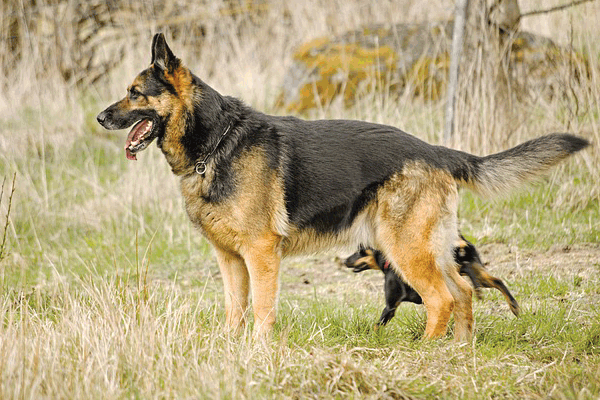
- Structure: This is the visible flap of the ear, composed primarily of flexible cartilage and covered in skin and fur. The size and shape of the pinna vary drastically between breeds (e.g., erect ears of a German Shepherd vs. pendulous ears of a Basset Hound).
- Function: Acts as a funnel to collect and localize sound. The vast array of muscles (about 18) attached to the pinna allows dogs to swivel their ears independently, precisely tracking the source of a sound without moving their head.
2. The Ear Canal (External Auditory Meatus)
- Structure: Unlike the relatively straight human ear canal, the dog’s canal is distinctly L-shaped (or V-shaped), featuring a long vertical section followed by a sharp turn into a horizontal section leading to the eardrum. This turn makes it difficult for foreign objects and moisture to exit naturally.
- Lining: The canal is lined with specialized skin containing hairs and glands (ceruminous and sebaceous) that produce earwax (cerumen).
- Clinical Significance: The deep, angled L-shape traps moisture and debris, making dogs highly susceptible to ear infections (Otitis Externa).
3. The Tympanic Membrane (Eardrum)
- Structure: A thin, delicate barrier located at the end of the external ear canal, separating it from the middle ear.
- Function: Vibrates in response to incoming sound waves.
B. THE MIDDLE EAR (Tympanic Cavity)
The middle ear is an air-filled chamber responsible for amplifying and transmitting sound vibrations from the eardrum to the inner ear.
1. The Auditory Ossicles
- Structure: Three tiny bones (the smallest bones in the body) arranged in a chain:
- Malleus (Hammer): Attached to the eardrum.
- Incus (Anvil): Connects the malleus and stapes.
- Stapes (Stirrup): Fits into the oval window of the inner ear.
- Function: These bones act as a mechanical lever system, amplifying the vibrations received by the eardrum and efficiently transferring the sound energy into the fluid-filled cochlea.
2. The Tympanic Bulla
- Structure: A large, bony, bubble-like structure that encases and protects the middle ear cavity.
- Clinical Significance: This area is often affected by chronic middle ear infections (Otitis Media), which are notoriously difficult to treat due to the dense bony protection.
3. The Eustachian Tube (Auditory Tube)
- Structure: A narrow tube connecting the middle ear cavity to the back of the throat (nasopharynx).
- Function: Equalizes air pressure between the middle ear and the outside environment, ensuring the eardrum vibrates properly.
C. THE INNER EAR (Labyrinth)
The inner ear is encased within the bone of the skull and is the site of two primary sensory functions: hearing and balance.
1. The Cochlea (Hearing)
- Structure: A delicate, spiraling, snail-shaped structure filled with fluid (endolymph and perilymph).
- Function: Contains the Organ of Corti, which houses thousands of tiny hair cells. When the fluid moves (triggered by vibrations from the ossicles), these hair cells bend, transducing the mechanical energy into electrical nerve signals. These signals are sent to the brain via the Cochlear Nerve (part of Cranial Nerve VIII) for interpretation as sound.
2. The Vestibular System (Balance and Equilibrium)
- Structure: Composed of three Semicircular Canals and two sacs (Utricle and Saccule), all filled with fluid and lined with sensory hairs.
- Function: This is the primary organ of balance.
- Semicircular Canals: Detect rotational movement (angular acceleration), allowing the dog to track spinning, turning, and nodding movements.
- Utricle and Saccule: Contain tiny calcium carbonate crystals (otoliths) that detect gravity and linear acceleration (up/down, forward/backward), informing the dog of its head position relative to the ground.
- Clinical Significance: Damage or infection in the vestibular system (vestibular disease) leads to severe signs: head tilt, loss of balance, staggering, and rapid, involuntary eye movements (nystagmus).
II. KEY FUNCTIONS OF THE CANINE EARS
1. Superior Hearing Acuity (The Primary Function)
Dogs possess a hearing range far superior to humans.
- Frequency Range: Humans hear pitches up to about 20,000 Hz. Dogs can hear up to 45,000–65,000 Hz (ultrasonic sounds). This explains why they react to high-pitched silent whistles.
- Sensitivity: Dogs can detect sounds at significantly lower volumes (softer), and are far better at rapidly locating the precise source of a sound (localization) due to the independent movement of their large pinnae.
2. Maintenance of Balance and Coordination
The vestibular system in the inner ear ensures the dog remains oriented and stable. It dictates the dog’s posture and helps coordinate eye, neck, and body movements, providing spatial awareness.
3. Communication and Expression
The position of the external ear is a vital non-verbal communication tool:
- Erect and Forward: Alertness, interest, or aggression.
- Flattened/Pasted Back: Fear, submission, or anxiety.
- Relaxed/Neutral: Calmness or contentment.
4. Thermoregulation (Secondary Function)
In breeds with very large, thin ears (like the African Basenji or the Fennec Fox, a close relative of the canine), the ears contain a high density of blood vessels close to the surface. By flushing blood into these vessels, the dog can dissipate heat into the environment, aiding in cooling the body.
III. CLINICAL CONSIDERATIONS & CARE
Due to the deep, L-shaped structure, the canine ear is prone to several common ailments:
- Otitis Externa: Inflammation or infection of the external ear canal. Often caused by bacteria, yeast (Malassezia), allergies (the most common underlying cause), excess moisture, or foreign bodies (like grass awns).
- Aural Hematoma: Caused by aggressive head shaking (usually due to severe itching from an infection), which ruptures blood vessels within the pinna, causing a large, blood-filled swelling.
- Deafness: Can be congential (often seen in white dogs lacking pigment, like Dalmatians), age-related (presbycusis), or acquired due to chronic infection leading to scar tissue, or exposure to ototoxic drugs.
Essential Care Tips
- Regular Inspection: Check the ear canal weekly for redness, discharge, excessive odor, or inflammation.
- Strategic Cleaning: Only clean the external part of the L-shaped canal, using veterinary-approved cleaning solutions. Never use cotton swabs, as they compact debris against the eardrum.
- Moisture Control: Always dry the ears thoroughly after swimming or bathing, especially in floppy-eared breeds (which naturally have poor air circulation).
#DogEars, #CanineAnatomy, #DogEarAnatomy, #EarStructure, #EarFunction, #DogHearing, #CanineHearing, #DogCommunication, #DogBalance, #CanineSenses, #Pinna, #EarCanal, #VestibularSystem, #AuralHealth, #DogHealth, #PetHealth, #DogFacts, #PetEducation, #VeterinaryAnatomy, #CaninePhysiology, #VetMed, #DogsOfInstagram, #DogLife, #LearnAboutDogs, #FascinatingFacts, #DogEars, #CanineAnatomy, #VetScience, #DogHealth, #PetCareTips, #DogFacts, #CanineOtolgy, #DogHearing, #VestibularSystem, #DogCareGuide, #AnimalAnatomy


Add comment