
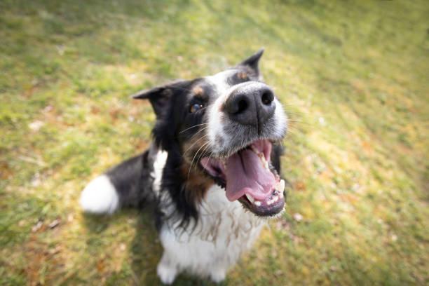
Elbow dysplasia (ED) is a complex and debilitating inherited condition that is one of the leading causes of forelimb lameness in medium to large and giant breed dogs. It refers to a collection of developmental abnormalities involving the elbow joint, which is comprised of three bones: the humerus (upper arm bone), the radius (larger forearm bone), and the ulna (smaller forearm bone). These abnormalities lead to incongruity, instability, and ultimately, degenerative joint disease (osteoarthritis) in affected dogs. Understanding this multifaceted condition is crucial for responsible pet ownership, early diagnosis, and effective management, ensuring the best possible quality of life for canine companions.
What is Elbow Dysplasia?
Elbow dysplasia is not a single disease but rather a syndrome encompassing several distinct, yet often co-occurring, developmental abnormalities within the elbow joint. These conditions typically lead to abnormal cartilage and bone development, joint incongruity, and often present early in a dog’s life. The primary components of elbow dysplasia include:
- Fragmented Medial Coronoid Process (FCP): This is the most common manifestation of ED. The medial coronoid process is a small bony projection on the ulna. In FCP, a piece of this process fails to properly attach or breaks off, leading to an intra-articular fragment that irritates the joint cartilage and causes pain and inflammation.
- Ununited Anconeal Process (UAP): The anconeal process is another bony projection on the ulna, important for elbow stability. In UAP, this process fails to fuse with the main part of the ulna during development (fusion typically occurs by 5 months of age). The ununited fragment can cause joint instability and significant pain.
- Osteochondrosis (OCD) of the Medial Humeral Condyle: Osteochondrosis is a disturbance in the normal process of endochondral ossification (the formation of bone from cartilage). In OCD, a thick flap of cartilage, or even bone and cartilage, may detach from the surface of the medial condyle of the humerus, leading to pain and inflammation within the joint.
- Elbow Incongruity: This refers to an abnormal fit between the articular surfaces of the humerus, radius, and ulna. It can manifest as either a “step” between the radius and ulna (where one bone is relatively shorter or longer than the other) or an ovalization of the trochlear notch of the ulna. This poor fit causes abnormal loading, increasing stress on the cartilage and potentially contributing to FCP or UAP.
- Ununited Medial Epicondyle (UMEC): Less common, this involves a failure of fusion of the medial epicondyle, a small bony prominence on the humerus where ligaments attach.
All these conditions contribute to the development of painful osteoarthritis, which is the inevitable consequence of elbow dysplasia if left untreated or poorly managed. The severity of the osteoarthritis often dictates the long-term prognosis.
Causes of Elbow Dysplasia
Elbow dysplasia is a multifactorial disease, meaning it results from a complex interplay of genetic predisposition and environmental factors.
1. Genetic Predisposition (Heredity)
Genetics play a significant role, making ED primarily an inherited condition. It is polygenic, meaning multiple genes contribute to its expression, rather than a single gene. This polygenic nature makes breeding for its elimination challenging, as carriers may not always show clinical signs. The heritability of ED varies among breeds but is generally considered moderate to high. Dogs inheriting genes for rapid growth, altered joint conformation, or defective cartilage development are more susceptible. Responsible breeding programs aim to reduce the incidence by screening breeding stock.
2. Rapid Growth and Over-nutrition
Overfeeding, particularly with diets high in calories, protein, and calcium, during the rapid growth phase of large and giant breed puppies (typically between 3 to 8 months of age), is a major contributing environmental factor. Rapid weight gain and bone growth place excessive stress on developing joints and can disrupt the delicate process of endochondral ossification. This can lead to conditions like OCD and exacerbate joint incongruity.
3. Diet and Nutrition
While related to rapid growth, specific dietary imbalances can predispose dogs to ED.
- Excessive Calories: Leads to rapid weight gain and bone growth, stressing immature joints.
- Calcium Imbalance: Too much or too little calcium, especially when supplemented inappropriately, can interfere with bone mineralization and development. Large breed puppy foods are formulated with precise calcium-to-phosphorus ratios to support slow, steady growth.
- Vitamin D: Both deficiencies and excesses can be detrimental to bone health.
4. Trauma and Exercise
- Excessive or Inappropriate Exercise: High-impact activities (e.g., jumping from heights, long-distance running, intense agility training) in young, rapidly growing puppies can put undue stress on developing joints, potentially triggering or worsening components of ED.
- Minor Repetitive Trauma: Everyday activities, when combined with underlying genetic predisposition and rapid growth, can contribute to the development of lesions like FCP. The elbow joint is particularly susceptible to rotational and compressive forces.
5. Asynchronous Growth of Radius and Ulna
This is a specific form of elbow incongruity. If the radius and ulna grow at different rates, even by a small amount, a “step” can occur in the joint surface.
- Short Radius Syndrome: The radius grows slower than the ulna, causing the ulna to bear excessive weight and stress, often leading to FCP.
- Short Ulna Syndrome: The ulna grows slower than the radius, leading to increased pressure on the medial coronoid process and the weight-bearing surface of the humerus, potentially causing FCP or OCD.
This asynchronous growth can be genetically predetermined or influenced by nutritional factors affecting growth rates.
Signs and Symptoms
The signs and symptoms of elbow dysplasia can vary widely depending on the severity of the condition, the specific lesions present, the dog’s age, and the extent of secondary osteoarthritis. They often manifest during the rapid growth phase, typically between 4 to 12 months of age, but can sometimes be more subtle initially and become pronounced later in life as arthritis progresses.
Early Signs (Puppies & Young Adults):
- Forelimb Lameness: This is the hallmark sign. It can be intermittent or persistent, often worse after rest (stiffness after sleeping) and after exercise. The lameness is typically exacerbated by strenuous activity. It may shift from one leg to the other if both elbows are affected, making diagnosis challenging.
- Stiffness: Dogs may appear stiff, especially when getting up after lying down or in the morning.
- Reluctance to Exercise/Play: Affected puppies may avoid jumping, running, or playing as much as their littermates. They may seem “lazy” or tire easily.
- Abnormal Gait:
- Inward Rotation of Paws: The dog may turn its paws outwards or inwards when standing or walking (often outward rotation of the lower limb).
- “Crab-like” Walk: The dog may have an unusual gait where it swings its leg out to the side to avoid bending the elbow fully.
- Shortened Stride: The dog may take shorter steps with the affected limb.
- Pain on Palpation/Extension/Flexion: Veterinary examination often reveals pain when the elbow joint is manipulated, particularly when extended fully or flexed. Crepitus (a crackling or grinding sound/sensation) may be felt.
- Swelling: While not always visible externally, the joint may feel subtly swollen or thickened due to inflammation and fluid accumulation.
Later Signs (Adult & Older Dogs, often due to Osteoarthritis):
- Chronic Lameness: The lameness may become more consistent and severe as osteoarthritis progresses, even with minimal activity.
- Decreased Range of Motion: The dog may struggle to fully extend or flex its elbow. This can be evident when they try to lie down or stand up, or when they can’t groom themselves easily.
- Muscle Atrophy: Due to disuse of the painful limb, muscles in the shoulder and forearm of the affected leg may waste away (atrophy), appearing thinner than the unaffected leg.
- Weight Shifting: Dogs may shift their weight to their hindquarters or an unaffected forelimb to relieve pressure on the painful elbow.
- Behavioral Changes: Chronic pain can lead to irritability, aggression, reluctance to be touched, depression, or changes in sleeping patterns.
- Licking at the Joint: Some dogs may compulsively lick at the painful elbow area.
It’s important to note that bilateral elbow dysplasia (affecting both elbows) is common (up to 70-80% of cases), which can mask lameness in one limb as the dog compensates by shifting weight equally to both painful elbows. This makes early detection challenging and underscores the need for a thorough veterinary examination.
Dog Breeds at Risk (with paragraph explanation)
Elbow dysplasia disproportionately affects medium to large and giant breed dogs. This is primarily due to their rapid growth rates and heavier body weight, which place increased stress on developing joints, coupled with a strong genetic predisposition.
- Labrador Retrievers: One of the most popular breeds globally, Labradors are unfortunately highly predisposed to elbow dysplasia, particularly FCP. Their strong genetic component, combined with their active nature and potential for rapid growth, makes careful breeding selection and controlled development crucial.
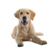
- Golden Retrievers: Similar to Labradors, Golden Retrievers are prone to ED, especially FCP and OCD. Their genetic background for orthopedic issues means breeders must meticulously screen parent dogs for elbow health, and owners must manage their growth and exercise carefully.
- German Shepherd Dogs: German Shepherds are well-known for their susceptibility to various orthopedic conditions, including elbow dysplasia (especially UAP and FCP). Their genetic lineage and structure, often bred for working capabilities, necessitate rigorous screening in breeding lines to mitigate the risk of ED.
- Rottweilers: These powerful dogs are at a significant risk for elbow dysplasia, frequently presenting with FCP. Their rapid growth and substantial body mass early in life contribute to the severity and incidence of the condition, emphasizing the importance of appropriate nutrition and controlled exercise.
- Bernese Mountain Dogs: A large, majestic breed, Bernese Mountain Dogs have a high incidence of elbow dysplasia, particularly FCP and OCD. Their rapid development as puppies and heavy build place considerable strain on their developing joints, making genetic screening and growth management vital.
- Newfoundlands: These giant, gentle dogs are highly susceptible to elbow dysplasia, often manifesting as FCP and UAP. Their immense size and rapid growth put them at considerable risk, necessitating strict adherence to large-breed puppy feeding guidelines and avoiding excessive activity in their formative months.
- Great Danes: As one of the largest dog breeds, Great Danes grow at an astonishing rate, predisposing them to various developmental orthopedic diseases, including elbow dysplasia (FCP and OCD are common). Managing their growth through appropriate diet and controlled exercise is paramount.
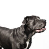
- Mastiffs (various types, e.g., English Mastiff, Bullmastiff): These colossal breeds share similar risks with Great Danes and Newfoundlands due to their extreme size and rapid development. Elbow dysplasia, often FCP, is a significant concern, requiring careful attention to genetics, diet, and exercise protocols.
- Basset Hounds: Although not a giant breed, Basset Hounds are included due to a notable predisposition to UAP. Their unique skeletal structure and growth patterns contribute to this specific elbow abnormality.
- Other Large/Giant Breeds: Breeds such as Siberian Huskies, Border Collies, American Staffordshire Terriers, Chow Chows, and many others, especially those with athletic builds or rapid growth, can also be affected, though often with lower incidence rates than the primary risk breeds.
Breeders of these at-risk breeds are encouraged to participate in official screening programs, such as those run by the Orthopedic Foundation for Animals (OFA) or the British Veterinary Association/Kennel Club (BVA/KC) schemes, to identify and selectively breed dogs with healthy elbows, thereby reducing the genetic prevalence of ED.
Affects Puppy, Adult, or Older Dogs
Elbow dysplasia is primarily a developmental disease, meaning it originates during the growth phase of a dog’s life.
- Puppies and Young Adults (4-12 months of age): This is the typical age range when clinical signs first become noticeable. The abnormalities in bone and cartilage development occur during this rapid growth period. Symptoms like lameness, stiffness, and reluctance to play are common as the lesions (FCP, UAP, OCD, incongruity) directly cause pain and inflammation in the immature joint. Owners often first notice issues between 5 to 8 months.
- Adult Dogs (1-6 years of age): If elbow dysplasia was mild or subclinical in puppyhood, or if the dog compensated well, overt signs might not appear until adulthood. By this stage, the primary lesions may have progressed, and significant secondary osteoarthritis would have developed. The chronic inflammation and wear-and-tear lead to cartilage degradation, bone remodeling, and pain. Lameness becomes more consistent and potentially more debilitating.
- Older Dogs (7+ years of age): In older dogs, elbow dysplasia almost invariably manifests as advanced osteoarthritis. Even dogs that underwent successful surgical intervention in their youth will likely develop some degree of arthritis as they age. The lameness may become chronic, severe, and significantly impact their quality of life. Management in older dogs primarily focuses on pain control, maintaining mobility, and supportive care for arthritis.
Therefore, while the underlying developmental abnormalities occur in puppyhood, the clinical manifestation and severity can evolve throughout a dog’s life, with osteoarthritis being the universal long-term consequence.
Diagnosis
Accurate and early diagnosis of elbow dysplasia is crucial for effective management and to potentially slow the progression of osteoarthritis. A veterinarian will typically employ a combination of clinical examination, imaging techniques, and sometimes advanced diagnostics.
1. Clinical Examination and Orthopedic Assessment
- History Taking: The vet will ask about the dog’s symptoms (onset, duration, severity, aggravating factors), breed, age, activity level, and diet.
- Gait Analysis: Observation of the dog walking, trotting, and sometimes turning reveals lameness, stiffness, or an abnormal gait (e.g., reluctance to fully extend the elbow, outward rotation of the paw). This can be subtle, especially if both elbows are affected.
- Palpation: The veterinarian will carefully palpate the elbow joints, feeling for swelling, thickening, pain response, and crepitus (a grinding or crackling sensation) when the joint is flexed and extended. Pain elicited on full extension or flexion is a common finding. Muscle atrophy in the affected limb may also be noted.
2. Radiography (X-rays)
X-rays are the initial imaging modality of choice for suspected elbow dysplasia.
- Views: Several specific radiographic views (e.g., mediolateral flexed, craniocaudal, mediolateral extended, oblique) are required to visualize the different components of the elbow joint. Sedation is often necessary to ensure proper positioning and minimize stress for the dog, as well as to achieve diagnostic quality images.
- Findings: X-rays can reveal:
- Osteoarthritis: The most consistent finding, characterized by osteophytes (bone spurs), joint space narrowing, and subchondral bone sclerosis.
- UAP: Visible as a distinct fragment of bone at the caudal aspect of the ulna.
- OCD: Appears as a flattened or irregular joint surface, sometimes with a visible cartilage flap.
- Elbow Incongruity: Can be assessed by visualizing the alignment of the humeral condyle, radial head, and trochlear notch of the ulna.
- FCP: Can be challenging to see directly on standard X-rays, as the fragment is often small and cartilaginous. However, secondary signs of osteoarthritis, particularly on the medial aspect of the joint, strongly suggest FCP.
- Limitations: X-rays are two-dimensional and have limited ability to visualize soft tissue or early cartilaginous lesions. They are not highly sensitive for detecting FCP directly.
3. Computed Tomography (CT Scan)
CT is considered the gold standard for diagnosing elbow dysplasia, particularly FCP and elbow incongruity.
- Mechanism: CT uses X-rays to create detailed cross-sectional images of the joint, allowing for a three-dimensional reconstruction.
- Advantages:
- Superior Detail: Provides much better visualization of bone abnormalities and joint incongruity compared to conventional X-rays.
- FCP Detection: Highly sensitive for detecting the fragmented medial coronoid process, which is often missed on plain radiographs.
- Pre-surgical Planning: Excellent for detailed surgical planning due to its 3D capabilities.
- Bilateral Assessment: Allows for efficient assessment of both elbows simultaneously.
- Disadvantages: Requires general anesthesia and is more expensive and less widely available than X-rays.
4. Magnetic Resonance Imaging (MRI)
MRI is less commonly used for primary diagnosis of ED unless soft tissue pathology (e.g., ligament damage, cartilage integrity) is suspected, or if CT is inconclusive for FCP.
- Mechanism: Uses powerful magnets and radio waves to create detailed images of soft tissues, including cartilage, ligaments, and fluid.
- Advantages: Excellent for soft tissue and cartilage assessment.
- Disadvantages: Very expensive, requires general anesthesia, and less detailed for primary bone abnormalities compared to CT.
5. Arthroscopy (Diagnostic and Therapeutic)
Arthroscopy involves inserting a small camera (arthroscope) into the joint through a small incision.
- Advantages:
- Direct Visualization: Allows direct visualization of the joint surfaces and lesions in real-time.
- Early Detection: Can detect subtle changes in cartilage and small fragments that might be missed on other imaging.
- Minimally Invasive: Less invasive than open joint surgery.
- Therapeutic Potential: Can often be used to treat lesions (e.g., remove FCP, OCD flaps) during the same procedure.
- Disadvantages: Requires general anesthesia, specialized equipment, and expertise.
A definitive diagnosis often involves a combination of these methods, tailored to the individual case and the suspected lesions.
Treatment
The treatment for elbow dysplasia aims to alleviate pain, improve joint function, and slow the progression of osteoarthritis. The approach depends on the dog’s age, severity of lameness, specific lesions identified, and the extent of secondary osteoarthritis. Treatment options are broadly categorized into conservative (medical) and surgical.
A. Conservative (Medical) Management
Conservative management is often the first approach for mild cases, very young puppies, or dogs with advanced osteoarthritis where surgery is no longer viable or beneficial. It’s also crucial as long-term management for nearly all ED cases, even post-surgery.
- Weight Management: Keeping the dog at an ideal body weight is paramount. Excess weight significantly increases stress on painful joints, exacerbating lameness and accelerating arthritis.
- Controlled Exercise:
- Activity Modification: Avoid high-impact activities (jumping, vigorous fetch, long runs, stairs) particularly in young dogs.
- Regular, Moderate Exercise: Short, leash-controlled walks on soft surfaces (grass, dirt) can help maintain muscle mass and joint mobility without overstressing the joint.
- Hydrotherapy: Swimming or underwater treadmill therapy is excellent for building muscle and improving range of motion with minimal joint impact.
- Pain Management (Pharmacological):
- Non-Steroidal Anti-Inflammatory Drugs (NSAIDs): Medications like carprofen, meloxicam, deracoxib, firocoxib are mainstays for pain and inflammation control. They should be used under veterinary supervision due to potential side effects (gastrointestinal, kidney, liver).
- Gabapentin/Amantadine: Neuropathic pain medications that can be used as adjuncts, especially for chronic pain.
- Opioids (e.g., Tramadol): Sometimes used for acute, severe pain, or in combination with other drugs for chronic pain that is not well-controlled.
- Joint Supplements (Chondroprotectants):
- Glucosamine and Chondroitin Sulfate: These are building blocks for cartilage and may help improve joint fluid quality and slow cartilage degradation.
- Omega-3 Fatty Acids (EPA/DHA): Potent natural anti-inflammatories found in fish oil.
- MSM (Methylsulfonylmethane): An organic sulfur compound believed to have anti-inflammatory and pain-relieving properties.
- Polysulfated Glycosaminoglycans (PSGAGs): Injectable chondroprotectants (e.g., Adequan) that can stimulate cartilage repair and reduce inflammation.
- Physical Rehabilitation and Physiotherapy:
- Therapeutic Exercises: Specific exercises to improve strength, flexibility, balance, and proprioception.
- Manual Therapy: Massage, joint mobilization.
- Modalities: Therapeutic laser, ultrasound, electrical stimulation (TENS/NMES) to reduce pain and inflammation.
- Regenerative Medicine:
- Platelet-Rich Plasma (PRP): Concentrated platelets from the dog’s own blood contain growth factors that can promote healing and reduce inflammation. Injected directly into the joint.
- Stem Cell Therapy: Adipose-derived (fat-derived) stem cells or bone marrow-derived stem cells can differentiate into cartilage cells and release anti-inflammatory factors. Injected into the joint. These are newer, advanced treatments with promising, but still evolving, evidence.
- Acupuncture: Can be effective for pain management and improving comfort in some dogs with chronic orthopedic pain.
B. Surgical Management
Surgical intervention is often recommended for young dogs (usually 6-18 months) with significant lameness and specific lesions that are amenable to repair, aiming to correct the underlying structural problem and ideally prevent or delay severe osteoarthritis.
- Fragmented Medial Coronoid Process (FCP) – Arthroscopic Removal/Subtotal Coronoidectomy:
- Procedure: The loose fragment(s) of the coronoid process are removed, and any abnormal cartilage is debrided. This is typically performed arthroscopically (minimally invasive).
- Goal: Eliminate the source of irritation and pain.
- Subtotal Coronoidectomy: In some cases, a larger portion of the medial coronoid process is removed (or coronoid osteotomy performed) to address persistent pressure or incongruity.
- Ununited Anconeal Process (UAP) – Fixation or Excision:
- Procedure:
- Lag Screw Fixation: If the dog is young (under 6-8 months) and minimal arthritis is present, the fragment can be reattached with a screw.
- Excision (Removal): If the fragment is large, displaced, or significant arthritis is present, the ununited anconeal process is often removed.
- Goal: Restore joint stability and eliminate the painful fragment.
- Procedure:
- Osteochondrosis Dessicans (OCD) – Flap Removal:
- Procedure: The loose cartilage flap is removed, and the underlying abnormal bone is debrided to stimulate fibrocartilage formation. This can be done arthroscopically.
- Goal: Remove the source of pain and irritation.
- Elbow Incongruity – Corrective Osteotomies:
- Procedure: If a significant step or incongruity is present, osteotomies (bone cuts) of the radius or ulna may be performed to lengthen or shorten one of the bones, thereby restoring congruity.
- Proximal Ulnar Osteotomy: A cut in the ulna allows it to shorten or lengthen, improving fit.
- PAUL (Proximal Abducting Ulnar Osteotomy): A specific procedure that rotates the ulna to offload the medial compartment of the elbow, often used for FCP cases where incongruity plays a role.
- Sliding Humeral Osteotomy (SHO): A more advanced procedure that alters the angle of the humerus to shift weight from the medial (damaged) compartment to the lateral (healthier) compartment of the elbow.
- Goal: Improve joint mechanics and reduce abnormal loading that contributes to FCP or OCD.
- Procedure: If a significant step or incongruity is present, osteotomies (bone cuts) of the radius or ulna may be performed to lengthen or shorten one of the bones, thereby restoring congruity.
- Bicipital Release / Ulnar Ostectomy for Flexor Tendon Release:
- Procedure: In some cases, the biceps tendon or surrounding soft tissues can contribute to chronic pain and pressure on the medial coronoid process. Releasing these structures can alleviate tension.
- Total Elbow Replacement (TER):
- Procedure: A salvage procedure for end-stage, severe osteoarthritis where other treatments have failed. The damaged joint surfaces are replaced with artificial components.
- Goal: Provide pain relief and restore function in severely arthritic elbows.
- Limitations: This is a complex, expensive surgery with potential complications (infection, loosening of implants). It is not widely available.
Post-Operative Care and Rehabilitation:
Regardless of the surgical procedure, strict post-operative care is crucial, including:
- Restriction of activity: Often for several weeks, gradually increasing.
- Pain management: Medications.
- Physical therapy: Essential for regaining strength, range of motion, and preventing muscle atrophy. This typically involves a structured program of passive range of motion, controlled exercises, and hydrotherapy.
The choice between conservative and surgical management, or a combination, should be made in consultation with a veterinary orthopedic specialist, considering the individual dog’s condition and owner’s commitment.
Prognosis & Complications
The prognosis for dogs with elbow dysplasia varies widely, depending on several factors: the specific type and severity of lesions, the age at diagnosis, the extent of secondary osteoarthritis, the chosen treatment, and the owner’s commitment to rehabilitation and long-term management.
Prognosis:
- Good for Pain Control, Not for Cure: It is important for owners to understand that elbow dysplasia cannot be truly “cured” because the joint’s development was abnormal. The goal of any treatment, whether conservative or surgical, is to manage pain, optimize joint function, and slow the progression of inevitable osteoarthritis.
- Early Intervention is Key: Dogs diagnosed and treated early (especially surgically for specific lesions) before severe osteoarthritis has developed generally have a better long-term outcome regarding comfort and function.
- Severity of Lesions:
- UAP: If surgically fixed in young dogs, the prognosis can be good. If severe arthritis is already present or removal is necessary, some discomfort may persist.
- FCP: Prognosis is fair to good for lameness improvement after arthroscopic fragment removal, but osteoarthritis will still progress. Dogs usually remain comfortable with good management.
- OCD: Good to fair after flap removal, but similar to FCP, some degree of arthritis will develop.
- Significant Incongruity: These cases often have a guarded prognosis, as correcting the fundamental fit can be challenging, and arthritis tends to be more severe.
- Osteoarthritis Progression: Regardless of treatment, some degree of osteoarthritis is almost guaranteed. The long-term prognosis is often related to how well this secondary arthritis can be managed. Many dogs lead comfortable lives with appropriate pain medication, joint supplements, weight control, and controlled exercise.
- Total Elbow Replacement (TER): While a salvage procedure for end-stage arthritis, it can offer a good prognosis for significant pain relief and improved function in carefully selected cases, but it carries higher risks.
Complications:
- Progression of Osteoarthritis: This is the most common and almost inevitable long-term complication. Even with successful treatment of the primary lesion, the joint has been compromised, leading to progressive cartilage degeneration and bone remodeling. This requires lifelong management.
- Persistent Lameness: Some dogs, despite surgery, may continue to exhibit some degree of lameness or discomfort, especially after strenuous activity or as arthritis advances.
- Surgical Complications:
- Infection: As with any surgery, there is a risk of post-operative infection, which can be particularly problematic in joints.
- Implant Failure: For procedures involving screws or plates (e.g., UAP fixation, osteotomies), implants can loosen, break, or migrate, requiring further surgery.
- Nerve Damage: Though rare, damage to nerves during surgery can lead to weakness or paralysis.
- Anesthetic Risks: All general anesthesia carries some inherent risk.
- Limited Range of Motion: Over time, the arthritic joint may develop fibrosis and osteophyte formation, leading to a reduced range of motion, making normal activities difficult.
- Non-Response to Conservative Management: Some dogs with severe or rapidly progressing ED may not respond adequately to medical management alone, necessitating more aggressive intervention or leading to chronic pain.
- Economic Burden: Managing elbow dysplasia, especially with surgery, rehabilitation, and lifelong medications, can be a significant financial commitment.
- Impact on Quality of Life: In severe, untreated, or poorly managed cases, chronic pain and lameness can severely diminish a dog’s quality of life, affecting its ability to play, exercise, and interact normally. In rare, intractable cases, euthanasia may be considered if pain cannot be controlled.
Close monitoring, regular veterinary check-ups, and a proactive approach to managing osteoarthritis are essential for maximizing the prognosis and minimizing complications in dogs with elbow dysplasia.
Prevention
Preventing elbow dysplasia focuses on responsible breeding practices and optimized environmental factors during a puppy’s critical growth period. Since ED has a strong genetic component, the most impactful prevention strategies start with breeders.
1. Responsible Breeding Programs (Genetic Screening):
- Parental Screening: This is the cornerstone of prevention. All dogs intended for breeding, especially those from at-risk breeds, should undergo official elbow screening programs to assess their elbow health.
- Orthopedic Foundation for Animals (OFA): In the US, OFA evaluates elbow radiographs and assigns a grade (Normal, Grade I, II, or III). Only dogs with “Normal” elbows (no evidence of ED) should be bred.
- British Veterinary Association/Kennel Club (BVA/KC) Scheme: In the UK, elbows are scored on a scale from 0 to 3, with 0 being normal and 3 being severe. The goal is to breed dogs with low scores (ideally 0/0).
- Breeding Only “Clear” Dogs: Only dogs that have been certified free of elbow dysplasia should be used for breeding. This helps reduce the incidence of ED in future generations.
- Considering Pedigree: Look at the elbow health of the parents, grandparents, and siblings. Even if a dog has clear elbows, if several relatives have ED, it may still carry the genes.
- Avoiding Breeding Affected Dogs: Dogs diagnosed with ED, even if treated, should not be bred.
2. Controlled Growth Rate and Appropriate Nutrition:
- Large Breed Puppy Food: Feed a high-quality commercial dog food specifically formulated for “large breed puppies.” These diets are designed to support slower, controlled growth rates. They typically have:
- Lower Calorie Density: To prevent rapid weight gain.
- Balanced Calcium and Phosphorus: Appropriate ratios (typically 1.1:1 to 1.4:1 calcium-to-phosphorus) to support bone development without excess. Avoid calcium supplementation unless specifically directed by a veterinarian.
- Moderate Protein Levels: Contrary to popular belief, high-protein diets do not cause ED, but excessive protein can contribute to higher overall calorie intake if not balanced.
- Avoid Overfeeding: Do not allow puppies to become overweight. Follow feeding guidelines on the food package, but adjust based on the individual puppy’s body condition score. Ribs should be easily palpable, but not visibly prominent.
- No Unnecessary Supplementation: Avoid supplementing with vitamins or minerals, particularly calcium, unless recommended by a veterinarian for a specific medical reason. Commercial large breed puppy foods are nutritionally complete and balanced.
3. Controlled Exercise and Environmental Management:
- Avoid High-Impact Activities in Growing Puppies: During the critical growth period (typically 3-12 months for large breeds), limit activities that put excessive stress on developing joints. This includes:
- Jumping (e.g., off furniture, out of cars, agility jumps).
- Long-distance running or forced exercise.
- Excessive stair climbing.
- Rough play with other dogs or humans that cause repetitive jarring or twisting of the joints.
- Provide Moderate, Consistent Exercise: Encourage regular, controlled exercise on soft surfaces (grass, dirt) like leash walks. This helps develop muscle strength and maintain joint health without overstressing.
- Provide Good Traction: Ensure puppies have good traction on indoor surfaces to prevent slips and falls, which can cause trauma. Use rugs on slippery floors.
- Monitor for Lameness: Pay close attention to any signs of lameness, stiffness, or reluctance to move. Early detection allows for earlier intervention.
4. Early Veterinary Check-ups:
- Regular puppy check-ups allow your veterinarian to monitor growth, body condition, and perform orthopedic examinations, potentially detecting subtle signs of joint problems early.
While genetic predisposition cannot be changed, adopting these preventative measures can significantly reduce the risk and severity of elbow dysplasia in at-risk breeds.
Diet and Nutrition
Diet and nutrition play a critical role in the development and management of elbow dysplasia, particularly during the rapid growth phase of large and giant breed puppies. Improper nutrition can exacerbate genetic predispositions and contribute to the expression of the disease.
1. For Growing Puppies (Prevention and Mitigation):
- Large Breed Puppy Formulation: This is the single most important dietary consideration. These diets are specifically engineered to promote a slower, controlled growth rate critical for proper skeletal development.
- Lower Calorie Density: Prevents rapid weight gain, which puts excessive stress on developing joints.
- Appropriate Calcium-to-Phosphorus Ratio: These diets maintain a tightly controlled calcium (1.1-1.4% dry matter basis) and phosphorus balance. Excess calcium is particularly detrimental, as it can disrupt normal bone formation and lead to conditions like OCD. Deficiencies are also harmful but less common with commercial diets.
- Moderate Fat and Protein: While protein itself doesn’t cause ED, high fat and protein levels can lead to excessive calorie intake, contributing to rapid growth if not carefully managed.
- Avoid Overfeeding (Maintain Lean Body Condition):
- Follow Feeding Guidelines: Start with the manufacturer’s recommendations for large breed puppy food, but adjust based on the individual puppy’s body condition.
- Body Condition Scoring (BCS): Regularly assess your puppy’s BCS. You should be able to easily feel, but not visibly see, its ribs. An overweight puppy is highly susceptible to orthopedic issues.
- No Unnecessary Supplementation:
- Calcium: Absolutely avoid supplementing calcium unless specifically instructed by a veterinarian for a diagnosed deficiency. Excess calcium is a known risk factor for developmental orthopedic diseases.
- Vitamins/Minerals: Commercial large breed puppy foods are complete and balanced. Adding extra vitamins or minerals without veterinary guidance can disrupt nutrient balances and be harmful.
- Omega-3 Fatty Acids (EPA & DHA): While not primarily for prevention, starting a supplement with Omega-3s (found in fish oil) during growth may offer mild anti-inflammatory benefits and support overall joint health. However, this should not replace a properly formulated large-breed puppy diet.
2. For Adult Dogs (Management of ED and Osteoarthritis):
- Weight Management: Maintaining an ideal body weight is crucial for any dog with elbow dysplasia or secondary osteoarthritis. Excess weight significantly increases the load on already compromised joints, accelerating cartilage degradation and exacerbating pain. Even a small amount of weight loss can lead to noticeable improvements in comfort and mobility.
- Joint Supplements (Chondroprotectants): These supplements aim to support cartilage health, reduce inflammation, and improve joint fluid quality.
- Glucosamine and Chondroitin Sulfate: These are precursors for glycosaminoglycans (GAGs), which are essential components of cartilage and joint fluid. They may help to slow cartilage breakdown and stimulate repair.
- MSM (Methylsulfonylmethane): An organic sulfur compound with anti-inflammatory properties often included in joint supplements.
- Omega-3 Fatty Acids (EPA & DHA): Potent natural anti-inflammatories. Supplementation with high-quality fish oil (e.g., salmon oil, krill oil) can help reduce joint inflammation and pain.
- Green-Lipped Mussel (GLM): Another source of GAGs and omega-3s, often used in joint supplements.
- Therapeutic Diets: Some prescription veterinary diets are formulated for joint health. These diets typically:
- Have controlled calorie levels for weight management.
- Are enriched with high levels of Omega-3 fatty acids.
- Contain added glucosamine, chondroitin, and other joint-supporting nutrients.
- May include antioxidants to combat oxidative stress in inflamed joints.
- Antioxidants: Vitamins E and C, selenium, and other antioxidants can help reduce oxidative stress and inflammation within arthritic joints. These are often included in therapeutic joint diets or can be supplemented.
Important Considerations:
- Consult Your Veterinarian: Always discuss your dog’s diet and any supplements with your veterinarian. They can provide tailored advice based on your dog’s specific needs, stage of ED, and overall health.
- Quality of Supplements: Not all supplements are created equal. Choose reputable brands with third-party quality assurance.
- Holistic Approach: Diet and nutrition are one component of a holistic management plan for elbow dysplasia, which also includes appropriate exercise, physical therapy, and potentially medication or surgery.
By carefully managing your dog’s diet and nutrition from puppyhood through adulthood, you can play a significant role in preventing the onset of elbow dysplasia and mitigating its progression once diagnosed.
Zoonotic Risk
Elbow dysplasia in dogs poses no zoonotic risk whatsoever.
Explanation:
- Definition of Zoonotic: A zoonotic disease is one that can be transmitted naturally from animals to humans. Examples include rabies, Lyme disease, salmonellosis, and toxoplasmosis.
- Nature of Elbow Dysplasia: Elbow dysplasia is a developmental orthopedic disease. It is a structural abnormality of the bones and cartilage of the elbow joint, resulting from a combination of genetic factors and environmental influences (like rapid growth, nutrition, and exercise). It is not caused by any infectious agent (bacteria, virus, parasite, fungus) that could be transmitted to humans.
- Not Transmissible: You cannot “catch” elbow dysplasia from your dog, nor can your dog transmit it to another species. It is specific to the skeletal development of the affected individual animal.
Therefore, owners and individuals interacting with dogs diagnosed with elbow dysplasia have no need to worry about any risk of contracting the condition themselves. Their only concern should be providing appropriate care and comfort for their canine companion.
#ElbowDysplasiaDogs #DogElbowDysplasiaExplained #CanineElbowProblems #DogJointDisease #PuppyGrowthProblems #LargeDogHealth #DogArthritisTreatment #VeterinaryOrthopedics #DogRehabVideos #ElbowDysplasiaSurgery #DogPainManagement #PreventingElbowDysplasia #DogHealthGuide #UnderstandingElbowDysplasia #DogCareTips #VetAdviceForDogs #HealthyDogJoints #DogMobilitySolutions #DogOwnerGuide #CanineWellness #ElbowDysplasia #DogHealth #CanineOrthopedics #JointCare #DogLameness #PuppyHealth #LargeBreedProblems #DogLife #VetMed #PetHealth #DogPain #ArthritisDogs #HealthyDogs #DogWellness #FCP #UAP #OCD #DogLove #InstaDogs #DogCareTips #OrthopedicDogs #VetAdvice #DogRehabilitation #DogNutrition #PreventionIsKey #ElbowDysplasia #CanineHealth #DogOrthopedics #DogJoints #PuppyCare #LargeBreedDogs #DogHealthAwareness #PetCare #VetMed #DogMom #DogDad #HealthyPaws #ArthritisInDogs #DogLameness #ElbowDysplasiaAwareness #VeterinaryAdvice #DogRehab #JointHealthForDogs #ResponsibleBreeding #PreventativeCare


Add comment