
The domestic cat (Felis catus) is a masterpiece of evolutionary design, a highly specialized predator whose anatomy reflects millennia of adaptation for stealth, agility, and precision hunting. While the cat’s musculoskeletal and digestive systems are optimized for a strictly carnivorous lifestyle, it is the sensory apparatus, particularly the visual system, that elevates the cat to its apex predator status in the nocturnal environment.
This elaborate guide will first provide a foundational overview of the cat’s general anatomy, establishing the context for its sensory functions, and then delve into a highly detailed examination of the nervous system and the intricate structure, function, and clinical significance of the feline eye.
I. THE FELINE ANATOMICAL BLUEPRINT: AN OVERVIEW
The cat’s body structure is characterized by flexibility and efficiency. Understanding the core systems is essential for appreciating how the visual system integrates with motor skills and environmental processing.
A. The Skeletal and Muscular Systems
The feline skeleton comprises approximately 244 bones (variation occurs due to tail length), significantly more than the human skeleton (206).
- Axial Skeleton: The spine is exceptionally flexible due to loosely articulated vertebrae and thick, elastic cushioning discs. Cats possess a floating clavicle, which is not connected to any other bone, allowing the thoracic limbs a wider range of motion, crucial for squeezing through tight spaces and achieving explosive bursts of speed.
- Appendicular Skeleton: The legs are built for running, jumping, and shock absorption. Cats walk digitigrade—on their toes—which increases their stride length and speed. The retractable claws, housed in specialized folds of skin and tendons when resting, are unique anatomical mechanisms allowing for silent stalking and preserved sharpness.
- Musculature: The musculature is dense and powerful. The massive development of fast-twitch muscle fibers facilitates rapid explosive movements (like pouncing), while the fine intrinsic muscles of the ears and eyes allow for precise sensory orientation.
B. Internal Organ Systems
Feline internal anatomy is fundamentally adapted for a high-protein diet.
- Digestive System: The cat is an obligate carnivore. Its short intestinal tract (optimized for rapid processing of animal protein) and lack of specific enzymes (like needed for synthesizing taurine or Vitamin A from plant precursors) necessitate a meat-based diet.
- Cardiovascular and Respiratory Systems: These systems are powerful and efficient, engineered to support bursts of intense activity. The heart-to-body size ratio is appropriate for sustaining high metabolic demands.
- Renal System: The kidneys are highly efficient at concentrating urine, a trait evolved to conserve water in arid environments, often resulting in a low thirst drive.
III. THE NERVOUS SYSTEM: THE CONTROL CENTER FOR VISION
The central nervous system (CNS)—the brain and spinal cord—is the processing unit that translates visual stimuli into actionable commands. The feline brain, while smaller relative to body size than the human brain, is highly developed in areas governing complex motor skills, balance, and, critically, sensory processing.
A. Major Brain Divisions
- Cerebellum: Highly prominent in cats, the cerebellum is responsible for coordinating complex movements, posture, and balance. Its large size directly reflects the cat’s need for precise acrobatic ability.
- Occipital Lobe (Visual Cortex): Located in the posterior part of the cerebrum, the visual cortex receives and interprets electrical signals transmitted via the optic nerve. Feline visual processing prioritizes motion detection above high-resolution detail.
B. The Optic Pathway: Cranial Nerve II
The visual process begins with the optic nerve (Cranial Nerve II).
In cats, the optic nerve fibers exhibit extensive crossover (decussation) at the optic chiasm—about 65% to 75% of fibers cross sides, compared to about 50% in humans. This means the left brain hemisphere receives slightly more input from the right eye, and vice-versa. This extensive crossing is characteristic of prey animals and animals with a wide visual field, although the cat’s frontal eye placement mitigates this somewhat, allowing for greater binocular overlap.
IV. THE SENSE OF SIGHT: AN EVOLUTIONARY MASTERPIECE
The cat’s visual system is a nocturnal specialist, unparalleled among common domesticated mammals. It is designed not for reading or appreciating a full spectrum of color, but for capturing maximum light in low-level conditions and detecting the slightest movement of prey.
The large size of the feline eye relative to the head enhances its light-gathering capability. Its structure is specialized to capture light 6 to 8 times more efficiently than the human eye.
V. DETAILED ANATOMY OF THE FELINE EYE (OCULUS)
The globe (eyeball) is housed within the bony orbit and protected by several accessory structures. The eye itself is composed of three primary layers (tunics): the fibrous tunic (outer), the vascular tunic (middle), and the neural tunic (inner).
A. External Protective and Accessory Structures
These structures ensure the health, moisture, and mechanical protection of the globe.
1. The Eyelids (Palpebrae)
The upper and lower eyelids are covered externally by skin and internally by the conjunctiva. They perform the crucial function of protecting the eye from trauma and distributing the tear film across the corneal surface during blinking. The margins contain specialized glands (Meibomian glands) that secrete an oily substance to prevent the evaporation of the tear film.
2. The Nictitating Membrane (Third Eyelid)
This structure, formally known as the Plica Semilunaris, is a T-shaped piece of cartilage covered in conjunctiva and located in the medial (inner) corner of the eye. It contains a significant accessory lacrimal gland (the gland of the third eyelid), contributing approximately 30-50% of the aqueous portion of the tears.
Function: The membrane sweeps diagonally across the cornea to remove debris and distribute tears. Unlike in humans, the third eyelid in cats is highly mobile and will partially or fully protrude when the cat is ill, deeply asleep, or heavily sedated—often serving as an indicator of general health status.
3. The Lacrimal Apparatus
The lacrimal system produces, distributes, and drains the tear film. Tears are essential for lubrication, nutrition of the avascular cornea, and protection (containing antibacterial agents like lysozyme). The tears drain through minute openings (puncta) into the lacrimal sac and down the nasolacrimal duct into the nasal cavity.
B. The Fibrous Tunic (Outer Layer)
This tough, protective layer maintains the shape of the globe.
1. The Sclera
The sclera is the opaque, white, tough, connective tissue layer that covers the majority of the globe. It provides the attachment points for the extraocular muscles. In cats, much of the sclera is covered by the conjunctiva; therefore, the visible white portion is minimal, making cats’ eyes appear larger and more concentrated.
2. The Cornea
The cornea is the transparent, curved anterior portion of the fibrous tunic. It is the primary structure involved in refracting (bending) light rays onto the retina, providing two-thirds of the eye’s total refractive power.
- Structure: The cornea is avascular (lacking blood vessels) and obtains oxygen directly from the air and nutrients from the aqueous humor and tear film.
- Clinical Significance: Because the cornea has a high concentration of pain receptors (trigeminal nerve), any scratch or foreign body causes immediate and intense pain.
C. The Vascular Tunic (Uvea)
Lying beneath the sclera, the uvea is rich in blood vessels and pigment cells (melanocytes). It is divided into three parts: the choroid, the ciliary body, and the iris.
1. The Choroid
This is the posterior portion of the uvea, highly vascularized to supply oxygen and nutrients to the outer layers of the retina. It is darkly pigmented to absorb stray light, preventing internal reflections—except in the area where the tapetum lucidum resides.
2. The Ciliary Body
The ciliary body is a ring of tissue located immediately behind the iris. It performs two critical functions:
- Accommodation: It contains the ciliary muscle, which changes the shape of the lens to focus on objects at different distances. Cats are generally considered slightly hyperopic (farsighted) compared to humans, and while they can change focus, their range is not as flexible as ours.
- Aqueous Humor Production: The ciliary processes secrete aqueous humor, a clear fluid that fills the space between the lens, iris, and cornea (the anterior and posterior chambers).
3. The Iris and Pupil
The iris is the colored, muscular diaphragm that surrounds the central opening, the pupil. Its melanocytes determine eye color (blue, green, copper, etc.).
- Function: The iris controls the amount of light entering the eye. It is composed of two muscle types:
- Sphincter Pupillae: Contraction causes the pupil to constrict (miosis).
- Dilator Pupillae: Contraction causes the pupil to widen (mydriasis).
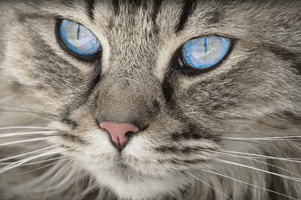
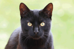
- Unique Feline Feature (Slit Pupil): The cat’s pupil is vertically elliptical (slit-shaped). This shape provides an enormous advantage, allowing for an incredibly fast and large range of aperture adjustment (up to 300-fold difference between constricted and dilated states, compared to 15-fold in humans). In bright light, the pinpoint vertical slit minimizes light entry; in darkness, it dilates to a nearly perfect circle to maximize light capture.
D. The Neural Tunic (Retina)
The retina is the light-sensitive inner layer of the eye where photoreception occurs. It is an extension of the brain itself and contains the complex neural circuitry for vision.
1. Photoreceptor Cells (Rods and Cones)
- Rods: Responsible for vision in low light (scotopic vision) and detecting motion. Cats have a significantly higher ratio of rods to cones (estimated 25:1 or more) compared to humans (4:1). This rod-heavy retina is the primary reason for a cat’s superior nocturnal vision. Rhodopsin, the pigment in rods, is crucial for maximum sensitivity.
- Cones: Responsible for color vision and high-detail central vision (photopic vision). While cats possess cones, their concentration is lower, limiting their color perception (believed to be dichromatic—seeing blues and yellows/greens, but perceiving red/green wavelengths poorly).
2. The Optic Disc (Blind Spot)
This is the location where the optic nerve and retinal blood vessels enter the eye. It contains no photoreceptors, creating a physiological blind spot, although the brain fills in this gap seamlessly.
3. The Fundus
The fundus is the posterior interior surface of the eye, visible with an ophthalmoscope. It displays the tapestry of the retina, vasculature, and the highly reflective structure known as the Tapetum Lucidum.
VI. SPECIALIZED STRUCTURES FOR NOCTURNAL VISION
The true mastery of feline sight lies in its specialized adaptations for hunting in dim light conditions.
A. The Tapetum Lucidum: The Eye Shine Phenomenon
The Tapetum Lucidum is a layer of reflective cells situated immediately behind the retina, within the choroid layer. It is this structure that causes the characteristic “eye shine” (or retinal glow) seen when a cat is exposed to a direct light source at night.
1. Mechanism
When light passes through the photoreceptor layer (retina) and is not immediately absorbed, it strikes the tapetum lucidum. The reflective cells bounce the light back through the retina a second time. This “double exposure” effectively means that photons that missed the rods the first time get a second chance to stimulate them.
2. Composition and Appearance
The feline tapetum is typically cellular (composed of layers of highly organized, crystal-like cells containing riboflavin or zinc cysteine). This crystalline structure gives the reflected light its characteristic green-yellow or blue-green color.
- Note on Albinos/Blue-Eyed Cats: In cats lacking pigmentation (such as Siamese or pure white cats with blue eyes), the tapetum may be obscured or structurally underdeveloped. These cats often show a unique reddish eye shine because the light reflects directly off the underlying blood-rich choroid instead of the tapetum.
B. Pupil Dynamics and Light Maximization
The combination of the vertical slit pupil and the tapetum lucidum provides maximum efficiency:
- The slit pupil allows an unprecedented amount of light into the globe when dilated in darkness.
- The rods maximize the detection of that scarce light.
- The tapetum lucidum re-uses any missed photons, almost doubling the light input efficiency.
This triad of adaptations allows the cat to see effectively in ambient light levels roughly one-sixth of what a human requires.
VII. PHYSIOLOGY OF FELINE VISION (HOW CATS SEE THE WORLD)
While renowned for their night vision, cats perceive the world differently from humans in terms of detail and color.
A. Field of View and Depth Perception
The cat’s eyes are positioned relatively far forward on the face, providing excellent binocular vision (the overlap of the visual fields from both eyes).
- Field of View: Cats have a total visual field of about 200 degrees (compared to 180 degrees in humans).
- Binocular Overlap: Their binocular field is approximately 130 degrees (compared to 120 degrees in humans). This extensive overlap is essential for superb stereopsis (depth perception). When pouncing, the cat requires highly accurate judgment of distance, a function directly reliant on this binocular overlap.
B. Visual Acuity (Sharpness)
A cat’s vision is optimized for sensitivity, not resolution. If human visual acuity is measured at 20/20, a cat’s acuity is estimated to be around 20/100 to 20/200. This means a cat must be much closer to an object to see it with the same detail a human would see from afar.
- Focal Preference: Cats are poor at focusing on very close objects (within 6 inches). They rely heavily on their whiskers (vibrissae) and sense of smell to interpret objects directly in front of their face.
- Motion Detection: Visual acuity drops off dramatically at the edges of the cat’s vision. However, the high density of rods makes them exquisitely sensitive to even the slightest movement in the periphery—a critical advantage when scanning for scurrying prey.
C. Color Perception
While once believed to be completely colorblind, research confirms that cats are not monochromatic. They possess cones that respond mainly to blue-violet and yellow-green wavelengths.
- Dichromatic Vision: Cats are primarily dichromats. They see the world in somewhat muted colors, likely perceiving grassy areas as shades of yellow, blue, and grey. Red and green distinctions, which rely on the third cone type present in humans (trichromats), are generally absent or poorly utilized.
- UV Sensitivity: Interestingly, the feline lens does not filter UV light as effectively as the human lens. This means cats may be able to perceive UV wavelengths, which could amplify the visibility of certain natural markings, scent trails, or prey items.
VIII. COMMON OCULAR HEALTH ISSUES AND CLINICAL SIGNIFICANCE
The delicate and highly specialized structure of the feline eye makes it susceptible to unique congenital, infectious, and degenerative diseases.
A. Congenital and Structural Anomalies
1. Entropion and Ectropion
- Entropion: A condition common in certain pedigree breeds (e.g., Persians, Maine Coons) where the eyelid rolls inward, causing the eyelashes and fur to constantly rub against the cornea. This leads to chronic irritation, pain, and corneal ulceration.
- Ectropion: The less common condition where the eyelid rolls outward, exposing the conjunctiva and predisposing the eye to dryness and infection.
2. Eyelid Agenesis (Coloboma)
A congenital defect where part of the eyelid (usually the upper outer portion) fails to develop completely, resulting in chronic dryness and corneal damage due to lack of protection.
3. Third Eyelid Gland Prolapse (“Cherry Eye”)
While less common in cats than dogs, the gland of the third eyelid can swell and protrude, appearing as a pink or red mass in the inner corner of the eye. This compromises tear production if not treated.
B. Infectious and Inflammatory Diseases
1. Conjunctivitis
Inflammation of the conjunctiva, the pink tissue lining the eyelids. This is one of the most common eye problems in cats, often presenting as redness, swelling, and discharge. It is frequently caused by infectious agents, notably Feline Herpesvirus-1 (FHV-1) and Chlamydophila felis.
2. Feline Herpesvirus (FHV-1)
This is arguably the most significant cause of ocular disease in cats. FHV-1 often causes upper respiratory tract infection but can lead to severe, chronic or recurrent eye issues, including:
- Dendritic Ulcers: Unique, branching corneal ulcerations pathognomonic for herpes infection.
- Keratitis: Chronic inflammation of the cornea, leading to scarring and vascularization (blood vessels growing onto the cornea, causing opacity and vision impairment).
3. Uveitis
Inflammation of the uveal tract (iris, ciliary body, and choroid). Uveitis is a serious, painful condition that can lead to permanent vision loss. In cats, uveitis is frequently linked to systemic infectious diseases, such as Feline Infectious Peritonitis (FIP), Feline Leukemia Virus (FeLV), Feline Immunodeficiency Virus (FIV), and toxoplasmosis.
C. Age-Related and Metabolic Diseases
1. Glaucoma
Glaucoma is characterized by elevated intraocular pressure (IOP), usually due to inadequate drainage of aqueous humor through the drainage angle (iridocorneal angle). The increased pressure damages the optic nerve and retina, leading to irreversible blindness.
- Feline Specificity: Unlike dogs, primary (inherited) glaucoma is rare in cats. The vast majority of feline glaucoma cases are secondary, caused by chronic inflammation (uveitis) or tumor growth blocking the drainage angle.
2. Cataracts
An opacity of the lens, preventing light from reaching the retina. While common in older humans and diabetic dogs, true cataracts are relatively uncommon in cats. When they occur, they are often linked to chronic ocular inflammation (uveitis) or genetic predispositions.
3. Progressive Retinal Atrophy (PRA)
A group of genetic, degenerative diseases that cause the photoreceptor cells (rods and cones) to slowly deteriorate. This leads to night blindness initially (due to rod loss) and progresses to complete blindness. Breeds like Abyssinians and Persians are genetically predisposed to specific forms of PRA.
IX. CONCLUSION
The cat’s anatomy is a testament to adaptive evolution. Every structure, from the flexible spine to the short digestive tract, is calibrated for efficiency as an ambush predator. However, the true marvel remains the eye.
The feline visual system is a highly specialized biological apparatus, prioritizing sensitivity, motion detection, and depth perception over fine detail and broad color range. The sophisticated interplay between the vertically slitted pupil, the high rod-to-cone ratio, and the remarkable light-amplifying mechanism of the tapetum lucidum ensures the cat’s dominance in low-light environments.
A comprehensive understanding of these anatomical intricacies is not only fascinating from a biological perspective but is also crucial for veterinary medicine, allowing for prompt recognition and effective management of the myriad conditions that threaten this essential sensory gateway. The cat’s eyes are not merely windows to the world; they are mirrors reflecting the animal’s extraordinary evolutionary journey.
#FelineAnatomy, #CatEyes, #VeterinaryOphthalmology, #FelineVision, #PetHealth, #CatScience, #TapetumLucidum, #NocturnalHunter, #CatAnatomyGuide, #EyeHealth, #AskAVet, #KittenVision, #FelineStructure, #BiologyOfCats, #GeniusOfCats, #AnimalEyes, #VetTech, #FelineAnatomyDeepDive, #SlitPupils, #CatFacts.


Add comment