
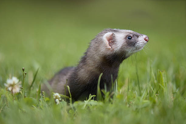
Tuberculosis (TB) is a chronic bacterial disease primarily known for affecting humans and cattle. In recent decades, however, an increasing number of cases have been reported in exotic mammals, including the domestic ferret (Mustela putorius furo). Ferrets occupy a unique niche as popular companion animals, research models, and, in some regions, fur‑producing livestock. Their close physical contact with owners and their propensity for respiratory secretions create a genuine zoonotic bridge for Mycobacterium tuberculosis complex (MTBC) organisms.
The significance of ferret TB can be captured in three overlapping domains:
- Animal Health – Untreated TB results in chronic wasting, organ failure, and high mortality.
- Public Health – Ferrets can act as a reservoir for human infection, especially in immunocompromised individuals.
- Veterinary Economics – Diagnostic and therapeutic costs, coupled with potential culling, burden breeders and owners alike.
Thus, veterinarians, pet owners, researchers, and public‑health officers must possess a deep understanding of the disease’s causation, clinical course, and control measures.
2. Etiology & Causative Mycobacteria
2.1 Mycobacterium tuberculosis (Mtb)
- Classification: Member of the Mycobacterium tuberculosis complex (MTBC).
- Primary Host: Humans, though it can infect a broad host range (cattle, elephants, non‑human primates, and ferrets).
- Transmission to Ferrets: Predominantly via inhalation of aerosolized droplets from infected humans or other animals, but also through contaminated bedding, food, or water.
2.2 Mycobacterium bovis (Mbo)
- Classification: Also part of MTBC; primarily a cattle pathogen.
- Ferret Infection Route: Contact with infected livestock, contaminated feed, or environmental exposure on farms where cattle TB is endemic.
- Zoonotic Importance: Mbo is inherently resistant to pyrazinamide, a key first‑line anti‑TB drug, which significantly influences treatment choices.
2.3 Nontuberculous Mycobacteria (NTM)
Several NTM species (e.g., Mycobacterium avium complex, M. marinum, M. fortuitum) can cause granulomatous disease in ferrets that mimics TB. While not classified as “tuberculosis” per se, they are part of the differential diagnosis and share many diagnostic pitfalls.
| NTM Species | Common Source | Typical Clinical Pattern in Ferrets |
|---|---|---|
| M. avium | Soil, water, plant material | Chronic diarrhea, weight loss, hepatic granulomas |
| M. marinum | Freshwater aquaria | Cutaneous nodules, ulcerative lesions on limbs |
| M. fortuitum | Tap water, biofilms | Rapidly progressive subcutaneous abscesses |
Key Point: Laboratory confirmation (culture, PCR) distinguishes MTBC from NTM, driving appropriate antimicrobial therapy.
3. Epidemiology in Ferrets
3.1 Global Distribution
- North America & Europe: Sporadic case reports, often linked to pet ferrets owned by individuals with active TB or working in TB‑endemic settings.
- Asia & Africa: Limited data; however, ferret farms in regions with high bovine TB prevalence (e.g., parts of China, South Africa) demonstrate higher seroprevalence.
- Surveillance Gap: No worldwide mandatory reporting for TB in ferrets, leading to underestimation of incidence.
3.2 Species‑Specific Susceptibility
Ferrets share immunologic characteristics with mustelids (e.g., mink, otters) that render them relatively susceptible to mycobacterial infections. Their high metabolic rate and the presence of abundant bronchial lymphoid tissue facilitate early colonization of inhaled bacilli.
4. Pathogenesis: How Mycobacteria Invade Ferret Hosts
- Inhalation or Ingestion: Bacilli reach alveolar macrophages or intestinal Peyer’s patches.
- Intracellular Survival: Mycobacteria inhibit phagosome‑lysosome fusion, allowing replication within macrophages.
- Granuloma Formation: Host immune response aggregates infected macrophages, epithelioid cells, and lymphocytes, forming caseating granulomas.
- Dissemination: Via lymphatic drainage or hematogenous spread, bacteria seed secondary organs (spleen, liver, kidneys, central nervous system).
- Latency vs. Active Disease: Ferrets, like humans, can harbor latent infection with low bacterial loads; immunosuppression or stress triggers reactivation.
5. Clinical Manifestations
5.1 General Signs & Symptoms
| Clinical Feature | Frequency in Ferrets | Description |
|---|---|---|
| Weight loss & cachexia | 80% | Progressive, despite normal appetite initially |
| Lethargy & reduced activity | 70% | Often first noted by owners |
| Fever (38‑40 °C) | 60% | Low‑grade, may be intermittent |
| Respiratory distress | 50% | Coughing, dyspnea, nasal discharge |
| Anorexia or reduced food intake | 45% | Secondary to systemic illness |
| Lymphadenopathy | 40% | Enlarged mandibular, retropharyngeal nodes |
| Ocular changes (conjunctivitis, uveitis) | 20% | Rare, indicate disseminated disease |
| Neurologic signs (tremors, ataxia) | 10% | Usually late-stage or CNS involvement |
5.2 Organ‑Specific Presentations
Respiratory System
- Chronic cough (dry or wet) with occasional hemoptysis.
- Radiographic Findings: Multifocal nodular infiltrates, hilar lymphadenopathy, cavitary lesions in advanced cases.
Gastrointestinal Tract
- Diarrhea (often with blood or mucus) when intestinal granulomas are present.
- Ultrasound: Hypoechoic hepatic lesions, splenomegaly.
Lymphatic System
- Palpable cervical and submandibular masses (granulomatous lymphadenitis).
- Fine‑needle aspirates reveal caseating necrosis and acid‑fast bacilli.
Central Nervous System
- Meningeal signs (head tilt, seizures) are uncommon but carry poor prognosis.
5.3 Subclinical Carriers
Ferrets can shed low numbers of bacilli for weeks to months without overt disease, especially when immunocompromised (e.g., on corticosteroids). Carrier status is often uncovered only through targeted testing of breeding colonies or post‑mortem examinations.
6. Diagnostic Approach
A systematic, multimodal strategy maximizes detection while minimizing false‑negatives.
6.1 History & Physical Examination
- Exposure Assessment: Contact with TB‑positive humans, cattle, or other infected animals.
- Vaccination Record: BCG vaccination (if performed) can affect interferon‑γ assay results.
6.2 Laboratory Tests
| Test | Purpose | Sample | Sensitivity/Specificity (approx.) |
|---|---|---|---|
| Acid‑Fast Stain (Ziehl‑Neelsen, Auramine) | Rapid detection of bacilli | Smear from sputum, tissue, or lymph node aspirate | 30‑50% (low) |
| Polymerase Chain Reaction (PCR) | Species‑specific identification (Mtb, Mbo, NTM) | Tissue, blood, or environmental swab | 70‑90% (high) |
| Mycobacterial Culture (Lowenstein‑Jensen, MGIT) | Gold standard, drug susceptibility testing | Sputum, tissue | 80‑95% (takes 2‑8 weeks) |
| Interferon‑γ Release Assay (IGRA) | Detect cell‑mediated immune response | Whole blood | 65‑80% (validated for ferrets in 2024) |
| Complete Blood Count (CBC) & Biochemistry | Assess systemic impact | Blood | — |
| Serology (ELISA for anti‑mycobacterial antibodies) | Screening tool (limited utility) | Serum | 40‑60% |
Sample Collection Tips
- Sputum/Tracheal Wash: Sedate ferret, instill sterile saline, aspirate.
- Tissue Biopsy: Use ultrasound‑guided core needle for accessible lesions.
- Safety: Perform all procedures in a biosafety level‑2 (BSL‑2) cabinet; wear N95 respirator or higher.
6.3 Imaging
- Thoracic Radiographs: Lateral & ventrodorsal views; detect nodular infiltrates, cavitations, mediastinal lymphadenopathy.
- Computed Tomography (CT): Provides detailed 3‑D mapping of pulmonary lesions, especially valuable pre‑surgical planning.
- Abdominal Ultrasound: Evaluates hepatic, splenic, and renal granulomas.
6.4 Differential Diagnosis Checklist
| Condition | Overlapping Feature | Distinguishing Test |
|---|---|---|
| Mycobacterium avium complex (MAC) | Chronic weight loss, granulomas | PCR/Sequencing (NTM) |
| Feline herpesvirus (in ferrets) | Respiratory signs | PCR for FHV‑1 |
| Aleutian disease | Lymphadenopathy, weight loss | Serology for Aleutian disease virus |
| Neoplastic lymphoma | Enlarged nodes | Cytology/histopathology (absence of acid‑fast bacilli) |
| Bacterial pneumonia (e.g., Pseudomonas) | Cough, fever | Culture, sensitivity |
7. Treatment Protocols
7.1 First‑Line Antimicrobials
| Drug | Class | Mechanism | Typical Dosage in Ferrets* | Duration | Key Monitoring |
|---|---|---|---|---|---|
| Rifampicin | Rifamycin | Inhibits RNA polymerase | 10‑15 mg/kg PO q24h | ≥6 months (minimum) | Liver enzymes, GI upset |
| Isoniazid | Isonicotinic acid hydrazide | Inhibits mycolic acid synthesis | 5‑10 mg/kg PO q24h | ≥6 months | Baseline & periodic hepatic panel, pyridoxine supplementation (10 mg/kg PO q24h) |
| Ethambutol | Ethambutol | Disrupts cell wall assembly | 15‑20 mg/kg PO q12h | ≥6 months | Visual acuity (ferrets have limited color vision—monitor for blindness) |
| Pyrazinamide | Pyrazinamide | Acidic environment activation → bactericidal | 25‑30 mg/kg PO q24h (Mtb only) | 2 months (intensive phase) | Hepatotoxicity, uric acid elevation |
*Dosage ranges reflect current pharmacokinetic studies (2023‑2024) in ferrets; adjustments may be required based on weight, liver function, and drug‑level monitoring.
Special Considerations for M. bovis
- Exclude pyrazinamide due to intrinsic resistance.
- Extend rifampicin‑isoniazid‑ethambutol therapy to 9–12 months.
7.2 Species‑Specific Pharmacokinetics & Dosing Strategies
- Absorption: Ferrets possess a rapid gastric emptying time; oral medications achieve peak plasma within 30 minutes.
- Metabolism: High hepatic CYP450 activity leads to faster clearance of rifampicin; therapeutic drug monitoring (TDM) is advisable if available.
- Excretion: Predominantly renal; monitor serum creatinine in prolonged therapy.
7.3 Adjunctive Therapies
| Adjunct | Rationale | Dosage/Administration |
|---|---|---|
| Corticosteroids (Prednisone) | Reduce severe inflammatory edema in CNS or airway obstruction | 0.5 mg/kg PO q24h for ≤7 days, then taper |
| Vitamin D3 (Cholecalciferol) | Enhances macrophage antimicrobial activity | 100 IU/kg PO q48h |
| Omega‑3 Fatty Acids (EPA/DHA) | Anti‑inflammatory, improves cachexia | 50 mg/kg PO q24h |
| Probiotics (Lactobacillus spp.) | Mitigate antibiotic‑associated dysbiosis | 10⁹ CFU/kg PO q24h |
7.4 Duration of Therapy & Monitoring for Relapse
- Standard Regimen (Mtb): 2‑month intensive phase (Rifampicin + Isoniazid + Pyrazinamide + Ethambutol) → 4‑month continuation phase (Rifampicin + Isoniazid).
- Extended Therapy (Mbo or Relapse): Minimum 9‑12 months; consider adding streptomycin (15 mg/kg IM weekly) if sputum remains positive after 2 months.
- Follow‑up Schedule:
| Timepoint | Assessment |
|---|---|
| Baseline | CBC, chemistry, thoracic radiographs, IGRA, culture |
| Week 2 | CBC, liver enzymes, clinical exam |
| Month 1, 2, 4, 6 | Radiographs, repeat cultures, drug plasma levels (if available) |
| End of Therapy | Full physical, imaging, negative culture and IGRA before discontinuation |
Relapse Criteria: Positive culture or IGRA after ≥4 months of treatment, or resurgence of clinical signs.
8. Prognosis & Complications
8.1 Factors Influencing Outcome
| Factor | Positive Impact | Negative Impact |
|---|---|---|
| Early Diagnosis | Higher cure rate (>80%) | — |
| Complete, Adherent Therapy | Reduces relapse risk | — |
| Absence of CNS Involvement | Better long‑term survival | CNS disease drops survival to <30% |
| No Concurrent Immunosuppression | Improves immune clearance | Steroid therapy, HIV‑like conditions deteriorate prognosis |
| Species (Mtb vs. Mbo) | Mtb responds to standard regimen | Mbo requires longer therapy, higher drug toxicity |
8.2 Common Complications
| Complication | Mechanism | Clinical Signs | Management |
|---|---|---|---|
| Drug‑Induced Hepatotoxicity | Rifampicin, Isoniazid metabolism | Jaundice, elevated ALT/AST | Dose reduction, switch to alternative drugs (e.g., fluoroquinolones) |
| Peripheral Neuropathy | Isoniazid‑induced pyridoxin deficiency | Hind‑limb paresis, ataxia | Pyridoxine supplementation (as noted) |
| Drug‑Resistant TB | Incomplete therapy or spontaneous mutation | Persistent positive cultures | Second‑line agents (e.g., moxifloxacin, bedaquiline) under specialist guidance |
| Granulomatous Obstruction | Enlarged lymph nodes compress airway | Dyspnea, stridor | Surgical debulking + adjunctive steroids |
| Secondary Bacterial Infection | Immunosuppression | Fever spikes, purulent discharge | Targeted antibiotics based on culture |
9. Prevention Strategies
9.1 Biosecurity in Home & Breeding Facilities
- Quarantine New Arrivals – Minimum 30 days with TB testing (IGRA & culture).
- Environmental Controls – Use HEPA filtration in ventilation systems; regular cleaning of cages with 0.5% chlorine solution.
- Personnel Hygiene – Hand washing, disposable gloves, and masks when handling sick animals.
- Rodent & Insect Management – Reduce wildlife reservoirs that could harbor NTM.
9.2 Vaccination
- BCG (Bacillus Calmette‑Guérin): Experimental use in ferrets (pilot 2022‑2023) demonstrated a 40‑50% reduction in disease severity but did not prevent infection.
- Novel Subunit Vaccines (e.g., Ag85A‑ESAT‑6 fusion): Phase I trials in ferrets (2024) showed robust interferon‑γ responses with minimal adverse events; still investigational.
Current Recommendation: Vaccination is not yet standard; consider only in high‑risk breeding colonies under veterinary supervision.
9.3 Testing, Culling, and Legal Obligations
- Routine Screening: Annual IGRA for breeding colonies in TB‑endemic regions.
- Positive Cases: Euthanasia is recommended for ferrets with confirmed active TB to prevent zoonotic spillover, especially in household settings with immunocompromised individuals.
- Regulatory Framework: In most countries, TB in ferrets is a reportable disease under the One‑Health surveillance program; owners must notify local veterinary public health authorities.
10. Dietary & Nutritional Support
10.1 Nutrient Requirements of Infected Ferrets
| Nutrient | Role in TB Management | Recommended Level |
|---|---|---|
| Protein (high‑quality animal protein) | Supports tissue repair and immune function | 30‑35% of metabolizable energy |
| Vitamin A (retinol) | Maintains mucosal integrity | 5000 IU/kg diet |
| Vitamin D3 | Enhances macrophage killing of mycobacteria | 100 IU/kg diet (supplement if low sunlight) |
| Zinc | Crucial for T‑cell function | 50 mg/kg diet |
| Iron | Needed for hemoglobin but excess can favor bacterial growth; maintain balance | 30 mg/kg diet (monitor serum ferritin) |
| Omega‑3 Fatty Acids | Anti‑inflammatory | 2–3% of diet as EPA/DHA |
10.2 Immune‑Modulating Foods & Supplements
- Ferret‑Specific Commercial Diets enriched with taurine and pre‑biotics.
- Cooked Lean Meats (chicken, turkey) – Provide digestible protein without excess fat.
- Bone Broth – Supplies collagen and electrolytes, aids hydration.
- Probiotic Powder – Containing Lactobacillus acidophilus and Bifidobacterium animalis (10⁹ CFU per day).
- N-Acetylcysteine (NAC) – Antioxidant that may reduce lung inflammation (10 mg/kg PO q24h).
10.3 Feeding Protocols During Antimicrobial Therapy
| Day | Feeding Strategy |
|---|---|
| Day 1‑3 | Offer palatable, moisture‑rich soft foods to offset early drug‑induced nausea (e.g., wet kitten food mixed with broth). |
| Day 4‑14 | Gradually re‑introduce standard dry ferret kibble; monitor stool consistency. |
| Week 2‑4 | Add high‑protein treats (boiled egg, lean meat) to boost caloric intake. |
| Beyond 4 weeks | Maintain a balanced diet; monitor weight weekly; adjust caloric density if >10% weight loss persists. |
11. Zoonotic Risk to Humans
11.1 Occupational & Household Exposure Scenarios
| Scenario | Transmission Route | Human Risk Level |
|---|---|---|
| Veterinarian handling infected ferret | Aerosolized droplets, direct contact with lesions | High (requires N95 respirator, gloves) |
| Pet owner with active TB ferret | Prolonged close contact, licking, shared bedding | Moderate‑High (especially if immunocompromised) |
| Breeder with multiple ferrets | Environmental contamination (cage dust) | Moderate (requires routine testing) |
| Research laboratory (ferret model for TB) | Inhalation of aerosolized bacilli during experiments | Very High (BSL‑3 containment mandatory) |
11.2 Human TB Diagnostic Pathways Linked to Ferret Exposure
- Contact Investigation: When a ferret case is confirmed, a public‑health investigation should be initiated to screen all household members using IGRA or tuberculin skin test (TST).
- Molecular Typing: Whole‑genome sequencing (WGS) of isolates from ferrets and humans can confirm transmission chains.
- Treatment of Human Cases: Follow standard WHO TB regimens, adjusting for drug susceptibility patterns identified in the ferret isolate.
11.3 One‑Health Recommendations
- Integrated Surveillance: Combine veterinary and human TB reporting databases to identify cross‑species clusters.
- Education: Provide owners and staff with clear guidance on PPE, hygiene, and early symptom recognition.
- Vaccination of High‑Risk Human Populations: Offer BCG (where applicable) to individuals with frequent ferret contact in endemic areas.
- Research Priorities: Development of rapid point‑of‑care PCR assays for ferret samples; studies on ferret‑specific TB vaccine efficacy.
12. Summary & Key Take‑Home Messages
| Point | Practical Implication |
|---|---|
| TB can infect ferrets – Do not dismiss chronic respiratory or systemic illness as “just a cold.” | |
| Early, multimodal diagnosis – Combine IGRA, PCR, culture, and imaging for highest yield. | |
| Standard anti‑TB regimen works – Rifampicin, isoniazid, ethambutol ± pyrazinamide (Mtb only) for ≥6 months, longer for M. bovis. | |
| Monitor liver function – Toxicity is common; adjust doses promptly. | |
| Zoonotic potential is real – Apply strict biosecurity and consider testing all household members. | |
| Nutrition matters – High‑quality protein, vitamins A/D, zinc, and omega‑3 fatty acids support recovery. | |
| Prevention > cure – Quarantine, routine screening, and environmental hygiene drastically lower outbreak risk. | |
| One‑Health approach essential – Collaboration between veterinarians, physicians, and public‑health officials protects both animal and human health. |
#FerretTBGuide #ZoonoticTB #FerretHealthTips #VeterinaryEducation #OneHealthSeries #TBPrevention #FerretNutrition #FerretMedicine #PetSafety #AnimalHealth #FerretTuberculosis #FerretHealth #Zoonosis #TBawareness #FerretLife #PetWellness #Mycobacterium #OneHealth #FerretFamily #VetLife, #FerretCommunity #FerretTB #ZoonoticDisease #PetHealth #OneHealth #TBPrevention #FerretCare, #Mycobacterium, #VeterinaryMedicine, #FerretLovers, #AnimalWelfare


Add comment