
Gallbladder Mucocele (GBM), often referred to by the broader term “gallbladder obstruction,” is a severe, life-threatening condition characterized by the abnormal accumulation of thick, gelatinous bile and highly viscous mucus within the lumen of the gallbladder. This pathological accumulation of inspissated material leads to progressive distension, compromised blood flow to the gallbladder wall, and, ultimately, a significant risk of biliary tract obstruction and subsequent life-threatening rupture and septic peritonitis.
Unlike simple cholelithiasis (gallstones), which are structural calcifications, a mucocele represents a fundamental dysfunction of the gallbladder’s mucosal lining, which begins overproducing tenacious mucin. This condition demands prompt recognition and aggressive management, typically involving surgical intervention (cholecystectomy).
I. DEFINITION AND PATHOPHYSIOLOGY
A. The Anatomy and Function of the Canine Gallbladder
The gallbladder is a small, hollow organ located between the liver lobes. Its primary function is the storage and concentration of bile produced by the liver. When a dog eats a meal, particularly one containing fat, the duodenum releases the hormone cholecystokinin (CCK), which signals the gallbladder to contract, releasing concentrated bile into the small intestine via the cystic and common bile ducts. Bile is essential for the emulsification and absorption of fats and fat-soluble vitamins (A, D, E, K).
B. Pathogenesis of Mucocele Formation
Canine Gallbladder Mucocele formation involves a complex interplay of genetic, metabolic, and hormonal factors, leading to a specific pathology:
- Mucin Hypersecretion: The primary event is the pathological proliferation of the mucosal cells lining the gallbladder (hyperplasia), resulting in an excessive production of neutral mucin (a gel-forming glycoprotein). This mucin is highly hydrophilic and combines with bile components to form a dense, sticky, gelatinous substance.
- Impaired Motility and Concentration: The presence of this thick, tenacious material impairs the normal contractility and emptying of the gallbladder. The bile stagnates, leading to further concentration and thickening of the mucocele.
- Obstruction and Ischemia: As the mucocele grows, it fills the entire lumen. The gel often extends into the cystic duct, causing complete or partial obstruction of the biliary outflow tract (Extrahepatic Biliary Obstruction – EHBO). The sheer volume and pressure exerted by the mucocele compromise the blood supply to the gallbladder wall (ischemia), causing necrosis (tissue death).
- Rupture: Continuous pressure and weakening of the ischemic wall lead to focal necrosis and eventual rupture, spilling infected bile and mucocele contents into the abdominal cavity, resulting in septic peritonitis—a catastrophic, highly fatal complication.
II. CAUSES AND ETIOLOGY (RISK FACTORS)
The exact etiology of Canine Gallbladder Mucocele is multifactorial and often linked to underlying systemic diseases that disrupt normal lipid metabolism and hormonal regulation.
A. Endocrine and Metabolic Dysregulation
The strongest associations for GBM development are with chronic endocrine disorders:
- Hyperadrenocorticism (Cushing’s Disease): This is perhaps the most significant modifiable risk factor. Excessive endogenous or exogenous glucocorticoids (cortisol) stimulate the gallbladder mucosa to overproduce mucin. Cortisol also alters lipid metabolism, potentially leading to increased cholesterol and lipid content in the bile, contributing to the mucocele matrix. Dogs with undiagnosed or poorly controlled Cushing’s are at exponentially higher risk.
- Hypothyroidism: Thyroid hormones play a crucial role in lipid metabolism and bile flow regulation. Hypothyroidism can lead to hypercholesterolemia and hyperlipidemia, promoting bile sludge formation and contributing to the environment necessary for mucocele development.
- Hyperlipidemia and Hypercholesterolemia: Elevated blood fats (lipids and cholesterol), regardless of the underlying cause (dietary or metabolic), are frequently observed in dogs with GBM. These lipids alter the physiochemical properties of the bile, promoting the precipitation of solids and mucin polymerization.
- Diabetes Mellitus: While not a direct cause, poorly controlled diabetes can lead to systemic metabolic derangements and lipid disorders that indirectly increase the risk.
B. Genetic Predisposition and Breed Links
Certain breeds possess a genetic predisposition, suggesting a defect in bile acid transport or mucin regulation. This genetic component likely involves the pathways of the biliary epithelium itself.
C. Primary Biliary Dyskinesia
This refers to a functional disorder where the gallbladder fails to contract properly (dysmotility), causing bile and mucus to stagnate, even without hormonal or metabolic disease.
D. Concurrent Conditions
While not primary causes, these conditions complicate and accelerate the disease:
- Chronic hepatitis or cholangitis (inflammation of the liver or bile ducts).
- Obesity (due to associated changes in lipid metabolism).
III. SIGNS AND SYMPTOMS (CLINICAL PRESENTATION)
The clinical signs of GBM can range from vague and intermittent to acute and rapidly fatal, depending on the degree of obstruction and whether rupture has occurred.
A. Early or Chronic Phase (Non-Obstructive Mucocele)
In the initial stages, the mucocele may be discovered incidentally during an ultrasound performed for other reasons. Signs may be subtle and related to the underlying disease (e.g., polyuria/polydipsia from Cushing’s).
- Mild Lethargy: A general decrease in activity level.
- Intermittent Anorexia: Fluctuating or partial refusal of food.
- Vomiting and Nausea: Occurring intermittently, often several hours after eating.
- Mild Abdominal Discomfort: Vague signs, sometimes manifesting as restlessness.
B. Acute or Obstructive Phase (Symptomatic GBM)
Once the mucocele causes significant biliary obstruction or the gallbladder wall becomes severely compromised, signs become pronounced and indicative of an emergency:
- Anorexia and Severe Lethargy: Complete refusal of food and profound weakness.
- Icterus (Jaundice): The hallmark sign of biliary obstruction. Yellow discoloration of the sclera (whites of the eyes), gums, and inner ear flaps occurs due to systemic retention of bilirubin, which can no longer be excreted via the blocked bile ducts.
- Abdominal Pain: Severe pain localized to the cranial (upper) abdomen. Dogs may exhibit the “praying posture” (front legs down, hind end up) in an attempt to alleviate pressure on the sensitive organs. Pain is constant and intense.
- Intractable Vomiting: Persistent, severe vomiting, often containing bile.
- Fever: Often associated with secondary bacterial infection (cholecystitis) or approaching rupture.
- Polyuria/Polydipsia (PU/PD): If the underlying cause is Cushing’s disease.
C. Septic Peritonitis (Gallbladder Rupture)
This is a life-threatening complication requiring immediate intensive care and surgery. Signs include:
- Collapse and Shock: Rapid deterioration, profound weakness, and pale mucous membranes.
- Severe Abdominal Distension: The abdomen becomes rigid, tense, and extremely painful.
- Sepsis: High fever followed by hypothermia, high heart rate (tachycardia), and rapid, shallow breathing (tachypnea).
- Disseminated Intravascular Coagulation (DIC): A severe systemic complication of sepsis, characterized by uncontrolled bleeding and clotting.
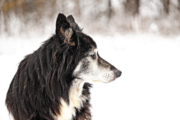
IV. DOG BREEDS AT RISK
Gallbladder Mucocele is overwhelmingly a disease of specific breeds, indicating a strong genetic predisposition. The disease typically affects middle-aged to older dogs (average age 9–11 years).
| Breed | Risk Level | Notable Features |
|---|---|---|
| Shetland Sheepdog (Sheltie) | Highest | Estimated 25x higher risk than other breeds; often present with mucocele concurrently with subclinical hypothyroidism. |
| Cocker Spaniel | High | Known for concurrent liver disease and metabolic issues. |
| Miniature Schnauzer | High | Prone to hyperlipidemia and pancreatitis, conditions that frequently increase mucocele risk. |
| Dachshund | Elevated | Known predisposition to various metabolic and endocrine disorders. |
| Chihuahua, Pomeranian, Beagle | Moderate | Increased risk compared to large or giant breeds. |
Explanation of Breed Predisposition
The heightened risk in specific breeds, particularly the Shetland Sheepdog, Cocker Spaniel, and Miniature Schnauzer, is hypothesized to be linked to genetic defects affecting the mucosal lining of the gallbladder and bile composition. In Shelties, researchers have investigated potential mutations in genes responsible for bile transport (such as the ABCB4 gene). Changes in these transport mechanisms may lead to altered bile acid concentrations or, more commonly, a dysregulation that promotes the hyper-secretion of mucin by the gallbladder epithelium. This results in the initial formation of the thick, gelatinous matrix that defines the mucocele. Furthermore, these breeds often have a higher incidence of metabolic disorders (like hyperlipidemia in Schnauzers) or endocrine diseases (like hypothyroidism in Shelties and Cocker Spaniels), which synergistically combine with the genetic susceptibility to trigger the mucocele formation. This genetic component suggests that GBM is often a primary biliary disease rather than solely a secondary complication of Cushing’s—though Cushing’s disease certainly exacerbates it.
V. AFFECTS PUPPY, ADULT, OR OLDER DOGS
Gallbladder Mucocele is predominantly a disease of middle-aged to older dogs.
- Puppies (Under 1 year): Mucocele formation is extremely rare in puppies. The underlying metabolic and hormonal conditions (Cushing’s, hypothyroidism, chronic hyperlipidemia) that drive mucocele development are diseases of aging.
- Adult Dogs (1–7 years): While less common than in seniors, GBM can occur in younger adult dogs, especially those with aggressive, early-onset endocrine disease or a severe genetic predisposition (like a Sheltie).
- Older/Senior Dogs (8+ years): This is the high-risk age bracket. The average age of diagnosis is around 9 to 11 years. The condition is often progressive, meaning the pathological thickening of the bile and mucus takes years to advance to the point of complete obstruction.
VI. DIAGNOSIS
Diagnosis of GBM requires a combination of clinical signs, bloodwork abnormalities, and definitive diagnostic imaging.
A. Laboratory Diagnostics (Bloodwork)
Blood tests indicate systemic stress, inflammation, and compromised liver/biliary function:
- Biochemistry Panel:
- Elevated Liver Enzymes: Alkaline Phosphatase (ALP) and Alanine Aminotransferase (ALT) are almost invariably elevated, sometimes dramatically. ALP elevation is particularly significant as it suggests cholestasis (bile flow obstruction).
- Hyperbilirubinemia: Elevated total bilirubin confirms extrahepatic biliary obstruction (bile cannot pass into the small intestine). This correlates clinically with jaundice.
- Hypercholesterolemia and Hypertriglyceridemia: Elevated lipids are common findings, often indicating the underlying metabolic issue contributing to mucocele formation.
- Complete Blood Count (CBC): May show leukocytosis (elevated white blood cells) if inflammation (cholecystitis) or infection (sepsis/peritonitis) is present.
- Coagulation Profile: Essential pre-operatively. Dogs with liver disease or sepsis are at high risk for inadequate clotting factor production or consumption (DIC). Prolonged PT (Prothrombin Time) and aPTT (Activated Partial Thromboplastin Time) indicate coagulopathy.
- Endocrine Testing: Screening for underlying Cushing’s (Low-Dose Dexamethasone Suppression Test, ACTH stimulation test) and hypothyroidism (Thyroid panel, T4, TSH) is crucial after stabilization, as managing these diseases is key to long-term prevention.
B. Diagnostic Imaging (The Gold Standard)
1. Abdominal Ultrasound: Ultrasound is the definitive diagnostic tool for GBM. The mucocele has a pathognomonic appearance, allowing for immediate identification:
- The “Kiwi Fruit” or “Starburst” Sign: The classic appearance is a non-mobile, stellate (star-shaped) or concentric, thickly striating pattern within the gallbladder lumen. This pattern represents the layers of inspissated mucin and is visually distinct from simple fluid bile or loose gallstones, which typically shift with gravity.
- Gallbladder Wall Thickening: The wall often appears thickened and sometimes irregularly layered (hyperechoic).
- Distension: The gallbladder is markedly enlarged (distended).
- Biliary Duct Assessment: The ultrasonographer assesses the patency of the cystic and common bile ducts. Dilation of these ducts proximal to the obstruction confirms EHBO.
- Peritoneal Fluid: The presence of free fluid in the abdomen (ascites) suggests possible leakage or rupture. Fluid sampling (abdominocentesis) may confirm septic peritonitis.
2. Radiography (X-rays): Radiographs are generally non-diagnostic for GBM itself, as the mucocele matrix is often non-calcified. However, X-rays may be useful for:
- Identifying underlying concurrent conditions (e.g., enlarged liver, calcifications associated with Cushing’s).
- Detecting free air or general loss of abdominal detail suggestive of peritonitis or rupture.
3. Contrast Studies/CT: While not typically required, advanced imaging can aid in complex surgical planning or if a mass (tumor) differential needs to be ruled out.
VII. TREATMENT
Treatment for a diagnosed, obstructive, or symptomatic Gallbladder Mucocele is primarily surgical. Medical management is reserved for stabilization or for non-obstructive, incidental findings in asymptomatic dogs.
A. Pre-operative Stabilization (Critical Period)
Due to the high risk of shock, sepsis, and coagulopathy, stabilization is mandatory before surgery:
- Fluid Therapy: Aggressive intravenous crystalloid fluid therapy to combat dehydration, shock, and optimize perfusion.
- Antibiotics: Broad-spectrum intravenous antibiotics are administered immediately, as bile is sterile, but obstruction often leads to bacterial proliferation (ascending infection, cholecystitis), and rupture guarantees sepsis. Antibiotics must cover enteric organisms (E. coli, Clostridium) and should be continued post-operatively for several weeks.
- Pain Management: Opioids (e.g., fentanyl, morphine) are used to control severe abdominal pain. NSAIDs are avoided pre-surgery due to their potential to worsen renal perfusion and increase bleeding risk.
- Coagulopathy Management: If a clotting disorder is detected, fresh frozen plasma (FFP) or packed red blood cells may be required. Vitamin K supplementation is critical, particularly pre-operatively, to aid in the production of functional clotting factors (as Vitamin K absorption is impaired by the lack of bile).
B. Surgical Treatment: Cholecystectomy
Cholecystectomy (surgical removal of the gallbladder) is the definitive treatment for symptomatic or severely obstructive GBM.
- Procedure: The entire gallbladder is carefully dissected from the liver bed (hepatic fascia) and removed, along with the cystic duct. The bile contents are aspirated and submitted for culture. A sample of the gallbladder wall and sometimes liver tissue is submitted for histopathology to confirm the diagnosis and check for malignancy or concurrent liver disease.
- Biliary Patency Check: After removal, the surgeon must ensure the remaining common bile duct (CBD) is patent and draining properly into the duodenum. If the CBD is obstructed (e.g., by migrating mucocele debris), a procedure to flush the CBD or place a temporary biliary stent may be required (choledochal tube placement).
- Abdominal Lavage: If rupture and peritonitis have occurred, the abdomen is repeatedly flushed with large volumes of sterile, warm saline to remove all contaminated bile and necrotic tissue.
- Post-Operative Drains: Abdominal drains (e.g., Jackson-Pratt) may be placed temporarily if there is significant contamination or risk of ongoing leakage.
C. Medical Management (Palliative/Incidental GBM)
Medical treatment is generally reserved for dogs that are completely asymptomatic with an incidental finding of a small mucocele, or dogs that are too unstable for surgery. This approach is fraught with risk, as the mucocele can become obstructive or rupture at any time.
- Ursodeoxycholic Acid (Ursodiol): This is the key medical agent. Ursodiol modifies the composition of bile, making it more fluid, easing the flow, and reducing the toxicity of endogenous bile acids to the liver. It helps prevent further sludge formation.
- Cholesterol and Lipid Control: Diet and medication (e.g., fibrates, statins) to manage hyperlipidemia.
- Antioxidants and Liver Support: S-Adenosylmethionine (SAMe) and milk thistle (silybin) may be used to support liver function.
- Management of Underlying Disease: Aggressive control of Cushing’s disease or hypothyroidism is essential to slow the progression of the mucocele.
VIII. PROGNOSIS & COMPLICATIONS
The prognosis for canine Gallbladder Mucocele varies dramatically based on two factors: the dog’s health status pre-surgery and whether the gallbladder has ruptured.
A. Prognosis
- Elective Cholecystectomy (Non-Ruptured, Stable Dog): Prognosis is generally good to excellent. Survival rates for stable dogs undergoing cholecystectomy are high, often exceeding 90%. Long-term quality of life is typically excellent, as the gallbladder is a non-essential organ.
- Emergency Cholecystectomy (Ruptured Mucocele/Peritonitis): Prognosis is guarded. Mortality rates are significantly higher, ranging from 20% to 40%, even with aggressive intensive care, due to the severe systemic septic shock and high risk of DIC.
B. Major Complications
- Gallbladder Rupture and Septic Peritonitis: The most devastating complication. Requires immediate intervention and carries the highest mortality risk.
- Disseminated Intravascular Coagulation (DIC): A severe, systemic complication often triggered by sepsis (from rupture) or severe inflammation. DIC causes micro-clots throughout the body, followed by massive hemorrhage, and is frequently fatal.
- Post-Surgical Bile Leakage: If the common bile duct is damaged or the suture line leaks, bile can continue to spill into the abdomen, requiring a second surgery.
- Biliary Stasis/Cholangitis: Even after gallbladder removal, the underlying conditions sometimes lead to inflammation or stasis in the remaining bile ducts (choledochus).
- Recurrence of Underlying Disease: If endocrine diseases (like Cushing’s) are not managed effectively post-surgery, they can still negatively impact overall lifespan and liver health.
IX. PREVENTION
While genetic predisposition is unavoidable, prevention efforts focus heavily on early detection and management of associated metabolic and endocrine disorders.
- Routine Monitoring for At-Risk Breeds: Shelties, Miniature Schnauzers, and Cocker Spaniels should undergo annual or bi-annual wellness screening, including bloodwork (checking for hyperlipidemia and elevated liver enzymes) and annual or bi-annual abdominal ultrasound screening starting around middle age (6–8 years). Ultrasound can identify a developing mucocele long before it becomes symptomatic.
- Aggressive Management of Endocrine Disease: Any dog diagnosed with Hyperadrenocorticism or Hypothyroidism must have their condition strictly controlled with appropriate medication (e.g., trilostane, levothyroxine).
- Dietary Modification for Hyperlipidemia: If the dog is prone to elevated triglycerides or cholesterol, a strict low-fat diet is essential to reduce the load on the liver and gallbladder.
- Proactive Ursodiol Therapy: In high-risk breeds showing only mild biliary sludge, some veterinary specialists recommend prophylactic treatment with Ursodiol to fluidize the bile and slow the mucocele’s progression.
X. DIET AND NUTRITION
Dietary management is paramount both in the prevention of GBM and in the recovery phase post-cholecystectomy, focusing on reducing fat intake and supporting liver function.
A. Pre-Operative and Maintenance Diet (For At-Risk Dogs)
The primary goal is to minimize the metabolic strain on the liver and reduce the amount of fat requiring emulsification by bile.
- Ultra Low-Fat Diet: Dogs prone to mucocele formation or those with subclinical mucocele MUST be transitioned to a prescription low-fat gastrointestinal or hepatic diet (e.g., Royal Canin Gastrointestinal Low Fat, Hills i/d Low Fat, Purina EN Low Fat). Dietary fat should typically be kept under 10% on a dry matter basis. Lipids stimulate CCK release, causing gallbladder contraction; a high-fat diet increases the workload on an already struggling gallbladder.
- High-Quality, Digestible Protein: Adequate protein is needed for liver regeneration and metabolic function, but should be highly digestible.
- Fiber: Controlled levels of soluble and insoluble fiber to support gastrointestinal health.
B. Post-Cholecystectomy Diet
Immediately following surgery, the dog may require temporary feeding via an esophageal or nasogastric tube if they are anorexic. Once stable and eating:
- Continuation of Ultra Low-Fat Diet: Even without a gallbladder, the liver still produces bile, but the storage reservoir is gone. The bile is constantly dripping directly into the small intestine. A low-fat diet ensures that the small, constant flow of bile is sufficient to emulsify the fat consumed, preventing maldigestion and diarrhea (steatorrhea).
- Vitamin K Supplementation: Post-operative supplementation with Vitamin K (often injectable initially, then oral) is crucial until liver function assessment is optimized, especially if there were pre-existing coagulopathies.
- B-Complex Vitamins: These water-soluble vitamins are often depleted during chronic illness and stress.
C. Nutritional Supplements
Liver support supplements containing SAMe (S-Adenosylmethionine), Silybin (Milk Thistle), and Vitamin E are recommended to reduce oxidative stress and aid in hepatocyte (liver cell) protection and regeneration. Taurine may also be beneficial in certain breeds.
XI. ZOONOTIC RISK
There is absolutely no zoonotic risk associated with Canine Gallbladder Mucocele.
Gallbladder Mucocele is a non-infectious, acquired disease stemming from the dog’s unique physiological and metabolic dysfunctions (hypersecretion of mucin, hormonal imbalances, lipid disorders). It is not caused by a virus, bacterium, or parasite that can be transmitted from the dog to human family members or other pets. The only associated infectious risk is bacterial cholecystitis, which is an endogenous infection (bacteria migrating from the GI tract into the biliary system) and poses no risk of horizontal transmission.
#GallbladderMucocele #DogHealth #CanineVeterinaryMedicine #VetMed #Cholecystectomy #DogGallbladder #SheltieHealth #CockerSpaniel #GallbladderSurgery #PetHealthGuide #DogWellness #VeterinarySurgery #CushingDisease #DogGIssues #CanineBiliaryObstruction #VetEducation #GallbladderRupture #DogCareTips


Add comment