
Derzsy’s disease, first described in the 1930s in Hungary by Dr. Károly Derzsy, is a highly contagious viral infection primarily of geese caused by Goose Parvovirus (GPV). Although the disease is most commonly associated with domestic and wild geese, the same virus (or very closely related strains) has been repeatedly isolated from ducks (Anas spp.) worldwide, especially in mixed‑species waterfowl operations.
In the decades following its discovery, GPV was identified as a member of the Parvoviridae family, subfamily Parvovirinae, genus Dependoparvovirus (formerly Parvovirus). The virus exhibits a high degree of genetic stability, yet minor antigenic drift has resulted in regional variants that differ in virulence and host‑range.
Understanding GPV in ducks is essential for several reasons:
- Economic impact – Duck farming contributes billions of dollars to the global poultry sector; an outbreak can decimate flocks, reduce egg production, and cause mortality up to 80 % in naïve ducklings.
- Animal welfare – The disease produces severe enteric and hepatic lesions, leading to prolonged suffering.
- Bio‑security – Mixed waterfowl operations provide a conduit for virus spread to adjacent poultry and wild bird populations.
This guide consolidates the most recent peer‑reviewed literature (2020‑2025), veterinary field manuals, and practical observations from commercial producers to furnish a single, exhaustive resource for veterinarians, farm managers, researchers, and students.
2. Etiology – The Virus and Its Characteristics
| Feature | Description |
|---|---|
| Taxonomy | Family Parvoviridae, subfamily Parvovirinae, genus Dependoparvovirus (formerly Parvovirus). |
| Genome | Single‑stranded DNA (~5.1 kb) encoding two major open reading frames (ORFs): non‑structural (NS) proteins (NS1, NS2) and capsid (VP1, VP2) proteins. |
| Virion | Non‑enveloped, icosahedral capsid (~25 nm diameter), highly resistant to environmental factors (temperature, pH, desiccation). |
| Stability | Survives for months in dry litter, water, and feces; resistant to many disinfectants (e.g., alcohol) but inactivated by 0.1 % sodium hypochlorite, 0.5 % phenolic solutions, or heat ≥ 60 °C for 30 min. |
| Replication | Strictly autonomous (no helper virus required) – replicates in rapidly dividing cells of the intestinal crypts, bone marrow, and lymphoid tissue. |
| Antigenic Variants | Two major clades: Classical GPV (European/Asian lineages) and Muscovy Duck Parvovirus (MDPV) – the latter is a divergent strain that can infect both Muscovy ducks and other Anseriformes. Cross‑neutralization is partial, which influences vaccine design. |
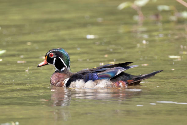
| Host Range | Primarily geese (Anser anser) and Muscovy ducks (Cairina moschata), but experimental infection demonstrates susceptibility of Pekin, Mallard, Runner, Khaki Campbell, and other domestic duck breeds. Wild waterfowl (e.g., Anas platyrhynchos) can act as reservoirs. |
The virus’s capacity to bind cellular receptors (predominantly α2‑3 sialic acid residues on enterocytes) and to subvert the host’s DNA‑damage response underpins its tropism for the gastrointestinal tract and bone marrow, which explains the characteristic hemorrhagic enteritis, pancytopenia, and immunosuppression observed clinically.
3. Transmission Pathways & Epidemiology
- Fecal‑Oral Route – The most efficient transmission mode. Infected ducklings shed 10⁸–10⁹ virions/g of feces for 7–10 days post‑infection. Contaminated water sources, feed, and litter act as vehicles.
- Vertical Transmission – Low but documented; embryonic infection leads to hatchability loss and “runting‑stunting” syndrome in chicks.
- Aerosol & Dust – Viable virus can be aerosolised from dried feces and litter, especially in poorly ventilated houses.
- Mechanical Vectors – Flies, beetles, and equipment (clippers, nets) can mechanically transport virions between pens.
- Wild Bird Interface – Migratory geese and ducks can introduce the virus into naïve farms, particularly during the autumn migration.
Epidemiologic Patterns
- Seasonality – Outbreaks peak in late spring to early summer, coinciding with the hatching of ducklings and higher moisture levels that favor virus stability in water.
- Geographical Hotspots – High incidence in Eastern Europe (Poland, Hungary, Romania), Southeast Asia (China, Vietnam, Thailand), and Northern United States (Midwest waterfowl farms) where intensive duck production co‑exists with geese.
Risk Factors
| Factor | Why It Increases Risk |
|---|---|
| High stocking density | Facilitates rapid fecal‑oral spread |
| Mixed‑species rearing (goose + duck) | Cross‑species infection via shared water |
| Inadequate litter turnover | Virus persists in dried droppings |
| Poor water sanitation | Virus remains infectious for weeks in stagnant water |
| Absence of vaccination program | Naïve flocks lack protective antibodies |
| Stress (handling, transport) | Immunosuppression augments susceptibility |
4. Duck Breeds at Risk – Who Is Most Vulnerable?
Overall Susceptibility
All domestic duck breeds can be infected, but genetic background, production purpose, and management practices create a hierarchy of risk.
| Breed | Typical Use | Relative Susceptibility | Rationale |
|---|---|---|---|
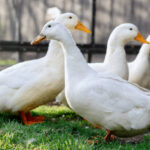
| Pekin (American Pekin) | Meat (broiler) | High | Fast growth, high stocking densities; early hatchlings are exposed to contaminated environments. |
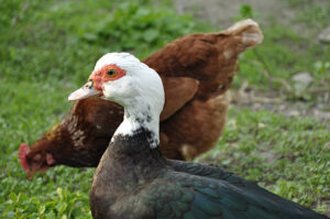
| Muscovy (Cairina moschata) | Meat, free‑range | Very High | Naturally more permissive to Muscovy Duck Parvovirus (MDPV) which often co‑circulates with GPV. |
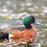
| Mallard (Anas platyrhynchos) | Game, ornamental | Moderate | Often kept in semi‑extensive systems with water bodies; exposure to wild carriers. |
| Runner (Indian Runner) | Egg production | Moderate‑High | Longer laying cycles mean they remain in the flock longer, increasing cumulative exposure. |
| Khaki Campbell | Egg production | High | High egg‑laying rates require intensive nutrition and housing, fostering virus persistence. |
| Aylesbury | Meat | High | Traditional large‑scale farms often share water sources with geese. |
| Native/Heritage Breeds (e.g., Saxony, Rouen) | Dual‑purpose | Variable | Typically reared on smaller farms with better bio‑security, but occasional contact with wild waterfowl raises risk. |
Paragraph Explanation
The Muscovy duck stands out as the breed most at risk because it is the natural host for Muscovy Duck Parvovirus (MDPV), a virus that is genetically close to GPV and often co‑circulates in the same environment. MDPV infection produces a clinical picture indistinguishable from classic Derzsy’s disease, yet Muscovy ducks show a higher mortality rate (up to 90 % in 1‑day‑old ducklings) due to a combination of immune naïveté, rapid viral replication, and severe bone‑marrow suppression.
The Pekin, being the global standard for duck meat production, is reared in high‑density, intensive systems where litter, water, and feed are shared among thousands of birds. The massive turnover of ducklings each season creates a perpetual pool of susceptible individuals, and the concentration of virus in the environment can reach epidemic levels within a few weeks.
In contrast, heritage and backyard breeds often benefit from lower stocking densities, more frequent litter changes, and limited contact with geese, thereby experiencing lower infection pressure. However, these flocks are not immune; wild waterfowl that frequent garden ponds or free‑range areas can act as carriers, delivering the virus directly to otherwise well‑managed domestic ducks.
Overall, any breed raised in proximity to geese or in environments where water is shared will face an elevated risk, and the age of the birds (especially the first two weeks of life) is the single most decisive factor in disease outcome.
5. Life‑Stage Susceptibility – When Are Ducks Most Vulnerable?
| Life Stage | Age Range | Immunological Status | Typical Clinical Outcome |
|---|---|---|---|
| Embryo (in‑ovo) | 0–21 days incubation | No immune system; dependent on maternal antibodies | Embryonic death, deformities, reduced hatchability |
| Day‑Old Duckling | 0–7 days | Immunologically naïve; maternal antibodies waning | Severe hemorrhagic enteritis, high mortality (60‑80 %) |
| Early Chick (1–3 weeks) | 8–21 days | Developing innate immunity; limited adaptive response | Diarrhea, stunted growth, moderate mortality (10‑30 %) |
| Grow‑out (3–8 weeks) | 22–56 days | Gradual maturation of lymphoid organs | Subclinical infections possible; occasional “runting‑stunting” |
| Adult (≥ 8 weeks) | > 56 days | Competent immune system, often with protective antibodies if previously exposed or vaccinated | Usually asymptomatic carriers; occasional recrudescence during stress or immunosuppression |
Key Points
- Maternal Antibodies – In flocks where breeders have been vaccinated against GPV, passive immunity can protect ducklings for the first 7‑10 days. However, antibodies decline rapidly, leaving a narrow protective window.
- Critical Window – The first 72 hours of life represent the period of greatest susceptibility. Viral replication in the intestinal crypts overwhelms the immature gut barrier, resulting in massive hemorrhage and dehydration.
- Age‑Related Clinical Spectrum – Younger birds display acute, fulminant disease with high case‑fatality rates, whereas older birds may experience milder, subclinical infections that nonetheless foster virus shedding and act as reservoirs.
6. Clinical Presentation – Signs & Symptoms
6.1. General Overview
Derzsy’s disease in ducks is an acute, highly contagious enteric infection that can progress to systemic involvement. The disease course can be divided into three phases:
- Incubation (2–7 days) – No outward signs; virus replicates subclinically in the intestinal crypts and bone marrow.
- Acute Phase (3–5 days post‑onset) – Sudden onset of clinical signs, rapid deterioration.
- Convalescent/Carrier Phase (≥ 7 days) – Survivors may appear normal but continue to shed virus.
6.2. Detailed Clinical Signs
| System | Specific Sign | Typical Onset | Comments |
|---|---|---|---|
| Gastrointestinal | Profuse, watery, often bloody diarrhea (dark red or tarry) | 2‑3 days after incubation | Feces may contain mucus and fibrin; dehydration is rapid. |
| Respiratory | Labored breathing (dyspnea), occasional nasal discharge | Often concurrent with diarrhea | Secondary bacterial pneumonitis common in later stages. |
| Skeletal/Locomotor | Weakness, staggering, “waddling” gait; occasional perosis (sloping legs) in survivors | Early to mid‑acute phase | Reflects bone‑marrow suppression and anemia. |
| Hepatic | Jaundice (icteric sclerae), enlarged liver on palpation | 3‑4 days | Hepatocellular necrosis contributes to coagulopathy. |
| Hematologic | Pale combs/skin (anemia), petechial hemorrhages on skin, cyanosis of extremities | Rapidly after diarrhea begins | Indicative of thrombocytopenia and hemorrhagic diathesis. |
| Neurologic | Tremors, ataxia, seizures (rare) | Late acute phase | Usually secondary to severe hypoxia or metabolic derangements. |
| General | Lethargy, inappetence, mortality (up to 80 % in naïve ducklings) | 1‑2 days after first signs | Mortality peaks at 48 h after clinical onset. |
| Post‑Recovery | Runting‑stunting syndrome, poor feather development, delayed sexual maturity | Weeks after infection | Survivors may become persistent carriers. |
Illustrative Case Example
A 3‑day‑old Pekin duckling flock (250 birds) presented with sudden onset of hemorrhagic diarrhea. Within 24 h, 60 % of the ducklings were dead, exhibiting pale mucous membranes, swollen livers, and extensive intestinal hemorrhage on necropsy. Surviving ducklings showed marked weight loss and delayed feathering for 3 weeks.
This scenario typifies the highly fatal nature of GPV in the first week of life and underscores the need for swift intervention.
6.3. Pathognomonic Findings
- Intestinal mucosal hemorrhage extending from the duodenum to the cloaca, with submucosal edema.
- Bone‑marrow aplasia on histology—marked depletion of erythroid and megakaryocytic lineages.
- Hepatocellular necrosis and focal hemorrhage.
These lesions are considered diagnostic when combined with appropriate laboratory confirmation.
7. Differential Diagnosis – Diseases That Mimic Derzsy’s Disease
| Disease | Key Distinguishing Features |
|---|---|
| Duck Viral Enteritis (DVE/DEAV) – caused by Anatid herpesvirus 1 | Typically older birds, with conjunctivitis, respiratory signs, and necrotizing enteritis; virus is a DNA herpesvirus, not a parvovirus. |
| Riemer’s Disease (Avian adenovirus) | Hepatomegaly, inclusion bodies in liver, transient immunosuppression; caused by adenovirus and usually seen in 2‑4 week‑old birds. |
| Bacterial Septicemia (e.g., Salmonella spp.) | Fever, purulent lesions, positive bacterial culture; diarrhea may be mucoid rather than hemorrhagic. |
| Coccidiosis (Eimeria spp.) | Diarrhea with white‑to‑yellow feces, presence of oocysts; lesions limited to intestinal epithelium without systemic hemorrhage. |
| Aflatoxicosis | Chronic weight loss, liver swelling, yellow discoloration; toxin analysis confirms exposure. |
| Nutritional deficiencies (e.g., Vitamin K deficiency) | Delayed clotting, hemorrhage but no intestinal lesions; diet history and coagulation profile help differentiate. |
| Muscovy Duck Parvovirus (MDPV) | Clinically identical to GPV; laboratory differentiation relies on PCR sequencing or serology. |
Approach to Differentials
- History – Age of affected birds, recent introductions of new flocks, vaccination status, feed changes.
- Clinical pattern – Rapid onset and high mortality in ducklings strongly favor GPV.
- Laboratory testing – PCR, virus isolation, histopathology, and serology are essential for definitive differentiation.
8. Laboratory & Field Diagnosis
8.1. Sample Collection
| Sample | Quantity | Collection Method | Recommended Transport |
|---|---|---|---|
| Feces | 1–2 g fresh | Direct scooping from cloaca or litter | Cold (4 °C) transport, ≤ 24 h |
| Intestinal tissue | 2–3 cm sections (duodenum, jejunum) | Post‑mortem aseptic excision | 10 % neutral buffered formalin for histology; separate portion in viral transport medium (VTM) for PCR |
| Liver | 1 g | Post‑mortem | Same as intestinal tissue |
| Bone marrow | Small core from femur | Post‑mortem | RNAlater or VTM |
| Serum | 0.5–1 ml | Blood from wing vein | Freeze at –20 °C for serology (ELISA) |
| Swab (cloacal) | One swab per 5 birds | Sterile cotton swab placed in VTM | Same as feces |
8.2. Diagnostic Techniques
| Method | Principle | Turn‑Around Time | Sensitivity / Specificity |
|---|---|---|---|
| Conventional PCR (targeting NS1 gene) | Amplifies virus‑specific DNA | 6–12 h | ≥ 95 % (detects as few as 10 copies) |
| Real‑Time qPCR | Quantitative detection with fluorescent probes | 3–6 h | Highest sensitivity (≤ 5 copies); allows viral load monitoring |
| Virus Isolation (primary duck embryo fibroblasts) | Cytopathic effect (CPE) – rounding, syncytia | 5–7 days | Gold standard but less sensitive; requires biosafety level 2 |
| Immunofluorescence (IF) / Immunohistochemistry (IHC) | Detect viral antigens in tissue sections | 24–48 h | Specific; useful for confirming lesions |
| ELISA (Serology) | Detects anti‑GPV antibodies | 4–6 h | Useful for flock immunity monitoring, not for acute diagnosis |
| Sequencing (Sanger / Next‑Gen) | Full‑genome typing, strain differentiation | 1–3 days | Critical for epidemiologic tracing and vaccine matching |
Interpretation Tips
- Positive PCR with compatible clinical signs = definitive diagnosis.
- Negative PCR does not rule out disease if sampling occurred after viral clearance; repeat testing 48 h later or use serology to detect seroconversion.
- Histopathology (hemorrhagic enteritis, marrow aplasia) strongly supports GPV infection even when molecular tests are equivocal.
9. Pathogenesis – How the Virus Hijacks the Host
- Entry & Attachment – GPV binds to α2‑3 linked sialic acid receptors on the apical surface of intestinal epithelial cells and bone‑marrow stromal cells.
- Endocytosis – The virus is internalized via clathrin‑mediated endocytosis and transported to the nucleus.
- Replication – The single‑stranded DNA is converted into a double‑stranded replicative form (RF) using host polymerases. The NS1 protein orchestrates viral DNA replication, induces DNA damage response, and triggers apoptosis.
- Cellular Damage – Massive replication in crypt cells leads to:
- Apoptosis of enterocytes → loss of barrier function → hemorrhagic diarrhea.
- Bone‑marrow aplasia → pancytopenia → anemia, thrombocytopenia, immunosuppression.
- Hepatocyte necrosis → impaired coagulation, jaundice.
- Immune Evasion – The virus lacks a capsid protein that is readily recognized by pattern‑recognition receptors, allowing it to replicate before innate immunity mobilizes.
- Systemic Spread – Viremia disseminates virus to liver, spleen, and kidneys, contributing to multi‑organ lesions.
- Shedding – Infected ducks excrete virus in feces, urine, and respiratory secretions for up to 10 days, maintaining a high environmental load.
The combined effect of gastrointestinal bleeding, severe anemia, and immunosuppression explains the rapid progression to death in naïve ducklings.
10. Treatment Options – Therapeutic Principles & Protocols
Note: No antiviral drug is licensed against GPV. Management is supportive and preventive. Prompt intervention can markedly reduce mortality.
10.1. Immediate Supportive Care
| Intervention | Details | Dosage / Frequency |
|---|---|---|
| Fluid Therapy | Rehydration with balanced electrolyte solution (e.g., Lactated Ringer’s or commercial avian electrolytes). | 30 ml/kg subcutaneously (SC) initially; repeat every 6–8 h as needed. |
| Oral Rehydration | 5 % glucose‑saline solution via drinking water; add electrolytes (Na⁺, K⁺, Cl⁻). | Provide ad libitum for 24‑48 h. |
| Antibiotics (Secondary Bacterial Infections) | Broad‑spectrum coverage (e.g., enrofloxacin 10 mg/kg IM/SC once daily or ampicillin 20 mg/kg IM/SC BID) until bacterial culture results. | 5‑7 days; adjust based on sensitivity. |
| Anti‑Inflammatories | NSAIDs (e.g., meloxicam 0.5 mg/kg IM/SC once daily) to reduce fever and inflammation. | 3‑5 days. |
| Vitamin K1 | Counteracts coagulopathy from hemorrhage. | 0.5 mg/kg SC daily for 3 days. |
| Probiotics / Prebiotics | Restore gut flora, reduce secondary enteritis. | Bacillus subtilis or Lactobacillus spp. 10⁸ CFU/kg feed. |
| Thermal Support | Maintain ambient temperature 30‑32 °C for ducklings < 2 weeks to reduce metabolic stress. | Continuous. |
10.2. Antiviral Research (Experimental)
- L‑nucleoside analogues (e.g., cidofovir) have shown in vitro inhibition of GPV replication, but toxicity precludes field use.
- RNA‑interference (siRNA) targeting NS1 is under experimental evaluation; no commercial product exists.
10.3. Nursing Strategies
- Isolate affected pens immediately; use dedicated footwear and equipment.
- Disinfect all surfaces with 0.1 % sodium hypochlorite or 2 % glutaraldehyde after removal of organic debris.
- Implement a “clean‑in‑place” (CIP) system for water lines – flush with heated water (≥ 70 °C) followed by chemical disinfectant.
10.4. Duration of Care
- Most ducklings that survive the first 72 h with intensive care recover within 5–7 days.
- Survivors should be monitored for 2 weeks for delayed sequelae (runting, anemia).
11. Prognosis & Complications – Expected Outcomes
| Scenario | Expected Mortality | Recovery Rate | Common Complications |
|---|---|---|---|
| Naïve ducklings (< 7 days) | 60‑80 % (up to 90 % in Muscovy) | 20‑40 % survive with intensive therapy | Persistent anemia, secondary bacterial sepsis, runting‑stunting, chronic immunosuppression |
| Older ducklings (1‑3 weeks) | 10‑30 % | 70‑90 % recover | Delayed growth, reduced feed conversion, occasional hepatic fibrosis |
| Adults (≥ 8 weeks) | < 5 % (often subclinical) | > 95 % survive | Carrier state (viral shedding), reduced egg production (5‑10 % drop) |
| Vaccinated flock | < 2 % (usually mild) | Near‑100 % recovery | Rare vaccine‑associated mild enteritis (transient) |
Key Complications
- Secondary Bacterial Septicemia – Common due to compromised gut barrier; E. coli and Salmonella are frequent culprits.
- Chronic Hematologic Deficits – Persistent pancytopenia can predispose to other infections.
- Runting‑Stunting Syndrome – Survivors may never reach normal market weight, impacting economic returns.
- Reduced Reproductive Performance – In layers, egg quality and hatchability can fall for 4‑6 weeks after an outbreak.
Overall, early detection and aggressive supportive therapy dramatically improve the prognosis, especially in the most vulnerable ducklings.
12. Prevention & Bio‑Security Strategies
12.1. Vaccination
| Vaccine Type | Composition | Administration | Schedule | Efficacy |
|---|---|---|---|---|
| Live Attenuated (LA) GPV vaccine | Attenuated GPV strain (e.g., GPV‑KX‑G). | Oral or eye-drop to breeding stock; subcutaneous to ducklings > 7 days. | Breeders: 2 doses, 3 weeks apart before lay; Ducklings: single dose at 7 days, booster at 21 days. | 85‑95 % protection against clinical disease; reduces viral shedding by ~80 %. |
| Inactivated (Killed) GPV vaccine | Formalin‑inactivated whole virus + adjuvant (oil‑based). | Intramuscular injection. | Breeders: 2 doses 4 weeks apart; Ducklings: 1 dose at 14 days, booster at 28 days. | 70‑80 % protection; safer for immunocompromised flocks. |
| Recombinant VP2 subunit vaccine (experimental) | Purified VP2 capsid protein expressed in baculovirus system. | Intramuscular or nasal spray. | 2 doses 2 weeks apart. | Preliminary data show 90 % seroconversion, pending regulatory approval. |
Vaccination Best Practices
- Cold‑chain maintenance – Keep vaccines at 2‑8 °C; discard if frozen.
- Batch testing – Perform serum neutralization on a subset of birds 2 weeks post‑vaccination to confirm seroconversion.
- Maternal antibodies – Avoid vaccinating ducklings before 7 days; maternal antibodies can neutralize live vaccines.
12.2. Environmental Hygiene
- Litter Management – Replace litter weekly; use absorbent, low‑dust materials (e.g., wood shavings).
- Water Hygiene – Clean water lines daily; use UV‑treated or chlorinated water (maintain ≤ 0.5 ppm free chlorine).
- Rodent & Insect Control – Deploy fly traps, insecticidal netting, and rodent baits to prevent mechanical vectors.
12.3. Quarantine & Movement Controls
- Isolation – New birds should be quarantined for minimum 30 days; test by PCR before integration.
- Personnel Protocols – Dedicated clothing, boots, and handwashing stations for each house.
- Equipment Disinfection – Immerse nets, feeders, and transport crates in 0.1 % hypochlorite for 10 min after each use.
12.4. Genetic Selection
Some breeding programs have identified lines with enhanced innate immunity (higher NK cell activity, stronger IFN‑α response). Select for these traits where possible, though commercial viability remains under study.
12.5. Monitoring & Early Warning
- Weekly fecal PCR screening of sentinel birds.
- Serological surveillance (ELISA) every 3 months to assess flock immunity.
- Automated water‑temperature & pH loggers – abnormal values may signal increased viral stability.
13. Nutrition & Dietary Management During Outbreaks
Proper nutrition supports immune function, intestinal integrity, and recovery.
13.1. Feed Formulation
| Nutrient | Recommended Level (per kg feed) | Rationale |
|---|---|---|
| Crude Protein | 22‑24 % for ducklings < 2 weeks; 20 % for growers | Provides amino acids for tissue repair and hematopoiesis. |
| Methionine + Cysteine | 0.9‑1.2 % (combined) | Sulphur amino acids are essential for glutathione synthesis, a key antioxidant. |
| Vitamin A | 12 000 IU | Enhances mucosal integrity and epithelial regeneration. |
| Vitamin E | 150 IU | Antioxidant; reduces oxidative stress from viral replication. |
| Vitamin C | 250 mg | Supports leukocyte function; can be added to drinking water during acute phase. |
| Vitamin D3 | 4 000 IU | Modulates innate immunity. |
| Zinc | 100 mg | Crucial for thymic function and wound healing. |
| Selenium | 0.3 mg | Works with vitamin E; deficiency worsens oxidative damage. |
| Probiotics | 10⁸ CFU/kg (Bacillus subtilis) | Restores gut flora after diarrhea. |
| Prebiotics (e.g., inulin) | 2 % | Fuels beneficial bacteria. |
13.2. Feeding Strategies During Illness
- Liquid Diets – Offer mesophilic gelatin‑based or soy‑based liquid feeds (e.g., 30 % protein) to ensure intake despite reduced appetite.
- Frequent Small Meals – Offer feed 4‑6 times daily rather than ad libitum to stimulate ingestion.
- Electrolyte‑Enriched Water – Add sodium bicarbonate (1 g/L) and potassium chloride (0.5 g/L) to compensate for losses.
13.3. Post‑Recovery Nutrition
- High‑Energy Supplements – Add corn oil (2 %) or fish oil (1 %) to improve weight gain.
- Bone‑Strengthening Additives – Calcium carbonate (1 %) and vitamin D3 to reverse potential osteopenia from marrow suppression.
13.4. Monitoring Nutritional Status
- Hematocrit & Hemoglobin – Weekly checks to assess anemia.
- Serum Protein (Total & Albumin) – Indicator of gut absorption.
- Body Condition Scoring (BCS) – Target BCS 3‑4 for optimal health.
14. Zoonotic Potential – Risks to Humans
Current Scientific Consensus (2020‑2025 literature) indicates that Goose Parvovirus is not zoonotic. The virus lacks the ability to infect mammalian cells; experimental inoculation of mice, rabbits, and humans has not produced infection or seroconversion.
- Occupational Exposure – While direct infection is unlikely, handlers may be exposed to secondary bacterial pathogens (e.g., Salmonella spp.) present in contaminated feces, presenting a food‑borne or occupational health risk.
- Protective Measures – Standard personal protective equipment (PPE) (gloves, boot covers, face masks) and hand hygiene are sufficient.
- Public Health Advisory – No specific public health alerts are required for GPV; however, proper cooking of duck meat eliminates any potential bacterial contaminants.
15. Future Directions – Vaccines, Research Gaps & Emerging Tools
| Area | Current Status | Research Needs |
|---|---|---|
| Universal GPV/MDPV Vaccine | Strain‑specific live attenuated vaccines dominate. | Development of a bivalent or multivalent subunit vaccine covering GPV, MDPV, and emerging variants. |
| Molecular Diagnostics | qPCR is gold‑standard. | Portable loop‑mediated isothermal amplification (LAMP) kits for field use; integration with smartphone read‑outs. |
| Genomic Surveillance | Limited to regional studies. | Global next‑generation sequencing (NGS) of field isolates to track viral evolution and vaccine escape. |
| Host Genetics | Preliminary identification of immune‑gene markers. | GWAS (genome‑wide association studies) to select for disease‑resistant lines. |
| Antiviral Therapies | No licensed drugs. | Screening of broad‑spectrum nucleoside analogues and RNAi constructs; evaluation of CRISPR‑Cas13 antiviral platforms. |
| One‑Health Integration | Focused on poultry. | Incorporate wild‑bird monitoring and environmental sampling into national surveillance programs. |
| Model Systems | In‑ovo and embryonic fibroblast cultures used. | Development of 3‑D organoid cultures of duck intestine for pathogenesis studies. |
Key Take‑Home Message – Investment in multidisciplinary research (virology, immunology, genomics, bio‑informatics) will be pivotal to curtail GPV’s economic burden and safeguard the growing global duck industry.
16. Key Take‑Home Messages
- Derzsy’s disease (GPV) remains a high‑mortality, fecal‑oral disease that primarily devastates ducklings < 7 days old.
- Clinical hallmark: hemorrhagic, watery diarrhea combined with severe anemia, pallor, and rapid death.
- Diagnosis relies on PCR/qPCR from feces or intestinal tissue, supported by histopathology and serology.
- Treatment is supportive (fluids, electrolytes, secondary‑bacterial control); no specific antivirals exist.
- Vaccination of breeders and ducklings (live attenuated or inactivated) is the most effective preventive measure, reducing mortality by > 80 %.
- Bio‑security (strict quarantine, water sanitation, litter management) and regular surveillance are essential for outbreak avoidance.
- Nutrition—high‑protein, vitamin‑rich, probiotic‑enhanced diets—optimizes immune competence and speeds recovery.
- Zoonotic risk is negligible, but secondary bacterial infections pose occupational hazards.
- Future advancements (universal vaccines, field‑ready diagnostics, genomic surveillance) will be crucial to control GPV long‑term.
By integrating vigilant monitoring, rigorous bio‑security, targeted vaccination, and prompt supportive care, duck producers can dramatically lower the impact of Goose Parvovirus and ensure the health, welfare, and productivity of their flocks.
#GooseParvovirus, #DerzsysDisease, #DuckHealth, #WaterfowlMedicine, #AvianVirology, #DuckFarming, #PekinDuck, #MuscovyDuck, #DucklingCare, #BirdBiosecurity, #VeterinaryScience, #AnimalWelfare, #FarmManagement, #AnimalNutrition, #Parvovirus, #ViralEnteritis, #DuckDiseases, #AvianMedicine, #DuckVaccination, #PCRDiagnostics, #FieldVeterinary, #LivestockHealth, #Zoonosis, #OneHealth, #EmergingDiseases, #FarmBiosecurity, #AnimalHusbandry, #DuckProduction, #VeterinaryEducation, #ScientificResearch, #FutureOfFarming, #SustainablePoultry.
Add comment