
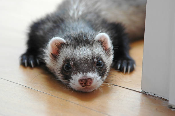
Ferrets (Mustela putorius furo) are popular pets due to their playful nature, intelligence, and affinity for human interaction. However, they are prone to a range of health issues, with gastrointestinal disorders being among the most common. One of the leading causes of vomiting and peptic ulcer disease (PUD) in ferrets is Helicobacter mustelae, a gram-negative, spiral-shaped bacterium that colonizes the gastric mucosa. This pathogen closely resembles Helicobacter pylori, the well-known causative agent of gastritis and ulcers in humans.
Understanding H. mustelae and its role in peptic ulcer disease is critical for both veterinary professionals and ferret owners. This comprehensive guide explores the etiology, clinical manifestations, diagnostic approaches, treatment strategies, prognosis, prevention, nutritional management, and zoonotic potential associated with H. mustelae infection in ferrets. The aim is to raise awareness, improve early diagnosis, and promote effective management of one of the most significant disease threats to ferret health.
What is Helicobacter mustelae?
Helicobacter mustelae is a microaerophilic, urease-producing bacterium first identified in the 1980s in the stomachs of ferrets suffering from chronic gastritis and gastric ulcers. It belongs to the Helicobacter genus, which includes over 40 species, many of which are known gastric or enterohepatic pathogens in mammals. H. mustelae is particularly significant because it is naturally pathogenic in ferrets, making the ferret one of the few animal models that develop naturally occurring peptic ulcer disease similar to that in humans.
This bacterium uses flagella to penetrate the gastric mucus layer and adheres to epithelial cells. Its urease enzyme breaks down urea into ammonia and carbon dioxide, neutralizing gastric acid in its microenvironment and enabling survival in the harsh acidity of the stomach. Once established, H. mustelae induces a chronic inflammatory response, leading to gastritis, mucosal damage, and ultimately ulceration.
Studies have shown that up to 90% of adult ferrets in captivity may be infected with H. mustelae, though not all develop clinical signs. The bacterium is highly adapted to its host and can persist for years, making it difficult to eradicate without targeted treatment.
Peptic Ulcer Disease in Ferrets: An Overview
Peptic ulcer disease (PUD) refers to erosions or ulcers in the mucosal lining of the stomach (gastric ulcers) or the first part of the small intestine (duodenal ulcers). In ferrets, gastric ulcers are more common than duodenal ulcers. PUD results from a disruption in the balance between aggressive factors (such as gastric acid, pepsin, and bacterial infection) and protective mechanisms (including mucus secretion, bicarbonate production, and mucosal blood flow).
H. mustelae plays a central role in breaking down these protective barriers. The ongoing inflammation caused by the bacterium leads to epithelial cell destruction, impaired healing, and ulcer formation. In severe cases, ulcers can penetrate deep into the submucosa or muscularis, increasing the risk of complications such as perforation, hemorrhage, and peritonitis.
PUD in ferrets can be acute or chronic. Acute ulcers may arise due to stress, trauma, or concurrent illness, while chronic ulcers are often associated with persistent H. mustelae infection. Young ferrets may show transient colonization, but clinical disease typically appears in adults over two years of age.
Causes and Risk Factors
While H. mustelae is the primary infectious cause of PUD in ferrets, other factors can contribute to or exacerbate ulcer development:
- Helicobacter mustelae infection: The most significant cause. Chronic colonization leads to persistent inflammation and mucosal erosion.
- Stress: Environmental stressors such as overcrowding, loud noises, changes in housing, or transport can increase gastric acid secretion and reduce mucosal blood flow, predisposing to ulcer formation.
- Dietary Factors: Poor diet, especially high-carbohydrate or low-protein diets, can disrupt gastrointestinal function. Ferrets are obligate carnivores, and diets not meeting their nutritional needs may impair mucosal integrity.
- Concurrent Illnesses: Conditions like insulinoma, lymphoma, adrenal disease, or renal failure can weaken the immune system or alter gastric motility, increasing susceptibility to ulceration.
- Medications: Non-steroidal anti-inflammatory drugs (NSAIDs), corticosteroids, and certain antibiotics can irritate the gastric lining or reduce protective mucus production.
- Age: Older ferrets, particularly those over 3 years, are more likely to develop clinical signs, likely due to prolonged bacterial exposure and age-related decline in mucosal repair.
- Genetics: Some evidence suggests a genetic predisposition, although this is not well-documented.
- Poor Hygiene and Living Conditions: Overcrowding, unclean cages, and poor sanitation can facilitate bacterial transmission.
- Immune Suppression: Conditions or treatments that compromise the immune system reduce the ability to control H. mustelae.
Signs and Symptoms
Clinical signs of H. mustelae-induced PUD in ferrets can be subtle in the early stages but become more apparent as the disease progresses. Common signs include:
- Vomiting: Often the first noticeable symptom. Vomit may be clear, mucus-laden, or contain partially digested food. In severe cases, it may appear coffee-ground in color, indicating the presence of digested blood (hematemesis).
- Anorexia: Loss of appetite is frequent, leading to weight loss.
- Weight Loss and Muscle Wasting: Resulting from chronic anorexia and malnutrition.
- Lethargy and Depression: Affected ferrets may become inactive and withdrawn.
- Bruxism (Teeth Grinding): A sign of abdominal pain, often heard as a chattering or grinding sound.
- Ptyalism (Excessive Salivation): Due to nausea and irritation of the esophagus and stomach.
- Black, Tarry Stools (Melena): Indicative of digested blood from upper gastrointestinal bleeding.
- Abdominal Pain: Ferrets may adopt a hunched posture, show discomfort when handled, or avoid abdominal pressure.
- Dehydration and Weakness: A consequence of prolonged vomiting and poor fluid intake.
- Pale Mucous Membranes: May indicate anemia due to chronic blood loss from ulcers.
In some cases, ferrets may present acutely with signs of gastric perforation, including sudden collapse, severe abdominal distension, and signs of shock. This is a life-threatening emergency requiring immediate intervention.
It’s important to note that some infected ferrets remain asymptomatic carriers and may intermittently shed the bacteria, posing a transmission risk to other ferrets.
Diagnosis
Diagnosing H. mustelae and associated PUD in ferrets can be challenging due to overlapping symptoms with other diseases. A combination of clinical signs, history, diagnostic imaging, and laboratory tests is typically required.
1. Clinical Evaluation and History
A detailed history including diet, environment, stress levels, and prior illnesses is crucial. The presence of chronic vomiting, weight loss, and bruxism raises suspicion.
2. Physical Examination
Veterinarians look for signs of dehydration, abdominal pain, weight loss, and pale mucous membranes. Palpation may reveal gastric tenderness.
3. Abdominal Ultrasound
Ultrasound can visualize gastric wall thickening, ulceration, or the presence of free fluid (suggesting perforation). It helps rule out other conditions like insulinoma or gastrointestinal lymphoma.
4. Endoscopy (Gastroscopy)
Considered the gold standard for diagnosis. It allows direct visualization of the stomach and duodenum. Endoscopic findings may include erythema, erosions, ulcers, and bleeding. Biopsies can be taken during the procedure.
5. Histopathology
Biopsy samples are examined under a microscope. Histological features include chronic gastritis with lymphoplasmacytic infiltration, epithelial damage, and the presence of spiral-shaped bacteria in the gastric mucosa. Special stains (e.g.,Warthin-Starry silver stain) enhance bacterial visibility.
6. PCR Testing
Polymerase chain reaction (PCR) assays can detect H. mustelae DNA in gastric biopsies or fecal samples with high sensitivity and specificity. This is valuable for confirmatory diagnosis and differentiating from other Helicobacter species.
7. Fecal Antigen Test
Although more commonly used for H. pylori in humans, similar tests for H. mustelae are under development and show promise for non-invasive screening.
8. Blood Tests
Complete blood count (CBC) may reveal anemia due to chronic blood loss. Biochemistry panel can assess organ function and detect abnormalities related to concurrent diseases.
9. Urease Breath Test
Not widely available for ferrets, but in research settings, it can detect active Helicobacter infection by measuring labeled carbon dioxide after ingestion of urea.
A definitive diagnosis usually requires histopathological confirmation combined with positive PCR or bacterial identification in biopsies.
Treatment
Treatment of H. mustelae-associated PUD aims to eradicate the bacterium, heal ulcers, relieve symptoms, and prevent complications. A multimodal approach is essential.
1. Antibiotic Therapy
Since H. mustelae is resistant to many antibiotics, combination therapy is required. Common regimens include:
- Amoxicillin (20–40 mg/kg PO q12h) + Metronidazole (10–20 mg/kg PO q12h) + Clarithromycin (7.5–15 mg/kg PO q12h) for 10–14 days. This triple therapy is similar to human H. pylori treatment.
- If clarithromycin resistance is suspected, Rifampin or Azithromycin may be substituted.
- Tetracycline has also been used in some cases, though it is less commonly favored due to side effects.
Treatment should be administered with food to minimize gastrointestinal upset.
2. Acid Suppression Therapy
Reducing gastric acidity promotes ulcer healing by minimizing acid exposure to damaged mucosa. Options include:
- Omeprazole (1–2 mg/kg PO q24h): A proton pump inhibitor (PPI), most effective for acid suppression.
- Famotidine (0.5–1 mg/kg PO q12–24h): An H2 receptor antagonist, less potent than PPIs but widely used.
Treatment duration is typically 4–6 weeks or longer, depending on severity.
3. Gastric Protectants
- Sucralfate (12.5–25 mg/kg PO q6–8h): Forms a protective coating over ulcers, shielding them from acid and pepsin. Best given on an empty stomach.
- Misoprostol: Occasionally used, though it may cause side effects.
4. Anti-emetics
To control vomiting and improve appetite:
- Maropitant (Cerenia) (1 mg/kg SC or PO q24h): An NK1 receptor antagonist effective for nausea and vomiting.
- Metoclopramide (0.2–0.5 mg/kg PO q8–12h): A prokinetic agent that enhances gastric emptying.
5. Nutritional Support
Force-feeding or assisted feeding may be necessary in anorexic ferrets. High-calorie, easily digestible diets can be administered via syringe.
6. Fluid Therapy
Subcutaneous or intravenous fluids are crucial for dehydrated ferrets to correct electrolyte imbalances.
7. Pain Management
If bruxism or abdominal pain is evident, analgesics such as buprenorphine may be prescribed.
8. Monitoring and Re-evaluation
Ferrets should be re-evaluated after 2–4 weeks. Repeat endoscopy or PCR testing may be needed to confirm bacterial eradication, especially in chronic or relapsing cases.
9. Treatment of Concurrent Diseases
Managing underlying conditions like insulinoma or adrenal disease is essential for full recovery.
Prognosis and Complications
The prognosis for ferrets with H. mustelae-induced PUD varies:
- Good Prognosis: With early diagnosis and appropriate treatment, many ferrets respond well and show significant improvement within weeks. Complete healing of ulcers and bacterial eradication are achievable.
- Guarded to Poor Prognosis: In advanced cases with severe ulceration, perforation, or comorbidities (e.g., cancer), the prognosis is less favorable. Mortality can occur due to complications.
Potential Complications:
- Gastrointestinal Hemorrhage: Ulcers may erode blood vessels, causing acute bleeding and anemia.
- Perforation: Full-thickness ulceration can lead to gastric or duodenal perforation, resulting in peritonitis—a medical emergency.
- Pyloric Obstruction: Chronic scarring and inflammation may narrow the gastric outlet, preventing food passage.
- Peritonitis: Follows perforation; signs include acute abdominal pain, fever, and shock.
- Chronic Weight Loss and Cachexia: Long-standing disease leads to malnutrition and muscle wasting.
- Recurrent Infections: Incomplete treatment or reinfection can lead to relapsing ulcers.
- Secondary Infections: Perforation increases the risk of sepsis.
Prompt recognition and aggressive management are key to preventing these outcomes.
Prevention
Preventing H. mustelae infection and PUD in ferrets involves multiple strategies:
- Hygiene and Sanitation:
- Clean cages regularly with non-toxic disinfectants.
- Replace bedding frequently.
- Wash food and water bowls daily.
- Quarantine New Ferrets:
- Isolate new arrivals for at least 30 days.
- Screen for H. mustelae via PCR or fecal testing if possible.
- Stress Reduction:
- Provide a calm, stable environment.
- Avoid overcrowding.
- Ensure adequate space, hiding spots, and enrichment.
- Proper Nutrition:
- Feed a high-quality, meat-based diet. Ferrets require 30–40% protein and 15–20% fat.
- Avoid sugary treats, fruits, vegetables, or high-carbohydrate foods.
- Offer small, frequent meals to maintain gastric buffering.
- Avoid NSAIDs and Steroids:
- Use medications only under veterinary supervision.
- Never administer human medications without approval.
- Regular Veterinary Check-ups:
- Annual or biannual exams help detect early signs of disease.
- Monitor weight and appetite closely.
- Treatment of Infected Ferrets:
- Prompt treatment reduces transmission risk.
- Consider treating all ferrets in a household if one is diagnosed.
- Minimize Exposure to Wildlife:
- Keep pet ferrets away from wild mustelids (e.g., weasels, minks) which may carry Helicobacter species.
While complete eradication from a colony may be difficult, these measures can significantly reduce prevalence and clinical disease.
Diet and Nutrition
Diet plays a crucial role in both managing and preventing PUD in ferrets. As obligate carnivores, their digestive physiology is adapted to a high-protein, high-fat, low-fiber diet.
Recommended Diet:
- Commercial Ferret Food: Choose premium brands with meat (e.g., chicken, turkey, or lamb) as the first ingredient.
- High Protein: Minimum 34–40% crude protein from animal sources.
- High Fat: 18–25% fat for energy.
- Low Fiber: Less than 3% crude fiber.
- No Carbohydrates or Sugars: Avoid grains, corn, soy, and fruits.
Feeding Practices:
- Free-Feeding: Most ferrets do well with food available at all times, though portion control may be needed for overweight individuals.
- Frequent Small Meals: Mimics natural feeding behavior and maintains gastric pH stability.
- Fresh Water: Always available; change daily.
Supplements:
- Probiotics: May support gut health during and after antibiotic therapy, though evidence in ferrets is limited.
- Vitamin B12 (Cobalamin): Can be beneficial in cases with chronic gastrointestinal disease.
- Omega-3 Fatty Acids: Possibly anti-inflammatory, but use cautiously.
Foods to Avoid:
- Milk and dairy (ferrets are lactose intolerant)
- Fruits and vegetables
- Sugary treats or human snacks
- Raw fish or pork
- Grains and legumes
In sick ferrets, a bland, easily digestible diet (e.g., meat-based baby food mixed with water) can be syringe-fed to maintain nutrition during recovery.
Zoonotic Risk
One of the most frequently asked questions about H. mustelae is whether it can infect humans.
Current Evidence:
- H. mustelae is highly host-specific and primarily infects ferrets.
- There are no well-documented cases of H. mustelae causing disease in humans.
- The bacterium is not considered a zoonotic pathogen under normal circumstances.
Precautions:
- While the zoonotic risk is extremely low, basic hygiene should be practiced.
- Wash hands after handling ferrets, especially before eating.
- Use gloves when cleaning cages or handling feces.
- Avoid face licking and kissing ferrets.
- Immunocompromised individuals (e.g., those with HIV, undergoing chemotherapy) should take extra precautions.
Comparison to H. pylori:
- H. pylori is a well-established human pathogen transmitted via oral-oral or fecal-oral routes.
- H. mustelae shares some genetic and pathogenic traits with H. pylori but does not appear to adapt to the human gastric environment.
In research settings, H. mustelae is used as a model for human H. pylori infection, but this does not imply transmissibility.
Thus, the consensus among veterinary and medical professionals is that H. mustelae poses negligible zoonotic risk to humans. Nonetheless, responsible pet ownership and hygiene are always recommended.
Conclusion
Helicobacter mustelae is a major pathogen in ferrets, responsible for a significant proportion of peptic ulcer disease and chronic vomiting cases. Its ability to establish persistent infection and induce chronic gastritis makes it a silent but serious threat to ferret health. Clinical signs such as vomiting, weight loss, bruxism, and melena should prompt immediate veterinary evaluation.
Diagnosis requires a multifaceted approach, with endoscopy and biopsy being the gold standard. Treatment involves a combination of antibiotics, acid suppressants, and gastric protectants, along with nutritional and supportive care. Prognosis is generally favorable with early intervention, but complications can be life-threatening.
Prevention focuses on hygiene, stress reduction, proper diet, and regular veterinary care. While the zoonotic risk of H. mustelae is considered negligible, practicing good hygiene remains essential.
As ferret ownership continues to grow, so must awareness of species-specific diseases like H. mustelae infection. By understanding this pathogen and its implications, owners and veterinarians can work together to ensure ferrets live longer, healthier lives.
#HelicobacterMustelae, #FerretHealth, #FerretUlcers, #PepticUlcerDisease, #FerretVomiting, #FerretCare, #FerretDiet, #FerretNutrition, #FerretDiseases, #FerretVets, #FerretOwners, #SickFerret, #FerretGastritis, #FerretStomachProblems, #FerretBruxism, #FerretMelena, #FerretAnorexia, #FerretWeightLoss, #FerretTreatment, #FerretMedicine, #FerretHygiene, #FerretPrevention, #FerretAntibiotics, #FerretUlcerTreatment, #FerretPCR, #FerretEndoscopy, #FerretLethargy, #FerretPain, #FerretZoonosis, #FerretBacteria, #FerretScience, #FerretResearch, #FerretWellness, #FerretLife, #FerretLove, #FerretCommunity, #FerretHealthTips, #FerretVetCare, #FerretEmergency, #FerretRecovery, #FerretRescue, #FerretSurgery, #FerretGutHealth, #FerretHighProteinDiet, #FerretObligateCarnivore, #MustelaPutoriusFuro, #FerretStress, #FerretOwnersGuide


Add comment