
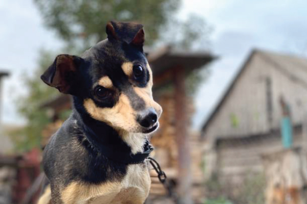
Intravenous Fluid (IV Fluids) Therapy in dogs is a critical and common medical treatment used to maintain hydration, correct electrolyte imbalances, support organ function, and deliver medications directly into the bloodstream. It’s a cornerstone of veterinary care for a wide range of conditions.
Here’s a detailed overview:
Why IV Fluids are Used (Indications)
IV fluid therapy is indicated for numerous conditions, including:
Dehydration: The most common reason. Dogs can become dehydrated from:
Vomiting and Diarrhea: Leading to significant fluid and electrolyte loss.
Anorexia (not eating/drinking): Especially if prolonged.
Heatstroke: Rapid fluid loss.
Kidney Disease: Impaired ability to conserve water.
Diabetes Mellitus: Excessive urination (polyuria).
Shock: To rapidly restore blood volume and blood pressure.
Hypovolemic Shock: Due to severe dehydration, hemorrhage, or trauma.
Septic Shock: Due to severe infection.
Anaphylactic Shock: Severe allergic reaction.
Support During Surgery and Anesthesia: To maintain blood pressure, prevent dehydration, and facilitate drug delivery.
Kidney Disease (Acute and Chronic): To flush toxins (diuresis), maintain hydration, and support kidney function.
Heart Disease: Used cautiously, but can be necessary to deliver medications or manage specific complications.
Pancreatitis: To maintain hydration, electrolyte balance, and perfusion of the pancreas.
Toxicities/Poisoning: To help flush toxins from the body and support organ function.
Electrolyte Imbalances: To correct deficiencies or excesses (e.g., low potassium, high sodium).
Medication Delivery: Allows for constant, controlled infusion of certain medications that cannot be given orally or intramuscularly.
Nutritional Support: In some cases, specific nutrients (e.g., dextrose) can be added to fluids, or full parenteral nutrition can be administered.
Types of IV Fluids
Veterinarians choose specific fluids based on the dog’s condition, the underlying cause, and the desired effect:
Crystalloids: These are salt solutions (electrolytes) that can freely cross membranes.
Isotonic Crystalloids: Most common. They have a similar osmotic pressure to blood, so they distribute fairly evenly between the intravascular space (blood vessels) and the interstitial space (between cells).
Examples: Lactated Ringer’s Solution (LRS), 0.9% Normal Saline (NaCl), Plasmalyte, Normosol-R.
Uses: General rehydration, maintenance fluids, shock.
Hypotonic Crystalloids: Lower osmotic pressure than blood. They cause fluid to move from the blood vessels into cells.
Examples: 0.45% Saline.
Uses: Less common for initial rehydration, sometimes used for hypernatremia (high sodium).
Hypertonic Crystalloids: Higher osmotic pressure than blood. They draw fluid from the interstitial and intracellular spaces into the blood vessels.
Examples: 7.5% Saline.
Uses: Used for rapid, temporary volume expansion in shock, or to treat cerebral edema (brain swelling). Given in small volumes.
Colloids: These are solutions containing large molecules that cannot easily cross cell membranes, so they primarily stay within the blood vessels, effectively increasing blood volume and maintaining oncotic (osmotic) pressure.
Natural Colloids:
Plasma: Contains albumin and other proteins, clotting factors. Used for low albumin, clotting disorders.
Whole Blood: Used for severe anemia, active bleeding.
Synthetic Colloids:
Examples: Hydroxyethyl Starch (HES) solutions.
Uses: Rapid volume expansion in shock, maintaining blood pressure in hypoproteinemic patients. Note: Use of HES has become more cautious due to potential risks like acute kidney injury and coagulopathy, especially in critically ill patients.
How IV Fluids are Administered
IV Catheter Placement: A sterile needle is used to insert a small, flexible plastic catheter (usually in a leg vein like the cephalic or saphenous vein, or sometimes the jugular vein in the neck) into the dog’s vein. The catheter is then secured with tape and protected.
Fluid Bag and Line: The catheter is connected to an IV fluid line, which leads to a bag of the selected fluid.
Fluid Pump: In most veterinary hospitals, a specialized IV fluid pump is used to precisely control the rate of fluid delivery (mL per hour). This ensures the dog receives the correct amount of fluid and prevents over or under-hydration.
Monitoring: The dog must be closely monitored throughout fluid therapy.
Monitoring During IV Fluid Therapy
Close monitoring is crucial to ensure the fluids are having the desired effect and to prevent complications. This includes:
Clinical Signs of Hydration:
Skin Turgor: How quickly the skin snaps back when gently tented.
Mucous Membranes: Moistness and color of gums.
Eye Position: Sunken eyes can indicate dehydration.
Capillary Refill Time (CRT): How quickly color returns to gums after pressing.
Vital Signs: Heart rate, respiratory rate, body temperature, blood pressure.
Body Weight: Daily weighing is critical to detect fluid retention or loss.
Urine Output: Measuring how much the dog is urinating.
Auscultation of Lungs: Listening for crackles or wheezes, which could indicate pulmonary edema (fluid in the lungs).
Blood Tests:
PCV/TS (Packed Cell Volume/Total Solids): To assess red blood cell concentration and protein levels, helping to gauge hydration status.
Electrolytes: Sodium, potassium, chloride, calcium, phosphorus.
Kidney Values: BUN (blood urea nitrogen), creatinine.
Blood Glucose: Especially in diabetic patients or those receiving dextrose.
Potential Complications
While life-saving, IV fluid therapy is not without risks:
Fluid Overload (Overhydration): The most serious common complication. Giving too much fluid too quickly can lead to:
Pulmonary Edema: Fluid accumulation in the lungs, causing difficulty breathing, coughing, and crackling sounds. This is a medical emergency.
Peripheral Edema: Swelling in the limbs or other body parts.
Cavitary Effusions: Fluid accumulation in the chest (pleural effusion) or abdomen (ascites).
Cardiac Strain: Especially in dogs with pre-existing heart disease.
Electrolyte Imbalances: Fluids can dilute existing electrolytes or introduce imbalances.
Catheter Complications:
Phlebitis: Inflammation of the vein.
Infection: Bacteria entering the bloodstream via the catheter.
Extravasation: Fluid leaking out of the vein into the surrounding tissue, causing swelling and discomfort.
Thrombosis: Blood clot formation in the vein.
Hypothermia: Large volumes of room-temperature fluids can lower a dog’s body temperature.
Dilutional Hypoproteinemia: Large volumes of crystalloids can dilute blood proteins, potentially exacerbating edema.
Conclusion
Intravenous fluid therapy is a powerful and versatile tool in veterinary medicine, often making the difference between life and death for critically ill or injured dogs. It requires careful assessment, appropriate fluid selection, precise administration, and vigilant monitoring by a trained veterinary professional. Never attempt to administer IV fluids to your dog at home without direct veterinary guidance.
========================================
#IVFluidsForDogs, #DogFluidTherapy, #CanineCare, #DogHealth, #PetWellness, #VeterinaryCare, #DogRecovery, #DehydratedDog, #SickDog, #PetCare, #AnimalHospital, #VetMed, #IntravenousFluids, #DogHydration, #GetWellSoonDog, #HealingPaws, #RoadToRecovery, #EmergencyVet, #CriticalCareVet, #PostSurgeryCare, #DogIllness, #SeniorDogCare, #PetLove, #FurryPatient, #DogsofInstagram, #SupportiveCare, #VetClinic, #BestFriendGetsBetter, #CanineHealth, #FluidTherapy


Add comment