

Lagenidiosis, often colloquially referred to as a “fungal infection,” is a rare but exceptionally severe and often life-threatening disease in dogs caused by an organism belonging to the genus Lagenidium. It is crucial to clarify from the outset that Lagenidium species are not true fungi; rather, they are oomycetes, commonly known as water molds. Despite this distinction, they are often discussed within the context of systemic mycoses in veterinary medicine due to their similar clinical presentations and the challenges they pose in diagnosis and treatment, which often overlap with severe fungal infections.
This devastating disease is characterized by aggressive, granulomatous inflammation that can affect various body systems, most commonly the skin and subcutaneous tissues, but also the gastrointestinal tract, and in severe cases, can disseminate throughout the body to vital organs. Lagenidiosis is particularly challenging due to the organism’s inherent resistance to many conventional antifungal medications, its rapid progression, and the difficulty in achieving an early and definitive diagnosis. The insidious nature of the disease, coupled with its often grave prognosis, makes it a significant concern for veterinarians and a source of profound distress for pet owners. Understanding the intricacies of Lagenidiosis—from its environmental origins to its complex diagnostic and therapeutic pathways—is paramount for effective management and, ultimately, for improving the outcomes for affected dogs. This comprehensive guide aims to shed light on every facet of this enigmatic disease, equipping dog owners and veterinary professionals with essential knowledge.
Causes (Etiology) of Lagenidiosis
Lagenidiosis in dogs is caused by infection with species from the genus Lagenidium, predominantly Lagenidium giganteum or closely related unnamed Lagenidium species. These organisms are eukaryotic microorganisms classified within the phylum Oomycota, making them distinct from true fungi (Phylum Fungi). While they share some morphological similarities with fungi, such as filamentous hyphae, their cell walls are composed primarily of cellulose, not chitin, and they reproduce asexually via motile zoospores and sexually via oospores, mechanisms distinct from those of true fungi. This fundamental biological difference has significant implications for diagnosis and, critically, for treatment, as many antifungal drugs targeting chitin synthesis or fungal ergosterol are ineffective against oomycetes.
The natural habitat of Lagenidium species is aquatic and semi-aquatic environments. They thrive in warm, stagnant, or slow-moving freshwater bodies rich in organic matter. Typical sources include ponds, lakes, swamps, marshes, irrigation ditches, and other poorly drained or standing water sources. The prevalence of the disease is higher in regions with tropical and subtropical climates, such as the southeastern United States (particularly the Gulf Coast states), and similar climatic zones in Central and South America, Australia, and parts of Asia. These warm, humid conditions provide an ideal environment for the proliferation of Lagenidium and its motile zoospores, which are the infective stage.
Dogs typically acquire Lagenidiosis through direct exposure to these contaminated aquatic environments. There are several primary modes of transmission:
- Cutaneous Inoculation: This is considered the most common route. Dogs that wade, swim, or play in contaminated water can sustain minor skin abrasions, cuts, or existing wounds. The motile zoospores present in the water can then penetrate these breaches in the skin barrier, gaining access to the subcutaneous tissues. Once inside, they transform into hyphal forms that invade and proliferate, leading to the characteristic granulomatous lesions. Activities like running through tall, wet grasses or dense vegetation bordering contaminated water bodies can also cause superficial skin trauma, increasing the risk of inoculation. Even seemingly insignificant skin nicks can serve as entry points for these aggressive organisms.
- Ingestion: Dogs may also become infected by ingesting contaminated water, aquatic vegetation, or animals (e.g., amphibians, fish) that have been exposed to or harbor Lagenidium. If ingested, the organisms can establish infection within the gastrointestinal tract, leading to severe inflammatory lesions, particularly in the stomach and intestines. This route is less common than cutaneous inoculation but can result in equally devastating, if not more rapidly progressive, disease due to the critical nature of the affected organ systems. Dogs with a habit of drinking from natural water sources during walks or consuming foreign objects from the environment are at a higher risk for this mode of transmission.
- Inhalation: While less frequently documented, it is theoretically possible for zoospores or aerosolized hyphal fragments to be inhaled, leading to primary respiratory tract infection. However, most respiratory involvement typically occurs secondary to systemic dissemination from primary cutaneous or gastrointestinal foci.
Unlike some opportunistic fungal infections, Lagenidiosis generally affects otherwise healthy, immunocompetent dogs. While a compromised immune system might exacerbate the disease, it is not a prerequisite for infection. The primary predisposing factor is direct environmental exposure. The organism’s inherent pathogenicity and ability to evade host immune responses are key to its establishment and progression, rather than a host’s weakened defenses. This highlights the importance of environmental management and prevention, as even robust dogs can succumb to this severe oomycete infection. The organism’s ability to resist phagocytosis and survive within host tissues contributes significantly to the persistent and destructive nature of the disease.
Signs and Symptoms (Clinical Manifestations)
The clinical presentation of Lagenidiosis in dogs is highly variable, depending on the site of initial infection and the extent of dissemination. Lagenidiosis is notorious for its aggressive and progressive nature, often mimicking other granulomatous diseases, which contributes to diagnostic delays. The disease often manifests in distinct forms, though overlap and progression from one form to another are common:
1. Cutaneous and Subcutaneous Form (Most Common)
This is the most frequently observed manifestation and often the first sign owners notice. Lesions typically appear on areas of the body that come into contact with water, such as the limbs (especially distal extremities), ventral abdomen, chest, and tail base.
- Lesions: Initially, these may present as small, firm nodules or plaques under the skin. Over time, they rapidly enlarge and become ulcerated, often with draining tracts that discharge serosanguinous (blood-tinged fluid) or purulent (pus-like) exudate. The skin around the lesions may be thickened, edematous (swollen), and hyperpigmented (darkened).
- “Kunkers”: A hallmark feature, particularly prominent in some oomycete infections like Pythiosis (a closely related disease), are necrotic cores or “kunkers.” These are often firm, yellowish-white, friable masses composed of necrotic tissue, inflammatory cells, and large numbers of oomycete hyphae. While classically associated with Pythiosis, Lagenidium lesions can also present with similar pyogranulomatous masses.
- Rapid Progression and Invasion: The lesions are characterized by their invasive and destructive nature. They tend to spread rapidly, often involving underlying fascia, muscle, tendons, and even bone. This deep invasion leads to significant tissue destruction and can result in severe lameness if joints or bones of the limbs are affected.
- Pain and Pruritus: Affected areas are often painful to the touch and highly pruritic (itchy), leading to self-mutilation and secondary bacterial infections. The constant irritation and discomfort significantly impact the dog’s quality of life.
- Lymphadenopathy: Regional lymph nodes draining the affected areas are frequently enlarged and firm due to the inflammatory response and potential spread of the organism.
2. Gastrointestinal (GI) Form (Less Common, Highly Severe)
When the organism is ingested, it can establish infection in the gastrointestinal tract, typically the stomach, small intestine, and/or large intestine. This form is particularly challenging and carries a grave prognosis due to the vital role of these organs.
- Vomiting and Diarrhea: Chronic, often intractable vomiting and diarrhea are common. The diarrhea may be mucoid, hemorrhagic (bloody), or melenic (dark, tarry stools, indicating digested blood), depending on the site and severity of mucosal damage.
- Weight Loss and Anorexia: Due to malabsorption, chronic inflammation, and discomfort, affected dogs often experience severe progressive weight loss, cachexia (wasting), and anorexia (loss of appetite).
- Abdominal Pain: Dogs may exhibit signs of abdominal pain, such as a hunched posture, reluctance to move, or guarding of the abdomen on palpation.
- Palpable Masses: In some cases, thickened intestinal loops or discrete granulomatous masses may be palpable on abdominal examination.
- Peritonitis: If the intestinal wall becomes severely compromised or perforated, it can lead to peritonitis (inflammation of the abdominal lining), which is a life-threatening emergency.
- Hypoproteinemia: Chronic protein loss through the damaged GI tract can lead to severe hypoproteinemia (low blood protein levels), resulting in generalized edema (swelling) or ascites (fluid accumulation in the abdomen).
3. Systemic/Disseminated Form (Rarest, Most Fatal)
In the most severe and advanced cases, Lagenidium can disseminate from the primary site of infection (cutaneous or GI) to distant organs via the bloodstream or lymphatic system. This leads to multi-organ involvement and rapidly progressive, often fatal, disease.
- Non-specific Signs: Initial signs of systemic dissemination can be vague and non-specific, including persistent fever, lethargy, generalized weakness, and severe weight loss.
- Organ-Specific Signs: The clinical signs will then reflect the organs involved:
- Lungs: Coughing, difficulty breathing (dyspnea), tachypnea (rapid breathing) if pulmonary granulomas develop.
- Lymph Nodes: Generalized lymphadenopathy (enlarged lymph nodes throughout the body).
- Bones: Lameness, pain, swelling over affected bones if osteomyelitis occurs.
- Eyes: Uveitis, glaucoma, or retinal detachment leading to vision impairment or blindness.
- Central Nervous System (CNS): Seizures, ataxia (incoordination), behavioral changes, or paralysis if brain or spinal cord lesions are present. This is a rare but devastating complication.
- Rapid Deterioration: Dogs with disseminated Lagenidiosis often experience rapid clinical deterioration despite aggressive symptomatic treatment, highlighting the grave prognosis associated with this form.
It is critical for owners and veterinarians to recognize that Lagenidiosis lesions often do not respond to conventional antibiotic or antifungal therapies, and may even worsen with time. A high index of suspicion based on the progressive nature of the lesions, history of aquatic exposure, and lack of response to standard treatments is crucial for prompting specific diagnostic investigations.
Dog Breeds at Risk (with a paragraph explanation)
While Lagenidiosis can technically affect any dog exposed to the causative oomycete, certain dog breeds appear to be overrepresented in reported cases. This increased susceptibility is generally not attributed to a specific genetic predisposition for the disease itself, but rather to a combination of their inherent behavioral traits, activity levels, and likelihood of environmental exposure to water sources where Lagenidium thrives. These breeds are typically active, outdoor-loving dogs that frequently engage in activities that increase their contact with potentially contaminated aquatic environments.
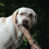
Among the breeds most commonly identified at higher risk are Labrador Retrievers, Golden Retrievers, German Shepherds, Great Danes, American Pit Bull Terriers, and Rottweilers. These breeds are often chosen for their athleticism, strong swimming abilities, and eagerness to retrieve or explore. Consequently, they are frequently taken to lakes, ponds, rivers, or marshy areas for recreation, training, or simply during walks. Their large size often means they spend more time submerged or wading in water, and their propensity for vigorous play can lead to minor skin abrasions or punctures, which serve as entry points for the Lagenidium zoospores. Furthermore, some of these breeds, particularly working dogs or those in rural settings, may have unsupervised access to stagnant water sources. Their curious nature may also lead them to ingest contaminated water or vegetation, increasing the risk of the gastrointestinal form of the disease. Therefore, the elevated risk in these breeds is primarily a reflection of their lifestyle and environmental exposure patterns, rather than a direct genetic susceptibility to the pathogen itself. Owners of these breeds residing in or traveling through endemic areas should be particularly vigilant about preventing exposure to potentially contaminated water bodies.
Affects Puppy, Adult, or Older Dogs
Lagenidiosis primarily affects young to middle-aged adult dogs, typically ranging from 1 to 6 years of age. This demographic accounts for the majority of reported cases. The reason for this age predilection is largely behavioral and environmental rather than physiological. Dogs in this age group are generally at their peak physical activity and energy levels. They are most likely to be engaged in outdoor activities such as swimming, retrieving, hunting, and exploring various natural environments, including ponds, lakes, marshes, and ditches where Lagenidium thrives. Their increased exposure to contaminated water sources, coupled with the likelihood of sustaining minor skin abrasions during vigorous play, significantly elevates their risk compared to other age groups.
While less common, puppies can also be affected, especially if they are introduced to outdoor environments and water bodies at a young age in endemic areas. Similarly, older adult dogs are not entirely immune; if they remain active and continue to have access to contaminated aquatic environments, they can also contract the disease. However, as activity levels often decrease with age, the incidence in senior dogs tends to be lower. The immune status of the dog does not appear to be a primary predisposing factor, as even healthy, immunocompetent animals can succumb to this aggressive oomycete. Therefore, regardless of age, the key determinant for infection remains environmental exposure to Lagenidium-laden water sources.
Diagnosis of Lagenidiosis
Diagnosing Lagenidiosis is notoriously challenging due to several factors: its rarity, non-specific initial clinical signs, the biological distinction of Lagenidium as an oomycete (not a true fungus), and its similarity to other pyogranulomatous diseases, including other oomycoses like Pythiosis. A high index of suspicion, especially in dogs from endemic areas with chronic, non-healing lesions unresponsive to conventional therapies, is critical to prompt appropriate diagnostic testing.
1. Clinical Suspicion and History
- Geographic Location: Dogs from or with a travel history to endemic regions (e.g., southeastern US, Gulf Coast) with warm, humid climates and abundant stagnant water sources.
- Environmental Exposure: A history of swimming, wading, or playing in ponds, lakes, marshes, or ditches.
- Clinical Signs: Persistent, progressive, often painful and pruritic cutaneous lesions (nodules, ulcers, draining tracts, often with “kunkers”), or chronic gastrointestinal signs (vomiting, diarrhea, weight loss) unresponsive to standard treatments.
- Failure of Prior Therapies: A key indicator is the lack of response to broad-spectrum antibiotics and conventional antifungal medications.
2. Sample Collection
The cornerstone of diagnosis involves obtaining appropriate samples for laboratory analysis. Deep tissue biopsies from the active margins of cutaneous lesions or from affected gastrointestinal tissue (endoscopic or surgical biopsies) are often necessary. Fine needle aspirates (FNAs) can sometimes be indicative but are less definitive than biopsies.
3. Histopathology
This is often the first definitive diagnostic step. Biopsied tissue samples are fixed in formalin, processed, sectioned, and stained (e.g., Hematoxylin and Eosin – H&E).
- Characteristic Microscopic Findings: Histopathology reveals a severe pyogranulomatous to eosinophilic granulomatous inflammatory response. Within these inflammatory infiltrates, the Lagenidium organisms can be observed. They typically appear as broad (5-10 µm), irregular, sparsely septate or aseptate hyphae with parallel walls. They often branch at irregular angles.
- Special Stains: Organisms are often difficult to visualize with H&E alone. Special fungal stains, such as Gomori Methenamine Silver (GMS) or Periodic Acid-Schiff (PAS), are crucial as they stain the cell walls of oomycetes (and fungi) strongly, making them more apparent.
- Differentiation: While histopathology can confirm the presence of an oomycete, differentiating Lagenidium from Pythium insidiosum (the cause of Pythiosis) purely on morphological grounds can be challenging due to their similar appearance in tissue.
4. Culture
Culturing Lagenidium is technically challenging and requires specialized laboratory conditions and media.
- Media Requirements: Lagenidium does not grow on standard fungal media like Sabouraud’s Dextrose Agar (SDA). It requires specific oomycete-selective media, such as cornmeal agar or potato dextrose agar, often supplemented with antibiotics to inhibit bacterial contamination.
- Slow Growth: Even on appropriate media, growth can be slow, sometimes taking weeks, and requires specific incubation temperatures.
- Expertise: Culture should only be attempted by laboratories with specific expertise in oomycete isolation and identification.
5. Molecular Diagnostics (PCR)
Polymerase Chain Reaction (PCR) is a highly sensitive and specific diagnostic tool for Lagenidiosis.
- Detection: PCR assays can detect Lagenidium DNA directly from fresh or frozen tissue samples (biopsies), formalin-fixed paraffin-embedded (FFPE) tissue, or even culture isolates.
- Differentiation from Pythium: Crucially, species-specific PCR primers can differentiate Lagenidium from Pythium insidiosum, which is vital for guiding treatment decisions as their therapeutic responses differ.
- Advantages: Rapid, highly accurate, and can identify the pathogen even when organisms are sparse or difficult to culture.
6. Immunohistochemistry (IHC)
Immunohistochemistry is another powerful tool if available in specialized laboratories.
- Mechanism: It uses specific antibodies that bind to Lagenidium antigens present in tissue sections. A secondary antibody linked to an enzyme or fluorochrome then makes the bound antigen-antibody complex visible under a microscope.
- Specificity: IHC staining can definitively identify Lagenidium and differentiate it from Pythium and other fungal organisms in tissue samples, making it a valuable adjunct to histopathology, especially when organism morphology is ambiguous.
7. Serology (ELISA)
Enzyme-Linked Immunosorbent Assay (ELISA) is available through some specialized diagnostic laboratories to detect antibodies against Lagenidium in a dog’s serum.
- Utility: A positive serological test suggests exposure to Lagenidium and the dog’s immune response to it. It can be useful for screening, supporting a presumptive diagnosis, or monitoring response to treatment.
- Limitations:
- False Negatives: Early in the infection, antibody levels may not be high enough for detection.
- False Positives: A positive test indicates exposure but not necessarily active disease, as some dogs may have been exposed without developing clinical infection.
- Cross-reactivity with other oomycetes or fungi can occur, though modern assays aim for high specificity.
- A negative result does not definitively rule out the disease, especially in chronic or localized cases.
8. Diagnostic Imaging (Radiography, Ultrasonography, CT/MRI)
Imaging studies are primarily used to assess the extent of the disease and guide biopsy collection, rather than for definitive diagnosis.
- Radiography: Useful for evaluating bone involvement in cutaneous forms (osteolysis, periosteal reaction) or assessing pulmonary lesions in disseminated cases.
- Ultrasonography: Abdominal ultrasound can reveal thickening of intestinal walls, mesenteric lymphadenopathy, or abdominal masses in the GI form of the disease. It can also guide real-time biopsy collection.
- CT/MRI: Computed tomography (CT) or Magnetic Resonance Imaging (MRI) provides detailed cross-sectional images, which are invaluable for defining the extent of soft tissue and bone involvement, especially for surgical planning, or for detecting neurological lesions in disseminated cases.
A definitive diagnosis often requires a combination of clinical suspicion, histopathology with appropriate stains, and confirmatory molecular (PCR) or immunohistochemical testing. The ability to distinguish Lagenidium from Pythium is particularly important, as while often grouped together due to similar pathology, treatment protocols may vary.
Treatment of Lagenidiosis
Treating Lagenidiosis is exceptionally challenging and often requires an aggressive, multi-modal approach due to the organism’s inherent resistance to many conventional antifungal agents and its invasive nature. The prognosis is generally guarded to poor, especially for advanced or disseminated forms of the disease. Early diagnosis and aggressive intervention offer the best chance of success, although recurrence remains a significant concern.
1. Surgical Excision (for Localized Cutaneous Lesions)
For localized cutaneous or subcutaneous lesions, radical surgical excision is considered the cornerstone of effective treatment.
- Wide Surgical Margins: It is absolutely critical to remove all infected tissue with wide, clean surgical margins. This often necessitates extensive resections, which can be disfiguring and may require reconstructive surgery, skin grafting, or even amputation of an affected limb. The goal is to obtain pathology reports with “clean margins” (no Lagenidium organisms at the edges of the excised tissue).
- Repeated Surgeries: In many cases, initial excisions may be incomplete, leading to rapid recurrence. Repeat surgeries, often even more aggressive, may be necessary.
- Limitations: Surgery is typically not feasible or effective for diffuse cutaneous lesions, extensive deep tissue invasion, gastrointestinal forms, or disseminated disease. For GI forms, resection of affected bowel loops may be attempted but is often associated with high morbidity and recurrence rates.
2. Antifungal/Oomycete-Specific Medications
Due to Lagenidium‘s classification as an oomycete, drugs targeting fungal ergosterol (like azoles) or chitin synthesis are often ineffective or less effective. However, some agents have shown limited efficacy or are used empirically, often in combination.
- Itraconazole: This is an azole antifungal often attempted first-line, but Lagenidium species are generally highly resistant. It may be used in combination therapy, but its sole use is rarely effective.
- Terbinafine: An allylamine antifungal that inhibits ergosterol synthesis. It has a different mechanism of action than azoles and has shown some in vitro activity against oomycetes. It is often used in combination with itraconazole or other drugs.
- Combination Therapy: The most common medical approach involves a combination of systemic drugs, most frequently Itraconazole in combination with Terbinafine. The rationale is to potentially achieve a synergistic effect or target different metabolic pathways, though evidence for consistent efficacy against Lagenidium specifically (as opposed to Pythium) is still developing. Often, high doses and prolonged treatment courses (many months to a year or more) are required.
- Amphotericin B: A polyene antifungal that disrupts fungal cell membranes. It is effective against a broad range of true fungi, but its efficacy against oomycetes like Lagenidium is variable and often poor. Furthermore, Amphotericin B is highly nephrotoxic (toxic to the kidneys) and can only be administered intravenously in a hospital setting for severe, disseminated cases where other treatments have failed. Newer lipid-formulated amphotericin B products are less toxic but still carry significant risks and are very expensive.
- Lufenuron: This is an insect growth regulator, not an antifungal. It interferes with chitin synthesis in insects. While Lagenidium (an oomycete) does not have chitin in its cell wall, Lufenuron has been anecdotally used by some veterinarians for Pythium and Lagenidium infections, possibly due to an undefined mechanism or a placebo effect. Its efficacy is not scientifically proven for oomycosis in dogs.
- Other Azoles (Fluconazole, Voriconazole, Posaconazole): While some specific azoles may have slightly broader spectrums, Lagenidium generally exhibits resistance to the azole class. They are typically not recommended as primary monotherapy.
3. Immunotherapy
- Autogenous Vaccines: In some specialized veterinary centers, autogenous vaccines (made from the specific Lagenidium isolate from the patient) or commercially available Pythium vaccines have been explored. The principle is to stimulate a robust cell-mediated immune response against the organism. However, the consistent efficacy of these vaccines specifically for Lagenidium in dogs is not yet well-established, and they are typically used as an adjunct to surgery and systemic medications, primarily for Pythium where there is more supporting evidence. They are not widely available and require specific laboratory capabilities.
4. Supportive Care
- Pain Management: Lagenidiosis lesions are often severely painful. Appropriate analgesia (e.g., NSAIDs, gabapentin, opioids) is crucial for the dog’s comfort and quality of life.
- Antibiotics: Secondary bacterial infections of ulcerated skin lesions or compromised GI tracts are common. Broad-spectrum antibiotics are often prescribed to manage these secondary infections, but they have no effect on the Lagenidium itself.
- Nutritional Support: Dogs with GI Lagenidiosis or severe systemic disease often suffer from anorexia and significant weight loss. Nutritional support, including highly digestible diets, appetite stimulants, or even assisted feeding (e.g., feeding tubes), may be necessary.
- Fluid Therapy: To manage dehydration, electrolyte imbalances, and hypoproteinemia, especially in GI forms.
5. Monitoring and Duration of Treatment
Treatment is typically prolonged, often lasting for many months to a year or more, even after clinical signs resolve. Regular monitoring of the dog’s response to therapy, general health, and potential side effects of medications (e.g., liver enzymes for azoles) is essential. Relapses are common, necessitating continued vigilance. Due to the high cost, prolonged nature of treatment, and often poor outcomes, euthanasia is a compassionate option considered by many owners, especially in advanced or disseminated cases.
In summary, treating Lagenidiosis demands a highly aggressive, multi-pronged approach combining radical surgery (where possible), prolonged and often combination systemic medication, and supportive care. Even with the most diligent and exhaustive treatment, the prognosis remains guarded, underscoring the severity and recalcitrance of this challenging oomycete infection.
Prognosis & Complications of Lagenidiosis
The prognosis for dogs diagnosed with Lagenidiosis is generally guarded to poor, reflecting the aggressive nature of the oomycete, its resistance to typical antifungal treatments, and the challenges in achieving complete eradication. The outcome heavily depends on several factors, including the location and extent of the infection, the promptness of diagnosis, and the aggressiveness and completeness of treatment.
Prognosis by Form:
- Localized Cutaneous Form: If the disease is detected very early and lesions are small and truly localized, with no evidence of deep invasion or dissemination, and if complete and radical surgical excision with wide, clean margins is achievable, the prognosis is guarded but offers the best chance for long-term survival. However, recurrence rates are still high, even with seemingly complete excision. Close monitoring and potentially prolonged adjunctive medical therapy are essential.
- Gastrointestinal Form: The prognosis for dogs with the gastrointestinal form is poor to grave. These infections are often extensive, leading to severe malabsorption, protein-losing enteropathy, perforation, and peritonitis. Surgical resection of affected bowel is often technically difficult, carries high risks of complications, and recurrence is almost inevitable. Medical management alone is rarely curative.
- Disseminated/Systemic Form: The prognosis for disseminated Lagenidiosis is grave to hopeless. Once the organism has spread to multiple internal organs (lungs, lymph nodes, bones, CNS), effective treatment becomes virtually impossible. These cases are usually fatal despite aggressive therapeutic attempts.
Complications:
- Recurrence of Lesions: This is one of the most common and frustrating complications. Even after aggressive surgery and prolonged medical therapy, Lagenidium has a strong tendency to recur, often developing at the margins of the previous surgical site or in new locations. This necessitates repeat surgeries and ongoing medical treatment.
- Dissemination: If the primary infection is not contained, the organism can spread to regional lymph nodes and eventually to distant organs via the bloodstream or lymphatic system. This leads to life-threatening systemic illness and organ failure (e.g., respiratory failure from lung involvement, neurological deficits from brain lesions).
- Severe Tissue Destruction and Disfigurement: The invasive nature of Lagenidium leads to significant destruction of skin, subcutaneous tissue, muscle, and bone. This can result in permanent disfigurement, chronic pain, lameness, and functional impairment, even if the infection is eventually controlled. Amputations may be necessary for limb lesions.
- Secondary Bacterial Infections: The ulcerated and necrotic lesions are highly susceptible to secondary bacterial infections, complicating treatment and worsening local inflammation and pain. These require additional antibiotic therapy.
- Debilitation and Cachexia: Chronic pain, inflammation, anorexia (especially with GI involvement), and the metabolic demands of fighting the infection often lead to severe weight loss, muscle wasting (cachexia), and general poor body condition.
- Adverse Effects of Treatment: The aggressive medical therapies used to combat Lagenidiosis often come with significant side effects. For example, azole antifungals can cause hepatotoxicity (liver damage), while Amphotericin B is notoriously nephrotoxic (kidney damage). Prolonged drug administration requires careful monitoring of organ function.
- High Cost of Treatment: The extensive surgical procedures, prolonged and expensive medications, frequent veterinary visits, and diagnostic tests mean that treating Lagenidiosis is financially demanding, often placing a significant burden on owners.
- Euthanasia: Due to the often poor prognosis, severe suffering, high recurrence rates, and the extensive financial and emotional toll, many owners ultimately opt for humane euthanasia, especially in cases of advanced or disseminated disease.
In summary, Lagenidiosis presents a formidable challenge in veterinary medicine. Even with optimal and aggressive management, the outcome is often bleak, and the disease typically leads to significant morbidity and mortality, making prevention paramount.
Prevention of Lagenidiosis
Given the severe nature, difficulty in treatment, and often poor prognosis of Lagenidiosis, prevention is by far the most effective strategy to protect dogs from this devastating disease. Prevention primarily revolves around minimizing exposure to the Lagenidium oomycete in its natural environment.
- Avoidance of Contaminated Water Sources:
- Restrict Access: The most critical preventive measure is to prevent dogs, especially those in high-risk breeds or living in endemic areas, from accessing stagnant or slow-moving freshwater bodies. This includes ponds, lakes, swamps, marshes, irrigation ditches, and areas with standing water, particularly during warm, humid weather.
- Supervision: Always supervise dogs when outdoors, especially near natural water sources, and actively deter them from entering or drinking from such environments.
- Awareness After Rainfall: Be particularly cautious after heavy rainfall, which can create new puddles and temporary shallow water bodies that may harbor the organism.
- Fencing: If you live near potential sources of contamination, consider secure fencing to prevent your dog from accessing them.
- Prompt Wound Care:
- Inspect and Clean: After any outdoor activity, especially if your dog has been in or near water, thoroughly inspect their skin for any cuts, abrasions, punctures, or open wounds.
- Disinfect: Cleanse any wounds promptly and thoroughly with an antiseptic solution (e.g., diluted povidone-iodine or chlorhexidine) and ensure they are covered or treated appropriately to prevent pathogen entry.
- Monitor: Pay close attention to any non-healing wounds or skin lesions that develop after potential exposure, and seek veterinary attention if they persist or worsen.
- Prevent Ingestion of Contaminated Material:
- Monitor Drinking Habits: Do not allow your dog to drink from natural water sources like ponds or ditches. Always provide fresh, clean drinking water.
- Deter Eating Aquatic Vegetation: Some dogs may chew on or ingest aquatic plants or debris found in or around water bodies. This behavior should be discouraged, as these materials can harbor Lagenidium.
- Geographical Awareness:
- Know Your Area: Owners living in or traveling to endemic regions (e.g., the southeastern United States, Gulf Coast states, other tropical and subtropical areas worldwide) should be acutely aware of the risk and implement stringent preventive measures. Consult local veterinarians for specific advice regarding endemic risks in your area.
- No Vaccine Available (Currently):
- Unlike Pythiosis, for which some experimental or commercially available vaccines exist and are used in high-risk animals, there is currently no widely available or proven effective vaccine specifically for Lagenidium infection in dogs. Research in this area is ongoing, but for now, environmental avoidance remains the cornerstone of prevention.
By adhering to these preventive strategies, especially restricting access to potentially contaminated water sources, dog owners can significantly reduce the risk of their beloved companions contracting Lagenidiosis. Vigilance and proactive measures are the most powerful tools against this severe oomycete infection.
Diet and Nutrition for Dogs with Lagenidiosis
While there is no specific “anti-Lagenidium” diet that directly treats the infection, proper diet and nutrition play a crucial supportive role in managing dogs with Lagenidiosis. Adequate nutrition is vital for bolstering the immune system, supporting tissue repair, maintaining energy levels, and helping the dog cope with the metabolic demands of fighting a severe, chronic illness and undergoing aggressive treatments.
General Nutritional Support:
- High-Quality, Highly Digestible Diet:
- Protein: Ensure the diet provides adequate levels of high-quality, easily digestible protein. Protein is essential for immune function, tissue repair (especially critical after surgery or in the presence of extensive lesions), and maintaining muscle mass, which is often compromised in chronic disease.
- Fat: Sufficient levels of healthy fats provide concentrated energy, which is important for debilitated dogs.
- Carbohydrates: Readily digestible carbohydrates are needed for energy without overburdening a compromised digestive system.
- Commercial Therapeutic Diets: Often, commercial therapeutic diets formulated for convalescence or highly digestible diets (e.g., prescription gastrointestinal diets) are beneficial.
- Caloric Density:
- Dogs suffering from Lagenidiosis, particularly those with GI involvement or systemic disease, often experience anorexia and significant weight loss. Providing a calorically dense diet in smaller, frequent meals can help meet energy requirements and prevent further wasting. Appetite stimulants may be necessary.
- Omega-3 Fatty Acids:
- Supplementation with omega-3 fatty acids (e.g., from fish oil) may be beneficial. Omega-3s possess anti-inflammatory properties that can help modulate the severe inflammatory response associated with Lagenidiosis, potentially reducing tissue damage and pain.
- Antioxidants and Vitamins:
- Ensure the diet is rich in antioxidants (e.g., Vitamins E and C, selenium) and B vitamins. Antioxidants help protect cells from oxidative stress during chronic inflammation and infection, while B vitamins are crucial for energy metabolism and overall health.
- A balanced, high-quality commercial diet should provide adequate levels, but supplementation might be considered under veterinary guidance.
- Hydration:
- Maintaining adequate hydration is paramount, especially for dogs experiencing vomiting or diarrhea (in the GI form) or those with draining cutaneous lesions. Encourage water intake and administer intravenous or subcutaneous fluids as necessary.
Specific Considerations for Gastrointestinal Lagenidiosis:
- Ultra-Digestible, Low-Fat Diets: For dogs with the GI form, a highly digestible, low-fat prescription diet is often recommended to minimize digestive workload, reduce vomiting and diarrhea, and improve nutrient absorption.
- Novel Protein/Hydrolyzed Diets: If gastrointestinal inflammation is severe or accompanied by concurrent food sensitivities, a novel protein or hydrolyzed protein diet might be considered to reduce potential immune reactions in the gut.
- Fiber: The role of fiber can be variable; some dogs with GI issues may benefit from moderate fiber for stool consistency, while others may require low-fiber diets to minimize GI transit time. This should be determined on a case-by-case basis.
- Appetite Stimulation: Anorexia is a significant problem. Mealtimes should be stress-free, and enticing, warmed foods may help. Medications to stimulate appetite (e.g., mirtazapine, capromorelin) may be prescribed by the veterinarian.
- Management of Hypoproteinemia: Dogs with protein-losing enteropathy due to GI Lagenidiosis may require specific management strategies, including specialized diets and potentially plasma transfusions in severe cases, to address dangerously low protein levels.
It is crucial to work closely with a veterinarian or a veterinary nutritionist to develop an individualized dietary plan for a dog with Lagenidiosis. The specific needs will vary based on the dog’s overall health, the form and severity of the disease, and the treatments being administered. Nutritional support is an integral component of comprehensive care, helping to support recovery and improve the dog’s quality of life during a very challenging illness.
Zoonotic Risk of Lagenidiosis
The zoonotic risk associated with Lagenidiosis is considered extremely low to negligible. This means that transmission of Lagenidium from an infected dog directly to a human is highly unlikely and has not been documented in scientific literature.
Here’s why the zoonotic risk is minimal:
- Specific Environmental Niche: Lagenidium species are primarily inhabitants of specific aquatic environments (stagnant warm freshwater). Humans, like dogs, would typically need exposure to these contaminated water sources for infection to occur.
- Mode of Transmission: In dogs, infection usually occurs through the direct inoculation of zoospores into a break in the skin barrier (e.g., a cut or abrasion) or through ingestion of contaminated water or vegetation. While humans could theoretically be exposed to environmental Lagenidium in a similar manner (e.g., swimming with an open wound, accidental ingestion), there is no evidence to suggest that contact with an infected dog facilitates this process or poses an additional risk.
- Host Specificity: While Lagenidium can infect various mammalian species, including dogs, cats, horses, and occasionally humans, direct animal-to-human transmission is not a recognized pathway. Human infections with Lagenidium or similar oomycetes (like Pythium) are exceedingly rare and typically occur in individuals with specific predisposing factors (e.g., immunocompromise, specific skin wounds exposed to contaminated water).
- No Direct Dog-to-Human Spread: There is no evidence or scientific basis to suggest that the lesions on an infected dog, or contact with their bodily fluids, can transmit Lagenidium to a human. The organism does not appear to survive or transmit efficiently in a way that would allow for direct zoonotic spread.
While Lagenidiosis is not considered a zoonotic disease, it is always a good practice to maintain general hygiene when handling any sick animal. This includes proper handwashing after contact with pets, especially after cleaning wounds or administering medications. However, there is no need for extreme isolation or specific zoonotic precautions beyond standard pet care hygiene for dogs diagnosed with Lagenidiosis.
Conclusion
Lagenidiosis represents one of the most severe and challenging infectious diseases encountered in canine veterinary medicine. Caused by the aggressive oomycete Lagenidium, this condition is characterized by its invasive nature, multiform manifestations (primarily cutaneous and gastrointestinal), and a disheartening resistance to conventional antifungal treatments. Its rarity, coupled with its often non-specific initial signs and the biological distinction of Lagenidium from true fungi, frequently leads to diagnostic delays that can critically impact prognosis.
The journey for a dog diagnosed with Lagenidiosis and their owner is invariably arduous. Treatment demands an exceptionally aggressive, multi-modal approach, typically involving radical surgical excision (often requiring extensive resections or amputations) combined with prolonged courses of often expensive and side-effect-laden systemic medications. Despite these heroic efforts, recurrence rates are high, and the prognosis remains guarded to poor, particularly for advanced or disseminated forms of the disease. The physical suffering endured by affected dogs, the emotional toll on their families, and the significant financial burden underscore the devastating impact of this illness.
Given the formidable challenges in treating Lagenidiosis, prevention emerges as the single most effective strategy. Owners, especially those with active dogs or breeds predisposed to outdoor activities in endemic regions, must exercise extreme vigilance. Restricting access to stagnant or slow-moving freshwater bodies, promptly cleaning and disinfecting any skin wounds after outdoor excursions, and preventing the ingestion of contaminated water or vegetation are paramount preventive measures.
Ultimately, Lagenidiosis serves as a stark reminder of the hidden dangers lurking in natural environments. A heightened awareness among dog owners and veterinary professionals, combined with a high index of suspicion and rapid diagnostic action, offers the best hope for early intervention. While the battle against Lagenidium is often an uphill one, knowledge, vigilance, and aggressive, targeted care can, in some fortunate instances, tip the scales towards a more favorable outcome for our canine companions.
#LagenidiosisInDogs, #DogFungalInfection, #Oomycosis, #WaterMold, #DogHealth, #VetMed, #PetCare, #DogDisease, #Lagenidium, #DogSkinIssues, #GIProblemsDogs, #DogBreedsAtRisk, #LabradorLife, #GermanShepherdHealth, #GoldenRetriever, #PreventLagenidiosis, #DogWellness, #SystemicMycosis, #VetEducation, #PetOwners, #CanineHealth, #OomyceteInfection, #RareDogDisease, #VeterinaryMedicine, #ProtectYourDog


Add comment