
Lead poisoning remains one of the most insidious, preventable, and widely reported causes of morbidity and mortality in both wild and domestic waterfowl. While the public often associates lead toxicity with human industrial exposure, its impact on ducks is profound because of the unique ways these birds interact with their environment—particularly through foraging on the surface of water bodies where lead fragments, pellets, and contaminated sediments accumulate.
In the United States alone, the U.S. Fish and Wildlife Service estimates that > 1 million waterfowl die annually from lead‑related causes, a figure that likely underrepresents the true global burden. Understanding the etiology, clinical presentation, and management of lead poisoning in ducks is essential for wildlife rehabilitators, veterinarians, conservationists, hunters, and enthusiastic backyard duck keepers.
This guide consolidates current scientific knowledge, field observations, and best‑practice recommendations into a single, user‑friendly resource. It is structured to be useful for both novices seeking an overview and seasoned professionals requiring detailed technical guidance.
2. What Is Lead Poisoning?
Lead poisoning (plumbism) is a systemic toxicosis resulting from the absorption of metallic lead (Pb) or its soluble compounds into the bloodstream. In ducks, the primary route is oral ingestion, although inhalation of lead‑laden dust and, rarely, transdermal exposure can contribute. Once absorbed, lead binds to erythrocytes, circulates systemically, and interferes with a cascade of enzymatic processes that are vital for cellular respiration, neurotransmission, and hemostasis.
Key biochemical mechanisms:
| Mechanism | Description | Consequence in Ducks |
|---|---|---|
| Inhibition of δ‑Aminolevulinic acid dehydratase (ALAD) | Blocks heme synthesis, leading to accumulation of δ‑ALA | Microcytic, hypochromic anemia; increased oxidative stress |
| Impairment of ferrochelatase | Prevents incorporation of iron into protoporphyrin IX | Further reduction in functional hemoglobin |
| Disruption of calcium signaling & synaptic transmission | Lead mimics Ca²⁺, displacing it from voltage‑gated channels | Neurological deficits, tremors, ataxia |
| Renal tubule toxicity | Direct tubular necrosis and interstitial fibrosis | Impaired excretion, secondary electrolyte disturbances |
| Immunomodulation | Suppression of lymphocyte proliferation | Increased susceptibility to secondary infections |
These pathophysiologic changes manifest clinically within hours to weeks, depending on dose, form of lead, and the bird’s overall health status.
3. Primary Causes of Lead Exposure in Ducks
| Source | Typical Form | How Ducks Encounter It | Geographic Hotspots |
|---|---|---|---|
| Lead‑based ammunition (shot pellets, slugs) | Small, spherical or cylindrical metal fragments | Ingestion while feeding on sediment, dabbling in shallow water, or scavenging carrion containing embedded pellets | Hunting regions, especially wetland game preserves |
| Fishing sinkers & jigs | Cast‑iron or lead‑coated metal | Swallowed accidentally during surface feeding or while preening | Lakes, reservoirs, coastal estuaries |
| Industrial waste & lead smelter runoff | Dissolved lead salts, contaminated silt | Direct ingestion of contaminated water or mud | Downstream of factories, mining districts |
| Lead‑containing paint chips | Flaked paint particles | Foraging in or near old structures, barns, or abandoned buildings adjacent to water | Rural farms, historic sites |
| Lead‑based batteries & automotive parts | Lead plates, granules | Dumped waste entering waterways; ducks ingest debris while foraging | Urban storm drains, landfill leachate |
| Old leaded gasoline residues | Leaded octane dust | Soil and water contamination near former high‑traffic areas | Urban wetlands, former highway corridors |
| Lead‑treated wood (e.g., railroad ties) | Creosote‑lead impregnated wood | Peeling litter enters ponds during decay | Riparian corridors, railway embankments |
Why lead persists: Lead does not biodegrade. Even minute fragments (< 2 mm) can remain bioavailable for decades. In water, lead may bind to organic matter, making it less visible but still ingestible.
4. Duck Breeds and Populations at Greatest Risk
4.1 Wild Waterfowl (Anas spp., Branta spp., Anser spp.)
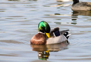
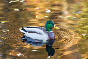
Mallard (Anas platyrhynchos) – The most ubiquitous North American duck, Mallards frequent both natural and artificial water bodies, often foraging in shallow, sediment‑rich areas where lead shot settles. Their opportunistic feeding behavior makes them a sentinel species for lead contamination.
Northern Pintail (Anas acuta) – Prefers cleaner, open water but will dabble where prey is abundant, including contaminated mudflats. Their longer necks and narrower bills can lead to deeper sediment ingestion.
Canada Goose (Branta canadensis) – Though not a true duck, Canada Geese are frequently included in waterfowl surveys. Their grazing on grasses near water brings them into contact with lead‑contaminated soils, especially in agricultural fields treated with lead‑based fertilizers historically.
4.2 Domestic & Semi‑Domesticated Breeds
Pekin (Anas platyrhynchos domesticus) – The most common commercial meat duck, raised in confinement. While indoor rearing reduces exposure, outdoor access for foraging or using lead‑containing water bowls can be hazardous.
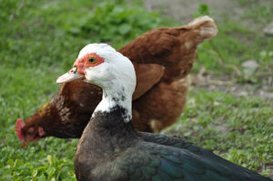
Muscovy (Cairina moschata) – Larger body mass and a propensity for ground foraging make Muscovies vulnerable when kept on pastures where lead shot is used for pest control.
Khaki Campbell – Renowned for prolific egg production, these ducks often have free‑range access to ponds that may be impacted by nearby hunting activities.
4.3 Why Certain Breeds Suffer More
- Feeding Ecology – Dabbling and surface feeding bring the bill and head close to the sediment where lead concentrates. Species that feed deeper (e.g., diving ducks) can also ingest lead, but the concentration in surface sediments is typically higher.
- Habitat Overlap with Human Activity – Ducks that inhabit or migrate through heavily hunted wetlands (e.g., the Mississippi Flyway) encounter higher lead densities.
- Body Size & Metabolism – Smaller ducks (e.g., teal) have a higher surface‑area‑to‑mass ratio, so a given dose of lead yields a higher blood concentration compared with larger species.
- Management Practices – Domestic breeds kept on farms that still use lead shot for rodents or hunting will be directly exposed, whereas those housed in lead‑free facilities are largely protected.
5. Life‑Stage Susceptibility
| Life Stage | Lead Sensitivity | Typical Exposure Scenarios | Clinical Implications |
|---|---|---|---|
| Embryo (egg) | Very high – developing organs are highly vulnerable | Maternal transfer of lead from contaminated diet; exposure via shell contamination | Reduced hatchability, skeletal malformations, impaired organogenesis |
| Hatchling (0‑4 weeks) | High – rapid growth demands iron and calcium, both disrupted by lead | Ingestion of contaminated brood‑minder water or feed; foraging in shallow puddles | Failure to thrive, severe anemia, neurologic deficits |
| Juvenile (4‑12 weeks) | Moderate‑high | Transition to outdoor ponds; exposure to lead shot on feeding grounds | Subclinical anemia, subtle behavioral changes (e.g., reduced flight) |
| Adult (≥ 12 weeks) | Moderate | Seasonal migration through hunting zones; feeding on agricultural runoff | Acute clinical disease if dose high; chronic low‑level exposure may impair reproduction |
| Senescent | Lower relative sensitivity (larger body mass) but impaired detoxification | Accumulation of lead over years; reduced renal function | Chronic renal disease, decreased immunity, higher mortality from secondary infections |
Key Insight: The critical window for lethal toxicity is often the early post‑hatch period, when a small absolute amount of lead can represent a massive proportion of body weight. Preventive measures should prioritize keeping hatchlings and brood chicks away from any lead‑contaminated water sources.
6. Clinical Signs & Symptoms
Lead poisoning is a multisystemic disease, and the clinical picture may be acute, sub‑acute, or chronic. Signs can be subtle, especially in wild birds that may hide weakness to avoid predation.
6.1 Neurologic Manifestations
- Ataxia & Wobbly Gait – Loss of coordination, especially evident when the bird attempts to take off.
- Tremors & Twitching – Often noted in the neck or wings.
- Seizure‑like Activity – Rare but may occur in severe intoxication.
- Behavioral Changes – Lethargy, reduced vocalization, delayed response to predators.
6.2 Hematologic & Respiratory Signs
- Pallor of the Comb, Bill, and Mucous Membranes – Indicative of anemia.
- Tachypnea – Rapid, shallow breathing compensating for reduced oxygen‑carrying capacity.
- Weakness & Exhaustion – Inability to sustain flight for even short distances.
6.3 Gastrointestinal & Renal Signs
- Anorexia & Weight Loss – Secondary to nausea and reduced appetite.
- Diarrhea or Constipation – Variable, occasionally fecal discoloration (dark, tarry).
- Polyuria/Polydipsia – Excessive drinking and urination due to renal tubule damage.
6.4 Musculoskeletal and Skin Findings
- Swollen Joints – Occasionally due to secondary infection.
- Subcutaneous Edema – Especially around the legs and vent.
6.5 Reproductive Effects (chronic exposure)
- Reduced Egg Production – Oviposition frequency drops by 30‑50 % in affected females.
- Thin‑Shell or Shell‑less Eggs – Impaired calcium metabolism.
6.6 “Red‑Heart” Phenomenon
A hallmark for many avian species (including mallards) is a bright red–purple discoloration of the heart and liver on necropsy, caused by the accumulation of lead‑induced porphyrins. While not visible in a live bird, it is a useful necropsy clue for pathologists.
7. Pathophysiology – How Lead Damages a Duck’s Body
- Absorption & Distribution – Approximately 90 % of ingested lead in ducks is absorbed via the gastrointestinal tract, especially when the metal is in a soluble form (e.g., lead nitrate from industrial effluents). Lead binds to erythrocytes (≈ 95 % of blood lead) and is transported to the liver, kidneys, bone, and brain.
- Heme Synthesis Interruption – Inhibition of ALAD and ferrochelatase blocks the conversion of δ‑ALA to porphobilinogen and the final insertion of iron into protoporphyrin IX, respectively. This results in ineffective erythropoiesis, macrocytic and microcytic anemia, and accumulation of δ‑ALA, a neurotoxic compound that further impairs neuronal function.
- Neurological Toxicity – Lead competes with calcium at voltage‑gated channels, disrupting neurotransmitter release (particularly glutamate and GABA). The resulting excitotoxicity manifests as tremors, ataxia, and seizures.
- Renal Damage – Lead accumulates in proximal tubular epithelial cells, causing oxidative stress, mitochondrial dysfunction, and eventual cell death. Clinically, this leads to proteinuria, polyuria, and progressive renal failure.
- Immune Suppression – Lead interferes with the proliferation of B‑ and T‑lymphocytes, diminishing antibody production and cellular immunity, which predisposes ducks to secondary bacterial and fungal infections.
- Bone Deposition & Re‑Release – Approximately 30‑40 % of absorbed lead is stored in the skeletal matrix. During periods of high calcium demand (e.g., egg‑laying, growth), stored lead can be mobilized back into circulation, causing delayed or recurrent clinical signs.
8. Diagnostic Approach
A thorough diagnosis combines history, clinical examination, laboratory testing, and, when possible, imaging.
8.1 History
- Geographic Location – Recent hunting season, proximity to shooting ranges, or known industrial sites.
- Feeding Routine – Access to open water, presence of lead shot in feed, use of lead‑based fishing gear.
- Observed Behavior – Sudden lethargy, abnormal gait, or visible ingestion of foreign material.
8.2 Physical Examination
- Full body condition score (BCS) assessment.
- Pale mucous membranes, respiratory rate, and neurologic evaluation (balance, proprioception).
- Palpation for abdominal masses or fluid accumulation.
8.3 Laboratory Tests
| Test | Sample | Expected Findings in Lead‑Poisoned Ducks |
|---|---|---|
| Blood Lead Concentration (BLC) | Whole blood (0.5‑1 mL) | > 0.3 ppm (30 µg/dL) = clinically significant; > 0.6 ppm (60 µg/dL) = severe toxicity. |
| Complete Blood Count (CBC) | Blood | Regenerative or non‑regenerative anemia, leukopenia, thrombocytopenia. |
| Serum Biochemistry | Blood | Elevated uric acid (renal dysfunction), increased AST/ALT (hepatic stress), low calcium/phosphate. |
| δ‑ALA Urine Test | Urine | Elevated δ‑ALA (> 2 mg/L) indicating ALAD inhibition. |
| Radiography | Whole‑body X‑ray | Visible lead fragments in gastrointestinal tract; may be obscured by fecal material. |
| CT/CT‑Angiography (advanced) | Head/abdomen | Precise mapping of metallic foreign bodies; useful for surgical planning. |
| Necropsy & Histopathology (post‑mortem) | Tissues | “Red‑heart” lesion, basophilic granules in hepatic Kupffer cells, renal tubular degeneration. |
Interpretation of Blood Lead Levels (BLC):
- < 0.1 ppm (10 µg/dL): Background environmental exposure, usually asymptomatic.
- 0.1‑0.3 ppm (10‑30 µg/dL): Sub‑clinical; may cause mild anemia.
- 0.3‑0.6 ppm (30‑60 µg/dL): Clinical disease – neurological signs common.
- > 0.6 ppm (60 µg/dL): Severe toxicity – high mortality risk without rapid intervention.
8.4 Differential Diagnosis
- Avian botulism (neuromuscular signs)
- Salmonella/West Nile virus (neurologic & systemic signs)
- Nutritional deficiencies (e.g., vitamin E/Se deficiency causing similar myopathy)
- Heavy metal co‑toxicity (e.g., mercury, cadmium)
Confirming lead as the primary toxin requires correlation of clinical signs with elevated BLC and, when possible, detection of lead fragments or histologic evidence of lead deposition.
9. Treatment Protocols
Effective therapy hinges on rapid reduction of systemic lead, supportive care, and prevention of secondary complications.
9.1 Immediate Stabilization
- Isolation – Place the bird in a quiet, low‑stress environment with minimal handling.
- Thermoregulation – Maintain ambient temperature (20‑25 °C) using heat lamps; hypothermia worsens metabolism of toxins.
- Fluid Therapy – Initiate subcutaneous or intravenous lactated Ringer’s solution (10‑20 mL/kg) to correct dehydration and improve renal perfusion.
9.2 Chelation Therapy
| Chelator | Dosage (per kg) | Route | Frequency | Duration | Notes |
|---|---|---|---|---|---|
| CaNa₂EDTA (Calcium Disodium EDTA) | 20‑30 mg/kg | Intravenous (slow bolus) | Every 12 h | 5‑7 days | Most effective for acute high‑dose lead. Monitor calcium & renal function. |
| D-penicillamine | 10‑15 mg/kg | Oral (via gavage) | Every 12 h | 7‑10 days | Useful for sub‑acute cases; may cause GI irritation. |
| British Anti‑Lewisite (BAL, Dimercaprol) | 0.1 mg/kg | Intramuscular | Every 8 h | 3‑5 days | Reserved for severe neuro‑toxic signs; watch for painful injection site. |
| Succimer (DMSA) – Experimental in avian species | 30 mg/kg | Oral | Every 24 h | 5‑10 days | Limited data; reported mild efficacy. |
Administration Tips:
- Pre‑hydrate the bird before chelation to avoid hypotension.
- Calcium supplementation (e.g., calcium gluconate 10 % solution, 50 µL/kg IV) mitigates EDTA‑induced hypocalcemia.
- Monitor BLC, CBC, and renal parameters daily for the first 48 h, then every 48 h until stable.
9.3 Supportive Care
- Nutritional Support: High‑protein, easy‑to‑digest diets (e.g., boiled egg yolk + commercial waterfowl starter) enriched with vitamin B complex, selenium, and vitamin E to counter oxidative stress.
- Antibiotics (if secondary infection suspected): Enrofloxacin 10 mg/kg IM daily for 5 days or Trimethoprim‑Sulfadiazine 30 mg/kg PO BID.
- Analgesia/Anti‑inflammatories: Meloxicam 0.5 mg/kg PO q24h for pain control (use cautiously with renal compromise).
9.4 Post‑Treatment Monitoring
- Repeat BLC every 3‑5 days to confirm declining trend.
- Weight and BCS documentation; aim for a 5 % weight gain within 2 weeks.
- Behavioral assessment – return of normal foraging and preening indicates neurologic recovery.
9.5 When to Consider Euthanasia
- Persistent BLC > 0.6 ppm despite aggressive chelation.
- Irreversible organ failure (e.g., end‑stage renal disease).
- Profound neurologic impairment with no improvement after 48 h of therapy.
10. Prognosis, Complications, and Long‑Term Sequelae
| Factor | Impact on Prognosis |
|---|---|
| Blood Lead Level | Lower initial BLC (< 0.3 ppm) → > 80 % survival with treatment; > 0.6 ppm → < 30 % survival if untreated. |
| Age | Hatchlings have higher mortality due to small body mass; adults fare better. |
| Time to Treatment | Initiation within 12‑24 h of exposure improves survival dramatically. |
| Concurrent Illness | Co‑infection (Salmonella, avian influenza) worsens outcomes. |
| Renal Damage | Chronic nephropathy may cause delayed death even after BLC normalizes. |
Common Complications:
- Renal Failure – Persistent proteinuria, reduced uric acid excretion leads to gout.
- Secondary Infections – Immunosuppression predisposes to bacterial pneumonia, septicemia.
- Reproductive Failure – Even after clinical recovery, females may lay thin‑shell eggs for several months.
- Bone Lead Reservoir – Stored lead may leach back during calcium‑intensive periods, causing relapse.
Long‑Term Management:
- Periodic BLC screening (every 3‑6 months) for previously affected birds.
- Calcium‑rich diet (e.g., crushed oyster shells) to reduce bone resorption.
- Environmental remediation to prevent re‑exposure.
11. Prevention Strategies
11.1 Habitat Management
- Lead‑Free Hunting Zones – Encourage adoption of non‑lead ammunition (e.g., steel, bismuth, tungsten) in wetland hunting areas. Many states have already mandated this.
- Shot‑Recovery Programs – Conduct post‑hunt water sampling and retrieve spent shot; dispose of safely.
- Sediment Removal – Periodic dredging of heavily contaminated ponds, followed by safe disposal of contaminated silt.
11.2 Policy & Regulation
- Ban on Lead Fishing Gear – Numerous European Union directives have prohibited lead sinkers; similar legislation can be advocated in North America.
- Industrial Discharge Standards – Enforce EPA‑mandated limits for lead in effluent entering waterways.
- Public Education – Outreach to hunters, anglers, and farmers about the hidden dangers of lead in water bodies.
11.3 Husbandry Practices (Domestic Ducks)
- Provide Clean Water Sources – Use filtered or rain‑collected water for drinking and bathing.
- Avoid Lead‑Based Feed Additives – Use certified lead‑free grain, corn, and supplement mixes.
- Restrict Access to Dump Sites – Secure trash areas and discourage foraging on lead‑contaminated soils.
11.4 Rehabilitation Center Protocols
- Lead‑Screening of Incoming Birds – Immediate BLC test for any wild waterfowl rescued.
- Isolation Units Free of Lead – Use stainless steel cages, avoid lead‑based paints.
- Decontamination of Equipment – Regular cleaning with chelating agents (e.g., EDTA solutions) for tools that may become contaminated.
12. Diet and Nutrition – Supporting Recovery and Reducing Risk
| Nutrient | Role in Lead Toxicity Mitigation | Practical Sources for Ducks |
|---|---|---|
| Calcium | Competes with lead for absorption; reduces gastrointestinal uptake; aids in bone deposition of lead. | Crushed oyster shells, limestone grit, calcium‑fortified waterfowl pellets. |
| Vitamin C | Antioxidant; assists in regeneration of glutathione, protecting against oxidative damage. | Fresh citrus (orange slices), strawberries, commercial Vitamin C‑enhanced supplements. |
| Vitamin E & Selenium | Protects cell membranes from lipid peroxidation caused by lead‑induced ROS. | Wheat germ oil, selenium‑fortified yeast, mixed grain with added vitamin E. |
| Protein | Supports hematopoiesis to replace lead‑induced anemia. | Boiled egg yolk, soybeans, mealworms (high‑protein live feed). |
| Fiber | Promotes gut motility aiding in passage of undigested lead fragments. | Chopped leafy greens, alfalfa hay. |
| Water Quality | Low lead content prevents ongoing exposure; adequate hydration supports renal excretion. | Rain‑water collection, reverse‑osmosis filtered water; test water regularly (lead < 0.01 ppm). |
Feeding Recommendations for Recovering Ducks:
- First 24 h: Offer soft, high‑energy foods (e.g., scrambled eggs, warm waterfowl starter) to encourage intake.
- Days 2‑5: Introduce calcium‑rich grit (2 % of diet) and vitamin C fruit slices.
- Beyond 5 days: Transition to balanced commercial pellet (≥ 28 % protein) mixed with fresh greens.
- Monitor feed intake and fecal output; any decline may indicate lingering GI irritation.
13. Zoonotic Considerations – Risks to Humans
While lead itself is not transmissible between species, human exposure can occur through handling contaminated birds or consuming meat/eggs from affected ducks.
| Exposure Pathway | Risk Level | Mitigation |
|---|---|---|
| Direct handling (rehabilitation, hunting) | Moderate – skin contact with lead fragments or contaminated feathers | Wear nitrile gloves, wash hands thoroughly, use protective clothing; avoid crushing pellets with hands. |
| Consumption of duck meat/eggs from infected birds | High – lead accumulates in muscle and viscera; bone marrow can contain high concentrations | Do not consume any part of a bird with known lead exposure; follow USDA guidelines for testing lead in game meat. |
| Inhalation of lead dust (during cleaning of contaminated enclosures) | Low‑Moderate | Use dust masks (N95), ensure good ventilation, wet‑clean surfaces to minimize airborne particles. |
| Secondary contamination (e.g., lead shot left in the environment) | Community‑wide | Participate in lead‑shot retrieval programs; educate local hunters. |
Special Note for Children and Pregnant Women: Lead exposure is especially hazardous to developing fetuses and young children. Those involved in wildlife rehabilitation or hunting should observe strict personal protective equipment (PPE) protocols and avoid bringing contaminated clothing home.
14. Key Take‑Home Messages
- Lead poisoning is a preventable, yet pervasive, threat to ducks that manifests through anemia, neurologic dysfunction, renal failure, and reproductive loss.
- Primary exposure routes are ingestion of lead shot, fishing sinkers, and contaminated sediments; the highest risk occurs in hunting wetlands and areas with legacy industrial pollution.
- Early detection via blood lead testing (≥ 0.3 ppm) and rapid chelation (EDTA‑based) dramatically improve survival, especially in hatchlings and juveniles.
- Comprehensive supportive care—fluid therapy, nutrition, and infection control—are essential adjuncts to chelation.
- Long‑term monitoring is required because lead stored in bone can be remobilized, causing relapse during high calcium demand periods.
- Prevention is more effective than treatment: transition to non‑lead ammunition, secure clean water sources, and educate stakeholders.
- Zoonotic risk to humans exists primarily through handling contaminated birds and consuming their tissues; proper PPE and avoidance of consumption are mandatory.
#LeadPoisoning #DuckHealth #WaterfowlConservation #AvianToxicology #LeadFreeHunting #WildlifeRehab #EcoFriendlyFishing #BirdVeterinary #ConservationScience #SaveOurDucks #NonLeadAmmunition #ProtectWildlife #EcoAwareness #EnvironmentalHealth #ZoonoticSafety #DuckNutrition #LeadPoisonTreatment #RehabilitationSuccess #WildlifePolicy #WetlandProtection
Add comment