
Introduction: Decoding Canine Seasonal Atopy
Canine allergies, specifically Atopic Dermatitis (Atopy), are one of the most common reasons pet owners seek veterinary care. While many people associate pollen or grasses with seasonal discomfort, the breakdown of organic matter—particularly fallen leaves—presents a unique and often overlooked complex of allergens that plague dogs, especially during the autumn and damp spring seasons.
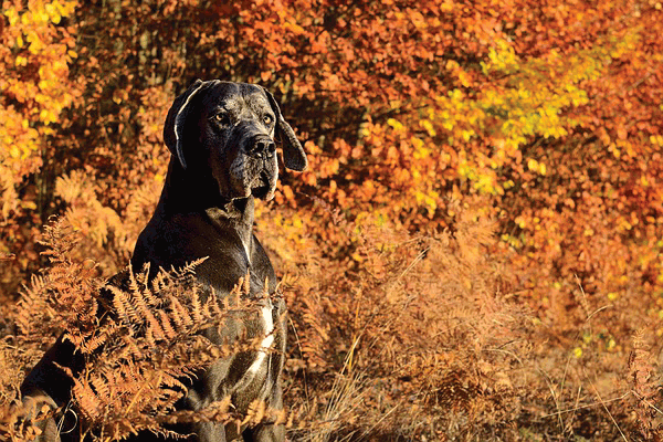
Leaves themselves are not the primary culprits. Instead, the danger lies in the cocktail of biological and chemical substances that accumulate on and within the decomposing foliage: microscopic molds, fungal spores, residual pollen, dust mites, and chemical fertilizers or pesticides. When a dog rolls in, walks through, or chews on a leaf pile, they are exposing their most sensitive skin areas (paws, belly, muzzle) to this concentrated blend of irritants.
This comprehensive guide delves into the mechanisms of leaf allergies, the specific irritants involved, detailed diagnostic pathways, and the multi-modal strategies necessary to manage and alleviate the chronic discomfort faced by affected dogs.
I. The Pathophysiology: Why Decomposing Leaves Cause Reaction
Canine atopic dermatitis is a genetically predisposed inflammatory and pruritic skin disease associated with IgE antibodies directed against environmental allergens. In simple terms, the dog’s immune system misidentifies harmless environmental substances as threats and launches a massive defensive response.
A. The Compromised Skin Barrier
A key factor in canine allergies is a defective epidermal barrier. Healthy skin acts like a brick wall, preventing invaders from entering and moisture from leaving. In allergic dogs, this “wall” is faulty. The skin is more permeable due to deficiencies in proteins (like filaggrin) and lipids (ceramides). This allows environmental allergens (like mold spores on leaves) to penetrate deeply into the skin layers rather than remaining on the surface.
B. The Allergic Cascade
- Penetration: Allergens (mold, pollen residue) enter the compromised skin.
- Recognition: Antigen-presenting cells (dendritic cells) capture the allergen and process it.
- Immune Activation: T-lymphocytes are activated, releasing inflammatory cytokines (chemical messengers).
- Mast Cell Degranulation: Cytokines signal mast cells (immune cells concentrated near the skin surface) to release potent inflammatory mediators, primarily histamine.
- Pruritus and Inflammation: Histamine causes blood vessels to dilate (redness/heat) and stimulates nerve endings, resulting in the relentless, maddening itch (pruritus).
C. The Unique Allergens of Decomposing Foliage
The primary allergens associated with leaves are not the cellulose structure but the biological matter that thrives on decay:
1. Leaf Mold and Fungal Spores
As leaves accumulate and become damp, they provide a perfect habitat for mold and fungi. These microscopic organisms release vast quantities of spores into the air and onto the ground. Common culprits include:
- Alternaria: Often associated with outdoor vegetation and soil.
- Cladosporium: Highly prevalent in decaying plant material.
- Aspergillus: Found widely in organic matter and dust. Inhaling these spores or having direct contact with them on the skin is often the core issue driving autumn allergies.
2. Persistent Pollen Residue
While tree and grass pollen peaks in spring and summer, some residual pollen remains trapped on foliage. When leaves fall and dry out, wind currents can redistribute this allergenic dust, leading to flare-ups even outside the traditional pollination season.
3. Mites in Organic Matter
Decomposing leaf piles are a haven for various mites, including storage mites and certain varieties of dust mites. These creatures shed waste particles that are potent allergens when inhaled or contacted.
4. Contact Irritants and Chemicals
Many property owners treat their lawns and gardens before winter, using fungicides, herbicides, or synthetic fertilizers. These chemicals adhere to the leaf surface. When a dog lies down or chews leaves, these caustic substances can trigger non-allergic (irritant) contact dermatitis, mimicking an allergic reaction.
II. Clinical Presentation: Spotting the Signs of Leaves Allergy
An allergy specifically related to leaves and decaying matter often manifests as contact dermatitis, meaning the symptoms are concentrated (though not exclusively) in areas that touch the ground.
A. Primary Clinical Markers (The Itch)
The hallmarks of canine atopy dictate where the irritation usually begins:
- Intense Pruritus (Itching): This is the cardinal sign. The dog may rub vigorously against furniture, scoot, or shake their head almost constantly.
- Pododermatitis (Paw Involvement): Because paws are the main point of contact, they are often the most affected. Signs include:
- Excessive licking, chewing, and gnawing of the feet.
- Moist, red skin between the digital pads and toes.
- Rust-colored staining (saliva containing porphyrins stains the fur red-brown).
- Swollen, “puffy” pads.
- Ventral and Axillary Erythema: Redness and rash in the armpits (axilla), groin, and abdomen (ventral areas), where the skin is thin and easily exposed when the dog lies on the ground.
- Aural Hematomas and Otitis Externa: Leaves and mold spores often trigger inflammation in the ear canals, leading to recurrent ear infections (otitis externa). Chronic scratching can cause severe head shaking, resulting in broken blood vessels in the ear flap (aural hematoma).
B. Secondary Symptoms (Infections)
Chronic skin inflammation creates a warm, moist, and compromised environment perfect for opportunistic pathogens:
- Yeast Dermatitis (Malassezia): Characterized by an overpowering, musty, rancid smell (often described as “corn chip” odor), greasy skin, and dark, thickened plaques (lichenification).
- Bacterial Pyoderma (Staphylococcus): Often presents as bumps, pustules, scabs, and hair loss (alopecia), requiring oral or topical antibiotics.
- Hot Spots (Acute Moist Dermatitis): Rapidly developing, intensely painful, wet sores caused by the dog obsessively licking or chewing one localized area following an acute flare-up.
C. Differentiating Leaves Allergy from Other Allergies
| Condition | Primary Trigger | Peak Season | Affected Areas | Key Indicators |
|---|---|---|---|---|
| Leaves/Mold Allergy | Mold spores, decomposing matter, persistent pollen. | Fall, Early Spring (damp seasons). | Paws, ventral abdomen, ears. | Symptoms directly correlating with time spent outdoors in humid, leafy areas. |
| Flea Allergy Dermatitis (FAD) | Flea saliva. | Year-round, often worse in warm months. | Rear end (tail base), hind legs, inner thighs. | Presence of flea dirt; immediate relief with rigorous flea control. |
| Food Allergy | Specific proteins (e.g., chicken, beef, dairy). | Year-round (non-seasonal). | Ears, paws, anal pruritus (scooting). | Requires strict elimination diet trial for diagnosis. |
III. The Diagnostic Journey: Pinpointing the Environmental Trigger
Diagnosing any canine allergy is a process of elimination known as a “diagnosis of exclusion.” The veterinarian must first rule out parasites (especially fleas and mites) and food sensitivities before confirming environmental atopy.
A. Initial Dermatology Work-Up
- Cytology: Swabs and scrapings are taken from the skin and ears to identify secondary infections (yeast and bacteria) that must be treated before allergy management can begin effectively.
- Skin Scrape: Performed to rule out parasitic mites (e.g., Sarcoptes, Demodex).
- Flea Control Trial: Even if fleas are not visible, a rigorous 6-8 week use of a prescription-strength flea preventative is standard practice to exclude FAD.
- Environmental History: The most crucial step for leaf allergy diagnosis is a detailed history. The owner must track when and where flare-ups occur:
- Do symptoms worsen after wet, muddy walks?
- Does the rash appear after playing in the yard post-raking?
- Did the symptoms disappear when the dog was boarded indoors for a week?
B. Allergy Testing for Specificity
Once atopy is confirmed, testing can determine the specific allergens (e.g., oak mold vs. maple pollen) to guide long-term management, particularly immunotherapy.
1. Intradermal Skin Testing (IDST)
- Procedure: Considered the gold standard. A veterinary dermatologist shaves a patch of the dog’s side and injects tiny amounts of various common local allergens (including tree molds, dust mites, grasses, and weed pollens) just beneath the skin.
- Results: Within 20 minutes, a positive reaction creates a raised, red wheal (hive-like lump), confirming sensitivity to that specific substance.
- Caveat: Requires withdrawal from certain oral medications (antihistamines, steroids) for weeks prior, which can be challenging for a severely itchy dog.
2. Serum Testing (RAST/ELISA)
- Procedure: A blood sample is analyzed in a lab to measure the concentration of IgE antibodies directed against various allergens.
- Pros: Less invasive, does not require shaving, and medication withdrawal is often shorter or unnecessary.
- Cons: Less sensitive and specific than IDST; can sometimes produce false positive or negative results because IgE levels in the blood do not perfectly correlate with the reaction occurring in the skin.
For leaf-related allergies, testing often shows strong positive reactions to specific fungal/mold panels and local tree pollen, confirming that the decomposing organic matter is the primary trigger.
IV. Treatment Strategies: The Multi-Modal Approach
Managing canine atopy, especially seasonal contact dermatitis associated with leaves and mold, requires a year-round, multi-modal strategy combining immediate symptom relief, controlling secondary infections, and modifying the immune response.
A. Immediate Symptomatic Relief (Short-Term Crisis Control)
For acute flare-ups caused by contact with heavy leaf matter, immediate veterinary intervention is necessary to break the itch-scratch cycle.
- Glucocorticoids (Steroids): Highly effective anti-inflammatories, often prescribed as a short course (e.g., Prednisone). They provide rapid relief but are not suitable for long-term use due to side effects (increased thirst, urination, appetite, and potential long-term issues like Cushing’s disease risk).
- Antihistamines: OTC options like Benadryl (diphenhydramine) or prescription options are often used adjunctively. However, antihistamines are generally effective in only a small percentage (around 20-30%) of canine allergy cases. They are often best used preventatively or in conjunction with other therapies.
B. Immune-Targeted Medications (Long-Term Maintenance)
These medications specifically target the pathways that cause the allergic reaction, offering powerful relief with fewer side effects than long-term systemic steroids.
1. Janus Kinase Inhibitors (Apoquel / Oclacitinib)
- Mechanism: Blocks the activity of specific Janus Kinase enzymes involved in transmitting the itch signal and inflammation.
- Pros: Starts working within hours (very rapid relief); safe for long-term use in most dogs; does not interfere with allergy testing.
- Cons: Must be given orally once or twice daily; can be costly; contraindicated in dogs under 12 months or those with certain active cancers.
2. Canine Atopic Dermatitis Immunotherapeutic (Cytopoint / Lokivetmab)
- Mechanism: This is an injectable monoclonal antibody. It specifically targets and neutralizes Canine Interleukin-31 (cIL-31), one of the primary cytokines responsible for signaling pruritus.
- Pros: Non-drug (biologic); very safe with virtually no systemic side effects; administered via injection every 4-8 weeks by a veterinarian; ideal for seasonal flare-ups.
- Cons: Only targets the itch (cIL-31), not the underlying inflammation or secondary infections; can be expensive.
C. The Gold Standard: Allergen-Specific Immunotherapy (ASIT)
ASIT (often called “allergy shots” or sublingual drops) is the only treatment proven to modify the immune system’s response, potentially leading to a permanent reduction in allergic symptoms.
- Mechanism: Based on the results of IDST or serum testing, custom-made serum containing minuscule, increasing doses of the dog’s specific allergens (e.g., local mold spores, cottonwood pollen) is created. Over time, this desensitizes the immune system to the trigger.
- Delivery: Can be given via subcutaneous injection (shots) or as drops placed under the tongue (sublingual).
- Timeline: ASIT is a commitment, requiring 9–12 months to see initial improvement and potentially years to reach maximum efficacy. However, success rates are high, often eliminating the need for daily medication.
D. Treating Secondary Infections
Aggressive treatment of pyoderma (bacteria) and Malassezia (yeast) is essential, as the infection itself causes itching, compounding the allergy symptoms.
- Antibiotics/Antifungals: Oral medications (e.g., cephalexin, fluconazole) are often required for widespread infections, usually given for 3–6 weeks.
- Topical Therapy: Medicated baths are crucial (see Section V).
V. Environmental Management and Protective Protocols
Since leaf allergies are contact-driven, the most effective management involves rigorous reduction of exposure and effective removal of allergens after contact.
A. The Post-Walk “Decontamination” Protocol
This strategy is paramount during peak fall/spring humidity when mold counts are highest.
- Immediate Wipe Down: After any walk, especially through parks, forests, or areas with thick leaf coverage, wipe the paws, muzzle, and ventral abdomen thoroughly.
- Use specialized hypoallergenic, chlorhexidine, or cleansing wipes specifically designed to remove allergens (e.g., wipes containing Phytosphingosine).
- Focus particularly on the areas between the toes, using warm water or a specialized paw cleanser to flush debris.
- Protective Gear: For severely affected dogs, protective gear is highly effective:
- Dog Booties: Excellent barrier protection for the paws, preventing mold and chemical contact. Ensure a proper fit to avoid rubbing or discomfort.
- Protective Vests/Coats: Specialized vests can prevent mold and pollen from settling directly onto the highly sensitive skin of the chest and abdomen when the dog rolls.
B. Therapeutic Bathing Regimens
Bathing is the single most effective method for immediate physical removal of allergens from the skin surface, interrupting the allergic cascade before it begins.
- Frequency: During a flare-up, medicated baths may be needed every 2–3 days. For maintenance, a weekly bath is often recommended.
- Product Selection:
- Medicated Shampoos: Look for shampoos containing Chlorhexidine (antibacterial) and Miconazole or Ketoconazole (antifungal) to manage secondary infections.
- Soothing/Barrier Shampoos: Oatmeal-based or ceramides-containing shampoos help repair the skin barrier and provide immediate itch relief.
- The 10–15 Minute Soak: Medicated shampoos require a minimum contact time of 10–15 minutes to be effective. Ensure the dog is thoroughly lathered, focusing on the paws and groin, before rinsing completely.
C. Indoor Environment Control
While outdoor exposure is the immediate trigger, spores and pollen travel inside on clothing, fur, and the air.
- HEPA Filtration: Use high-efficiency particulate air (HEPA) filters in the rooms where the dog spends the most time (bedroom, living room). These filters trap mold spores and dust mites.
- Laundry: Wash dog bedding weekly in hot water (ideally 130°F/54°C) to kill mites and spores.
- Vacuuming: Use a vacuum cleaner equipped with a HEPA filter to minimize recirculation of microscopic allergens from carpets.
D. Yard Maintenance Adjustments
The goal is to minimize the decomposition zone where mold thrives:
- Raking and Disposal: Rake leaves frequently and dispose of them immediately, ensuring no large, damp piles remain. Allowing dogs to play in raked leaf piles exposes them to the highest concentration of spores.
- Chemical Review: If possible, minimize the use of commercial lawn chemicals, especially systemic fungicides and herbicides, which can be contact irritants.
VI. Nutritional Support and the Skin-Gut Axis
While diet rarely causes leaves/mold allergies, nutritional adjustments and supplements play a vital supportive role in improving skin barrier function and modulating the immune system.
A. Omega-3 Fatty Acids (The Anti-Inflammatory Powerhouse)
Supplements containing large, therapeutic doses of Eicosapentaenoic Acid (EPA) and Docosahexaenoic Acid (DHA), sourced mainly from marine fish oil, are essential.
- Mechanism: Omega-3s compete with inflammatory Omega-6 fatty acids (abundant in many commercial pet foods) in the body’s metabolic pathways. They reduce generalized inflammation, decrease pruritus, and are vital components in repairing the skin’s lipid barrier.
- Dosage: Effective treatment requires veterinary-grade supplements at high concentrations, often exceeding the amounts found in standard maintenance food.
B. Probiotics and Gut Health
Recent research highlights the strong connection between the gastrointestinal microbiome and skin health (the “skin-gut axis”).
- Mechanism: A healthy, diverse gut flora helps regulate the systemic immune response. Supplementing with specific strains of probiotics (e.g., Lactobacillus and Bifidobacterium species) can reduce overall inflammation and may temper the immune system’s overreaction to allergens.
- Recommendation: Use veterinary-specific probiotics, as human-grade products may not colonize the canine gut effectively.
C. Skin Barrier Supplements (Ceramides and Essential Oils)
Specific supplements and spot-ons are designed to physically repair the faulty epidermal barrier.
- Ceramides: These are the key lipids that hold skin cells together (like mortar between bricks). Topical therapy (found in sprays, mousse, or shampoos) can replenish ceramides, strengthening the skin’s defense against mold spores and environmental irritants.
VII. Identifying and Avoiding Toxic Foliage (A Critical Distinction)
While most leaves cause problems due to mold or contact irritation, certain foliage is inherently toxic or acutely irritating due to natural chemical compounds. It is essential to distinguish between allergy (immune response) and toxicity (poisoning/chemical irritation).
A. Plants Causing Irritant Contact Dermatitis
Dogs are generally less sensitive to the oil (urushiol) found in poison ivy, oak, and sumac than humans, as their dense fur provides protection. However, direct contact with thin-skinned areas (groin, ear flaps) or oil transfer from the dog’s coat to human skin can cause severe reactions.
- Poison Ivy/Oak/Sumac: If exposure is suspected, bathe the dog thoroughly with a degreasing dish soap to remove the oil immediately.
B. Common Foliage Toxicities (Ingestion Risk)
The ingestion of certain leaves can lead to systemic illness, independent of seasonal allergy:
| Plant/Foliage | Toxic Principle | Clinical Signs |
|---|---|---|
| Oak (leaves/acorns) | Tannins (gallic acid) | Vomiting, diarrhea (often bloody), lethargy, kidney failure (rare, but serious). |
| Maple (especially Red Maple) | Unknown toxin (usually dried/wilted leaves) | Lethargy, refusal to eat, dark urine, anemia (primarily a threat to horses, but ingestion should be avoided in dogs). |
| Rhubarb Leaves | Oxalates | Vomiting, diarrhea, tremors, kidney damage. |
| Black Walnut | Juglone (Toxins also found in the hulls) | Vomiting, tremors, seizures (often associated with decaying wood/hulls). |
Any time foliage is ingested, especially if the dog shows signs beyond skin irritation (e.g., vomiting, tremors, depression), immediate veterinary attention is warranted.
VIII. Long-Term Prognosis and Pet Owner Commitment
Canine atopy, including sensitivity to leaves and mold, is a chronic, lifelong condition. It is manageable but rarely curable. Successful management hinges entirely on the diligence and commitment of the pet owner.
A. Monitoring and Relapse Management
Owners must learn to recognize the subtle early signs of a flare-up (e.g., increased paw licking, a slight ear scratch) rather than waiting until the infection is established and severe pruritus is present.
- Trigger Avoidance: Management is often seasonal. Owners must be prepared to intensify grooming (more frequent medicated bathing, daily wiping) and potentially restart immune-modulating drugs (like a short course of Apoquel) as soon as the leaves begin to drop and humidity rises.
- Regular Veterinary Check-ups: Allergic dogs require check-ups every 3–6 months to monitor skin health, adjust medication dosages, and ensure secondary infections are not taking hold silently.
B. Quality of Life Assessment
The ultimate goal of allergy management is a good quality of life (QOL) score for the dog. Ideally, an allergic dog should be comfortable approximately 80% of the time, with symptom severity low enough that their behavior is not dominated by scratching or licking. If QOL is constantly poor, the treatment protocol needs escalation (e.g., transition from sole immune suppressants to ASIT).
Conclusion
Leaves allergies in dogs are complex, driven predominantly by the mold and fungal spores associated with decomposing organic matter. This seasonal challenge requires far more than simple topical creams. It demands a holistic, dedicated approach that integrates veterinary diagnostics, state-of-the-art medications (like Apoquel or Cytopoint), long-term immune modification (ASIT), and rigorous environmental control. By understanding the unique triggers of the seasonal environment and implementing focused post-exposure protocols, pet owners can significantly reduce their dog’s suffering, turning a miserable season into a manageable one.
#DogAllergies #SeasonalAllergies #DogDermatology #LeafAllergy #CanineAtopy #DogHealth #PetCare #MoldAllergy #ItchyDog #DogTips #VeterinaryAdvice #Apoquel #Cytopoint #DogSkinProblems #AutumnAllergies #PetWellness #DogMomProblems #PawLicking #FungalAllergy #VetLife #ImmunotherapyForDogs


Add comment