
Leishmaniasis is one of the most significant and geographically widespread parasitic diseases affecting companion animals globally. Endemic across vast regions of the Mediterranean Basin, parts of Asia, Africa, and the Americas, this complex zoonotic disease poses a substantial challenge to veterinary medicine and public health. Caused by protozoa of the genus Leishmania and transmitted by phlebotomine sand flies, Canine Leishmaniasis (CanL) typically presents as a chronic, debilitating systemic illness—a form known classically as Visceral Leishmaniasis (VL)—but it can also manifest purely cutaneously.
In dogs, the primary causative agent is Leishmania infantum (sometimes referred to as L. chagasi in the Americas). Dogs are considered the principal domestic reservoir host for human infection, making the effective diagnosis, treatment, and prevention of CanL paramount for both animal welfare and human disease control.
I. CAUSES, ETIOLOGY, AND PATHOPHYSIOLOGY
Leishmaniasis is fundamentally a disease driven by a complex interplay between the parasite, the sand fly vector, and the host’s immune system.
A. The Causative Agent
The protozoan parasites belong to the genus Leishmania. In dogs, the species of major concern is Leishmania infantum. This parasite is digenetic, meaning it requires two different hosts to complete its life cycle: an invertebrate vector (the sand fly) and a vertebrate mammal (the dog or human).
B. The Vector
Transmission occurs exclusively through the bite of infected female sand flies (tiny, hairy flies much smaller than mosquitoes, typically $2-3$ mm long). The specific genera responsible are Phlebotomus (in the Old World: Europe, Asia, Africa) and Lutzomyia (in the New World: North and South America). Sand flies are generally crepuscular or nocturnal, meaning the period of highest risk for transmission is from dusk until dawn, though they may also bite during shaded daylight hours.
C. The Life Cycle and Pathogenesis
- Infection: An infected sand fly takes a blood meal, injecting the infective stage (the flagellated promastigote) into the dog’s skin.
- Internalization: Once in the host, promastigotes are rapidly engulfed by macrophages and other phagocytic cells of the immune system.
- Replication: Inside the macrophage, the promastigote transforms into the non-flagellated, multiplying form (the amastigote). Amastigotes replicate extensively until the macrophage ruptures, releasing thousands of parasites to infect adjacent cells.
- Dissemination: Infected macrophages travel through the lymphatic system and bloodstream, disseminating the parasite throughout the body, primarily targeting organs rich in macrophages (the Reticuloendothelial System, or RES): spleen, liver, lymph nodes, bone marrow, and skin.
D. The Role of the Host Immune Response
The fate of the infected dog hinges entirely on the type of cell-mediated immune response mounted:
- Protective Immunity (Th1 Response): A robust Th1 (Type 1 Helper T-cell) response generates interferon-gamma ($\text{IFN-}\gamma$) and interleukin-2 ($\text{IL}-2$). This activates macrophages to produce nitric oxide, which effectively kills the amastigotes inside the cell. Dogs with a strong Th1 response may remain subclinically infected (asymptomatic carriers) or naturally clear the infection.
- Non-Protective Immunity (Th2 Response): A weak Th1 response coupled with a predominant Th2 (Type 2 Helper T-cell) response leads to the chronic, symptomatic disease. Th2 cells produce interleukins ($\text{IL}-4, \text{IL}-10$) that suppress macrophage activity, allowing unimpeded parasite replication. This response is characterized by massive, non-protective antibody production (hyperglobulinemia), which is a diagnostic hallmark but fails to clear the infection. These circulating immune complexes are responsible for the severe kidney damage (glomerulonephritis) that ultimately leads to death.
II. SIGNS AND SYMPTOMS (CLINICAL MANIFESTATIONS)
The incubation period for CanL is highly variable, ranging from three months to several years. Clinical presentation is diverse, earning Leishmaniasis the moniker “the great imitator” in veterinary medicine. Approximately $50%$ to $70%$ of infected dogs remain asymptomatic, but those that develop clinical signs usually present with one of three forms: generalized visceral disease, localized cutaneous disease, or a combination of both.
A. Systemic and Generalized Signs (Visceral Form)
These signs reflect the systemic dissemination of parasites and the chronic inflammatory status of the host:
- Weight Loss and Muscle Atrophy: Progressive, often severe cachexia despite a normal or even increased appetite.
- Lethargy and Exercise Intolerance: Due to chronic inflammation, anemia, and organ damage.
- Lymphadenomegaly: Swollen, non-painful superficial lymph nodes (prescapular, popliteal) are the most common finding.
- Splenomegaly and Hepatomegaly: Enlargement of the spleen and liver due to massive macrophage infiltration.
- Fever: Intermittent or recurrent fever spikes that often do not respond well to standard antibiotics.
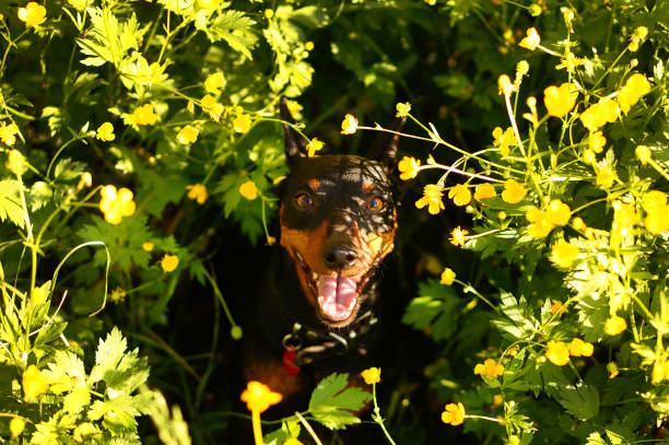
B. Cutaneous and Dermatological Signs (Most Common)
Dermatological signs are present in over $80%$ of symptomatic cases and are often the first reason a dog is presented to the veterinarian:
- Exfoliative Dermatitis: Symmetrical, non-pruritic (non-itchy) scaling and severe dry skin, especially over the head, ears, and limbs. This is often described as “moth-eaten” or “dandruff-like.”
- Alopecia: Hair loss, particularly around the eyes (the classic “spectacles” or periocular alopecia), the bridge of the nose, and the ears.
- Nail Abnormalities (Onychogryphosis): Abnormal, excessive, and sometimes painful growth, thickening, and curling of the claws. This sign is highly suggestive of CanL.
- Ulcerative Lesions: Painless ulcers that fail to heal, typically found over bony prominences (elbows, hocks) or the mucocutaneous junctions (muzzle, ear margins).
- Nodules and Papules: Hard, firm lumps in the skin, often associated with diffuse infiltration.
C. Ocular and Musculoskeletal Signs
- Ocular Disease: Uveitis (inflammation of the pigmented layer of the eye), conjunctivitis, or keratitis, often caused by immune complex deposition.
- Polyarthritis and Myositis: Inflammation of multiple joints (polyarthritis) causing stiffness, pain, and reluctance to move, or inflammation of muscle tissues (myositis).
D. Renal and Cardiovascular Signs (The Fatal Complications)
The most significant and often terminal complication of Leishmaniasis is Glomerulonephritis (kidney inflammation).
- Protein-Losing Nephropathy: Chronic circulation of antibody-antigen immune complexes that deposit in the kidney’s filtering units (glomeruli), leading to severe protein loss in the urine (proteinuria).
- Renal Failure: As the disease progresses, the damage leads to chronic kidney failure, presenting as Polyuria/Polydipsia (PU/PD – excessive urination and thirst), uremia, vomiting, and ultimately, death.
- Epistaxis (Nosebleeds): Recurrent nasal hemorrhage, usually due to vasculitis (inflammation of blood vessels) or sometimes related to kidney failure complications (azotemia-induced platelet dysfunction).
III. DOG BREEDS AT RISK
While any dog living in an endemic area is at risk of exposure, significant data confirms that certain breeds possess a genetic predisposition, exhibiting a higher likelihood of developing severe clinical disease once infected. This susceptibility is rooted in genetically controlled immune responses, specifically the inability to mount an effective Th1 response.
| Breed | Risk Level | Explanation of Susceptibility |
|---|---|---|
| Boxer | High | Boxers appear to have a genetic defect related to the major histocompatibility complex (MHC) class II region, which plays a pivotal role in initiating the appropriate Th1 immune response. They are prone to developing severe visceral forms quickly. |
| German Shepherd Dog (GSD) | High | GSDs often display a biased Th2 response, resulting in excessive antibody production that is non-protective. This leads to heavy parasitic loads and a rapid progression to severe systemic illness. |
| Rottweiler | Moderate to High | Similar to GSDs, Rottweilers exhibit immune profiles that favor non-protective humoral immunity (antibody-driven), making them highly susceptible to chronic, severe disease. |
| Ibizan Hound (Podenco) | Highest (in endemic areas) | Podencos, particularly in Spain and the Mediterranean, show a high prevalence and a tendency toward disseminated visceral disease. Their genetics are thought to predispose them to systemic failure early in the infection course. |
| Cocker Spaniel | Moderate | Cocker Spaniels are noted in some studies for having a higher incidence of clinical disease onset following infection compared to mixed-breed dogs in the same region. |
Elaborate Explanation of Genetic Susceptibility: The susceptibility observed in these breeds is often linked to polymorphisms (variations) in genes that regulate cytokine production (MHC I and II regions, and genes for $\text{IL}-4, \text{IFN-}\gamma,$ and $\text{TNF-}\alpha$). Genetically susceptible breeds, upon infection, are intrinsically more likely to skew their immune response toward the ineffective Th2 pattern. This means even if they receive a small infective dose, their body fails to control the parasitic replication within the macrophages of the internal organs. Conversely, genetically resistant breeds (often local indigenous breeds in endemic areas) rapidly develop the protective Th1 response, limiting the disease to a subclinical or self-limiting localized cutaneous infection.
IV. AGE AND STAGE OF LIFE AFFECTED
Leishmaniasis can affect dogs of any age, but the symptomatic presentation often follows a specific timeline due to the slow nature of the parasite and the long incubation period.
Puppies (Congenital and Rapid Onset)
While the primary route is vector-borne, vertical (mother-to-puppy) transmission is possible, though less common. Puppies infected in utero or very early in life may present with rapidly progressive, severe, generalized disease (systemic lymphadenopathy, severe lethargy, and failure to thrive) because their immune systems are still immature and unable to mount any effective Th1 response.
Adult Dogs (3 to 5 Years Old)
The majority of clinically ill dogs are young adults. This timeframe reflects the typical incubation period: a dog is bitten as a puppy or young adult, and the clinical signs start to emerge one to five years later when the chronic immune response failure and organ damage (especially the kidneys) become apparent.
Older Dogs
Older dogs that have lived their entire lives in endemic areas without showing signs may present with Leishmaniasis if their immune systems become compromised (e.g., due to concurrent illness, stress, or immunosuppressive medication). This is often a case of the existing, subclinical infection reactivating into full-blown visceral disease.
V. DIAGNOSIS OF CANINE LEISHMANIASIS
Diagnosis requires combining clinical suspicion (signs, history of travel/residence in endemic areas) with specific laboratory confirmation. No single test is perfect, and a multi-modal approach is often necessary, especially for monitoring treatment.
A. Non-Specific Clinical Pathology and Biochemistry
General blood work often reveals non-specific but highly suggestive abnormalities related to the chronic inflammatory state:
- Hematology: Non-regenerative normocytic, normochromic anemia (due to chronic disease/bone marrow involvement), and occasionally thrombocytopenia (low platelets).
- Biochemistry: Hyperglobulinemia (increased total protein due to massive, non-protective antibody production) and often hypoalbuminemia (low albumin due to kidney loss or inflammation). A reversed albumin/globulin ($\text{A}/\text{G}$) ratio (where globulin is higher than albumin) is a strong indicator of chronic Leishmaniasis.
- Renal Parameters: Increased Blood Urea Nitrogen (BUN) and creatinine (azotemia) if renal failure has developed. Urine Protein:Creatinine ($\text{UPC}$) ratio is crucial for quantifying proteinuria and assessing the severity of glomerulonephritis.
B. Serological Tests (Antibody Detection)
Serology screens for the dog’s antibody response to the parasite. It is the most common diagnostic method for screening and monitoring.
- Indirect Fluorescent Antibody Test (IFAT): The historical gold standard for antibody titration. It provides a semi-quantitative measure (a titre) of the dog’s antibody level. A very high titre strongly indicates active clinical disease, while low titres can suggest early infection or asymptomatic status.
- Enzyme-Linked Immunosorbent Assay (ELISA): Easier to use, faster, and now widely commercialized, offering quantitative or semi-quantitative results. ELISA is excellent for screening large populations.
Interpretation Challenges in Serology:
- False Negatives: Can occur in very early infection (before antibodies develop) or in severely immunosuppressed dogs (who fail to produce antibodies).
- False Positives: Rare, but can occur due to cross-reactivity with other parasites.
- Asymptomatic Dogs: Seropositivity only means the dog has been infected and is mounting a humoral response; it does not confirm active clinical disease.
C. Direct Parasite Detection (Definitive Diagnosis)
Finding the amastigotes confirms infection definitively and is necessary when serology is equivocal or if rapid confirmation is required.
- Cytology/Histopathology: Tissue aspirates (sampled via fine-needle aspiration) are taken from sites rich in macrophages:
- Lymph Nodes: Most common and easiest site.
- Bone Marrow: High sensitivity, often used when other sites are negative.
- Spleen Puncture: High sensitivity, but carries a small risk and requires careful handling.
- The sample is stained (e.g., Giemsa) and examined microscopically for the characteristic oval amastigotes, often clustered within macrophages.
- Polymerase Chain Reaction (PCR): PCR detects the genetic material ($\text{DNA}$) of the parasite.
- Qualitative PCR: Confirms the presence of Leishmania $\text{DNA}$ in blood, tissue, or conjunctival swabs.
- Quantitative PCR ($\text{qPCR}$): Measures the parasite load (how many parasites are present). This is highly valuable for asymptomatic dogs and for monitoring the efficacy of treatment (a successful treatment should significantly reduce parasite load).
VI. TREATMENT AND MANAGEMENT
The goal of CanL treatment is clinical remission and reduction of parasite load, not sterilization (complete eradication). Because amastigotes are intracellular, it is nearly impossible to completely eliminate them, and the disease must be managed as a chronic, relapsing condition.
A. Combination Therapy (The Standard Protocol)
Treatment usually involves a combination of a fast-acting leishmanicidal drug paired with a slower-acting leishmanistatic drug:
1. First-Line Leishmanicidal Drugs (Initial Knockdown)
These drugs aim to rapidly kill the majority of the circulating parasites and those within the organs:
- Meglumine Antimoniate (Antimonials): Historically a cornerstone of treatment, administered by injection (subcutaneous or intramuscular) for a course of 28 to 30 days. It is highly effective but requires careful monitoring due to potential cardiotoxicity and nephrotoxicity.
- Miltefosine: An oral liquid formulation that is highly effective. It is often preferred for ease of administration and reduced side effects compared to antimonials. It is typically given daily for 28 days.
2. Second-Line/Maintenance Leishmanistatic Drugs
These drugs do not kill the parasite directly but inhibit its replication, making them ideal for long-term maintenance and synergistic use:
- Allopurinol: A purine analogue, administered orally, usually started concurrently with Miltefosine or Meglumine Antimoniate and continued for 6 to 12 months, often indefinitely. It inhibits the enzyme needed by the parasite to synthesize nucleic acids.
B. Management of Complications
The severity of the disease dictates the necessary supportive care, which is often more critical than the antiparasitic drugs themselves:
- Renal Failure Management: If glomerulonephritis and renal failure are present, the kidney disease must be managed aggressively regardless of the parasite load. This involves:
- Specific renal diet (low in phosphorus, restricted protein).
- Angiotensin-Converting Enzyme ($\text{ACE}$) inhibitors (e.g., benazepril) to reduce proteinuria and protect kidney filtering function.
- Fluid therapy.
- Control of Secondary Infections: Antibiotics are used to treat secondary bacterial skin infections that often accompany chronic dermatitis.
- Anti-Inflammatory Agents: Systemic inflammation may be managed with careful use of supportive drugs, though strong immunosuppressants (like high-dose corticosteroids) should generally be avoided as they may facilitate parasite replication.
C. Monitoring Treatment Efficacy
Monitoring should be performed clinically (improvement in lethargy, skin lesions, weight gain) and through lab work:
- Initial Follow-up: 1 month after the end of the leishmanicidal phase.
- Long-term Follow-up: Every 3 to 6 months indefinitely.
- Key Lab Parameters: Monitor kidney function (BUN, Creatinine, $\text{UPC}$ ratio) and serology/qPCR. While serological titres may take months or years to drop, persistence of high titres or rising titres indicates poor response or relapse. $\text{qPCR}$ is often the best objective measure of parasite load reduction.
VII. PROGNOSIS AND COMPLICATIONS
A. Prognosis
The prognosis for Canine Leishmaniasis is highly variable and depends almost entirely on the presence and severity of renal damage at the time of diagnosis and the dog’s response to initial treatment.
- Excellent Prognosis: Dogs diagnosed accidentally (asymptomatic carriers) or those presenting only with mild, non-ulcerative cutaneous signs and no evidence of kidney damage. These dogs usually achieve long-term clinical remission with appropriate treatment.
- Guarded/Fair Prognosis: Dogs with generalized, chronic skin signs, lymphadenopathy, and laboratory changes (hyperglobulinemia) but normal kidney function (no azotemia). These dogs require strict, lifelong monitoring due to the risk of relapse.
- Poor Prognosis: Dogs presenting with severe, progressive renal failure (azotemia, severe proteinuria, and hypoalbuminemia). Glomerulonephritis is often irreversible once advanced, and this complication is the leading cause of death in infected dogs. Even if the parasite is controlled, the kidney damage is usually terminal.
B. Complications of the Disease
- Glomerulonephritis: The critical, life-limiting complication. Immune complexes destroy the kidney tissue, leading to Chronic Kidney Disease ($\text{CKD}$) and eventual failure.
- Relapse: Since sterilization is rare, relapse is common, often triggered by stress, concurrent illness, or waning drug efficacy. Relapses usually require repeating the initial leishmanicidal drug course.
- Drug Resistance: Especially in high-prevalence areas, parasites may develop resistance to antimonials or allopurinol, complicating treatment protocols.
- Co-infections: Dogs in endemic areas often carry other vector-borne diseases (e.g., Ehrlichiosis, Anaplasmosis) that can exacerbate Leishmaniasis symptoms and complicate diagnosis and treatment.
C. Complications of Treatment
- Meglumine Antimoniate Toxicity: Potential for injection site pain, pancreatitis, cardiotoxicity, and worsening of pre-existing kidney disease.
- Allopurinol-Related Xanthine Urolithiasis: Allopurinol interferes with purine metabolism. If the dog is fed a diet high in purines (e.g., organ meats, certain fish), it can lead to the formation of xanthine stones in the urinary tract, causing obstruction. This is a critical consideration in nutritional management.
- Miltefosine Side Effects: Typically gastrointestinal upset (nausea, vomiting), which usually diminishes after the first week of therapy.
VIII. PREVENTION STRATEGIES
Prevention focuses on controlling the vector (the sand fly) and boosting the dog’s immune system to cope with potential exposure.
A. Vector Control and Management
Since sand flies are required for transmission, reducing exposure to bites is the most critical element of prevention:
- Topical Insecticides/Repellents: Use topical spot-ons, sprays, or collars containing synthetic pyrethroids (e.g., deltamethrin, permethrin, flumethrin). Deltamethrin-impregnated collars have proven highly effective in endemic areas by repelling the sand fly before it can bite. These products must be applied regularly during the sand fly season (warmer months).
- Environmental Management: Sand flies are weak flyers and prefer calm, sheltered, warm environments. Minimize the dog’s exposure outdoors between dusk and dawn (peak biting times). If dogs must be kennelled outdoors, use fine-mesh netting treated with insecticide.
- Indoor Measures: Use screened windows and air conditioning, as sand flies avoid drafts and cool temperatures.
B. Vaccination
Vaccination is a powerful tool available in regions where Leishmaniasis is endemic (e.g., Europe, Brazil). Vaccines do not prevent infection, but they significantly reduce the risk of developing clinical disease.
- Available Vaccines (e.g., CaniLeish, Leish-Tec): These vaccines typically contain specific parasite antigens combined with an adjuvant designed to skew the immune response forcefully toward the protective Th1 pathway.
- Protocol: Dogs must be tested and confirmed seronegative before vaccination. A primary course involves several injections, followed by annual boosters.
- Limitations: Vaccines are highly recommended for dogs living in or traveling to endemic regions but should always be combined with stringent vector control.
C. Chemoprophylaxis
In some high-risk scenarios, pharmacological prevention may be considered, although this is less common than vaccination. Certain drugs (like anti-parasitic moxidectin combined with imidacloprid in certain preparations) may have ancillary activity against the sand fly vector or the early stages of the parasite, offering an added layer of protection.
IX. DIET AND NUTRITION IN MANAGING LEISHMANIASIS
Nutritional support is paramount, especially since Leishmaniasis is a chronic, wasting disease often complicated by severe inflammation and kidney failure. Diet must be tailored to the specific clinical stage of the dog.
A. General Nutritional Goals
- Counter Cachexia: Provide a highly palatable, easily digestible, and calorie-dense diet to combat weight loss and muscle atrophy.
- Immune Modulation: Utilize dietary components that support immune function and help control systemic inflammation.
- Renal Protection: Modify protein and phosphorus levels if kidney disease is present.
B. Specific Dietary Considerations
1. Managing Allopurinol Therapy (Purine Content)
This is the most critical dietary modification for treated dogs:
- Restriction of High-Purine Foods: Allopurinol inhibits xanthine oxidase, which is necessary to break down purines. Dogs on long-term allopurinol must avoid foods extremely high in purine to prevent the formation of xanthine bladder stones.
- Foods to strictly limit or eliminate: Organ meats (liver, kidney), game meats, certain seafood (sardines, anchovies), yeast extract, and some high-purine legumes.
- Recommended: Encourage the feeding of easily digestible proteins like egg or white meat poultry.
2. Renal Disease Management
If the dog is azotemic (elevated BUN/Creatinine) due to glomerulonephritis, the diet must shift to a veterinary-specific renal formula:
- Protein Control: Moderate restriction of protein (not elimination) to reduce the metabolic load on the kidneys. The protein must be of very high biological value (highly digestible) to maintain muscle mass.
- Phosphorus Restriction: Phosphorus is highly correlated with the progression of $\text{CKD}$, thus the diet must contain restricted and highly regulated phosphorus levels.
3. Anti-Inflammatory and Antioxidant Support
Leishmaniasis generates systemic chronic inflammation (oxidative stress). Dietary augmentation can help mitigate this:
- Omega-3 Fatty Acids ($\text{EPA}$ and $\text{DHA}$): High levels of marine-sourced Omega-3s are anti-inflammatory and can help reduce the destructive inflammation associated with glomerulonephritis and skin lesions.
- Antioxidants: Supplementation with Vitamin E, Vitamin C, and Zinc can help combat oxidative stress caused by chronic immune activation.
- B Vitamins: Essential B vitamins may be depleted due to systemic illness and kidney loss and should be supported.
X. ZOONOTIC RISK AND PUBLIC HEALTH SIGNIFICANCE
Leishmaniasis is a classic and significant zoonotic disease. This means it is naturally transmitted between animals (including dogs) and humans. The presence of Canine Leishmaniasis in an area is the primary determinant of risk for human infection.
A. The Risk Pathway
Crucially, direct transmission of Leishmania from an infected dog to a human does not occur (e.g., through kissing, petting, or dog feces). Transmission to humans requires the intermediate sand fly vector.
- Dog as Reservoir: The infected dog acts as the primary reservoir host, harboring massive numbers of parasites in its skin and internal organs.
- Sand Fly Infection: A female sand fly bites the infected dog and ingests parasite-laden macrophages.
- Human Infection: The now-infected sand fly bites a human, transmitting the parasite.
B. At-Risk Human Populations
While any person living in an endemic area is susceptible, two groups face disproportionately high risk:
- Children: Due to their close proximity to the ground and developing immune systems, children in endemic regions are particularly vulnerable to Visceral Leishmaniasis (Kala-azar).
- Immunocompromised Individuals: People with weakened immune systems, particularly those with $\text{HIV}/\text{AIDS}$ or those receiving immunosuppressive therapies, are highly susceptible and frequently develop severe, opportunistic Visceral Leishmaniasis if exposed.
C. Public Health Control
Controlling the canine reservoir is critical for reducing human cases. Public health strategies rely on:
- Case Detection: Widespread screening of dogs in endemic areas.
- Vector Control: Mass use of insecticide-impregnated collars on dogs and residual spraying of homes.
- Treatment: Treating clinically ill dogs to reduce their parasite load and, therefore, their infectivity to sand flies.
- Euthanasia Controversy: In some regions, historical control measures involved the culling of seropositive dogs to eliminate the reservoir. Modern veterinary and public health consensus now favors treating seropositive dogs (especially if symptomatic) and implementing aggressive prevention, recognizing that effective treatment can reduce infectivity significantly without resorting to widespread euthanasia.
#CanineLeishmaniasis, #DogHealth, #Leishmania, #ParasiteInfection, #VeterinaryMedicine, #DogParasites, #VisceralLeishmaniasis, #PetHealth, #EndemicDisease, #SandFlyDanger, #LeishFighter, #DogCare, #Zoonotic, #CanineCKD, #VetGuidance


Add comment