
Ducks (family Anatidae) are among the most prolific poultry species kept for meat, eggs, feathers, and breeding stock. While adult duck health often dominates the conversation, embryonic health is equally vital. One of the most common, yet frequently overlooked, embryonic problems is malposition—the abnormal orientation of the developing chick inside the egg. Malposition can reduce hatchability, produce weak or deformed ducklings, and, if severe, cause complete embryonic loss. This guide consolidates the latest scientific knowledge, field observations, and practical hatchery experience into a single, exhaustive reference for duck producers, veterinarians, and avian researchers.
2. What Is “Malposition” in Duck Embryos?
Malposition refers to any orientation of the duck embryo that deviates from the normal head‑down, longitudinal position that facilitates an unhindered climb out of the egg during pipping and hatching. In a correctly positioned embryo, the air cell sits opposite the large end of the egg; this provides a breathable pocket that the chick uses to inflate its lungs before breaking the shell. When an embryo lies side‑up, head‑up, or twisted, the air cell may be obstructed, the chorioallantoic membrane may be compressed, or the embryo may become trapped against the shell membranes, all of which hinder normal hatching.
Typical malpositions observed in duck eggs include:
| Malposition Type | Description | Typical Consequence |
|---|---|---|
| Head‑up | Embryo’s head points toward the air cell in the large end. | Difficult pipping; possible internal trauma. |
| Side‑up (lateral) | Embryo lies on its side, often with the dorsal region contacting the shell. | Reduced access to air cell; higher mortality. |
| Twisted/Spiral | Embryo rotates multiple times along its longitudinal axis. | Incomplete development of circulatory systems; high mortality. |
| Back‑down | The embryo’s back faces the air cell, causing the yolk sac to block the airway. | Respiratory failure, poor hatching success. |
Malposition can be primary (occurring during the first few days of incubation) or secondary (developing later as a result of external stressors). Recognizing the type and timing of malposition is crucial for effective intervention.
3. Primary Causes of Embryonic Malposition
- Inadequate Egg Rotation (Turning) During Early Incubation
- Mechanism: Turning distributes the embryo’s mass evenly and keeps the yolk sac fluid, preventing the embryo from settling in a fixed abnormal orientation.
- Critical Window: Days 1‑6 of incubation for most duck species; some larger breeds (e.g., Muscovy) may require turning through day 10.
- Improper Storage Temperature and Duration
- Cold‑Shock: Eggs stored below 10 °C for > 24 h can cause the embryo to freeze‑freeze and lose its natural orientation.
- Warm‑Shock: Prolonged storage > 15 °C can accelerate yolk membrane changes, increasing the likelihood of the embryo “rolling” into a side‑up position once incubation begins.
- Excessive or Insufficient Humidity
- Low Humidity (< 45 % RH): Excessive water loss diminishes the air cell size, pulling the embryo toward the air pocket and potentially forcing a head‑up position.
- High Humidity (> 65 % RH): Over‑hydration expands the air cell, causing the embryo to drift away from its optimal spot.
- Genetic Predisposition
- Certain lines of domestic ducks display higher rates of embryonic rotation anomalies, possibly linked to egg shape (more elongated eggs are prone to rotation) and shell thickness (thin shells allow more internal movement).
- Mechanical Shock / Rough Handling
- Dropping, jostling, or vibrating the eggs during collection, transport, or placing in the incubator can tumble the embryo before it has firmly adhered to the inner shell membrane.
4. Secondary and Predisposing Factors
| Factor | Why It Matters | Practical Example |
|---|---|---|
| Air Cell Size & Position | A small or misplaced air cell limits the breathing space for a malpositioned embryo. | Eggs from older hens often have smaller air cells; adjusting incubation humidity can compensate. |
| Egg Shape (Aspect Ratio) | More elongated eggs (high length‑to‑width ratio) have a greater tendency to rotate within the incubator. | Mallard eggs are relatively elongated; they often need more frequent turning. |
| Shell Porosity | Highly porous shells lose moisture quickly, altering internal pressure that can shift the embryo. | Duck eggs from free‑range hens in dry climates often exhibit high porosity. |
| Nutrient Deficiencies in the Yolk | Deficiencies (especially vitamin E and selenium) impair membrane integrity, making the embryo more susceptible to movement. | Breeder diets lacking in antioxidant micronutrients correlate with higher malposition incidence. |
| Inadequate Ventilation in the Incubator | Poor gas exchange can cause the embryo to drift toward areas of higher CO₂, resulting in abnormal orientation. | Over‑crowded incubators with blocked airflow create CO₂ “hot spots.” |
| Infection (Bacterial or Mycotoxin) | Pathogenic invasion can weaken membranes, causing the embryo to detach and rotate. | Salmonella‑contaminated shells increase internal pressure and membrane compromise. |
Understanding these secondary variables helps fine‑tune management practices beyond the “standard” incubation protocol.
5. Signs & Symptoms in the Egg and After Hatching
In‑Egg Indicators
| Observation | Typical Timing | Interpretation |
|---|---|---|
| Abnormal Candling Pattern – embryo appears off‑center or asymmetrically thickened. | Day 3‑7 | Early malposition; may be corrected with turning. |
| Large, irregular air cell that is not opposite the large end of the egg. | Day 10‑14 | Suggests embryo drift; may cause head‑up position. |
| Excessive “jelly” movement when gently rocked. | Any stage | Indicates fluid accumulation in the yolk sac (possible secondary malposition). |
| Failure to pip despite normal embryo growth. | Day 24‑26 (for Pekin) | Classic sign of obstructed airway due to malposition. |
Post‑Hatch Manifestations
- Weak or “floppy” ducklings that cannot maintain balance.
- Asymmetrical or malformed legs/feet (often due to compression against shell membranes).
- Incomplete beak development when head‑up orientation prevented normal beak formation.
- Reduced vigor and poor feeding response within the first 24 h.
- High early‑mortality rates (often within the first 48 h).
Veterinarians should assess the chick’s posture, beak symmetry, and ability to stand as quick proxies for potential embryonic malposition.
6. Duck Breeds at Greatest Risk
While all domesticated ducks can suffer from embryonic malposition, certain breeds consistently show higher incidence rates due to their egg morphology, breeding practices, and genetic background.
6.1 Pekin Ducks
Pekins are the most widely commercialized meat duck. Their eggs are relatively large (≈ 70 g), elongated, and high in yolk proportion. The large yolk mass can cause the embryo to “float” more easily, especially if turning is insufficient. Commercial operations that store eggs for > 7 days before incubation often report a 5‑8 % increase in malposition-related hatch failures.
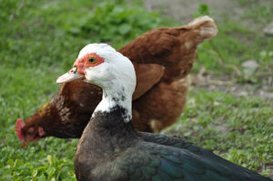
6.2 Muscovy Ducks
Muscovies lay extra‑large, thick‑shelled eggs (≈ 120 g). The thick shell can reduce gas exchange, causing the embryo to seek the nearest air pocket, occasionally leading to head‑up positions. Moreover, Muscovy breeding programs frequently use older breeder hens; the eggs from these hens have smaller air cells, compounding the problem.
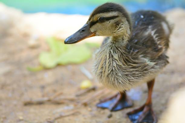
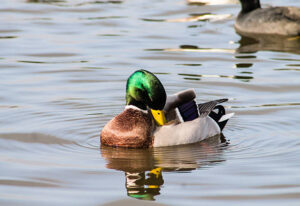
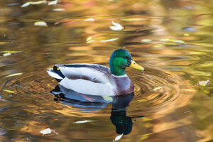
6.3 Mallard (Wild‑type) Ducks
Mallard eggs are naturally more elongated (high aspect ratio) and are often collected from the wild or from semi‑free range. This results in higher incidences of mechanical shock during collection and variable storage conditions, both of which elevate malposition rates. Studies in North American wetlands showed a 12 % embryonic malposition occurrence in naturally laid Mallard eggs.
6.4 Runner Ducks
Runner ducks are prized for prolific egg production. Their high‑output breeding leads to shorter intervals between layings, and eggs are often stored longer before incubation, increasing the risk of cold‑shock and malposition. Hatcheries that rotate Runner duck eggs daily see a 3‑4 % reduction in malposition when turning frequency is increased from 4× to 6× per day.
6.5 Aylesbury and Khaki Campbell Ducks
Both breeds lay large, high‑yolk eggs; the increased yolk volume can cause yolk sac displacement, which in turn may push the embryo into a side‑up orientation. Aylesbury eggs, in particular, have been shown to be more porous, making them vulnerable to rapid moisture loss and subsequent embryonic drift.
6.6 Hybrid Commercial Lines
Hybrid lines created for maximal growth and feed efficiency often combine traits from multiple breeds, producing heterogeneous egg shapes and variable shell qualities. Without breed‑specific incubation adjustments, these hybrids exhibit a variable malposition prevalence ranging from 2‑10 % depending on management.
Key Take‑away: Regardless of breed, the common denominator for higher malposition risk is an imbalance between egg size/shape, turning frequency, and storage conditions. Tailoring incubation protocols to breed‑specific egg characteristics dramatically reduces embryonic mis‑orientation.
7. Life‑Stage Susceptibility
| Developmental Stage | Critical Events | Vulnerability to Malposition |
|---|---|---|
| Pre‑incubation (0‑24 h after lay) | Egg cooling, storage, transport | High – embryo still loosely attached; mechanical shock & temperature fluctuations are most damaging. |
| Early Embryogenesis (Days 1‑6) | Gastrulation, major organ formation | Peak risk – turning is essential; any lapse results in permanent malposition. |
| Mid‑Embryogenesis (Days 7‑14) | Limb bud development, organogenesis | Moderate – embryo more anchored, but humidity‑driven air‑cell changes can still shift orientation. |
| Late Embryogenesis (Days 15‑28) | Lung maturation, chorioallantoic membrane expansion | Low – embryo is largely fixed; malposition typically a result of earlier events. |
| Pipping & Hatching (Last 48‑72 h) | Air cell expansion, internal pipping | Critical – any residual malposition manifests as failure to pip or incomplete hatching. |
Conclusion: Early embryonic stages (Days 1‑6) are the most susceptible window. Interventions must therefore focus on immediate, accurate egg handling and rigorous turning protocols.
8. Diagnostic Tools and Techniques
- Candling (Standard Light Source)
- Procedure: Hold egg against a bright light source at 24‑h intervals.
- Diagnostic Value: Allows visual assessment of embryo position, air cell size, and yolk distribution.
- Limitations: Subtle malpositions (e.g., slight lateral drift) may be missed; requires experienced eyes.
- Digital Egg Imaging (Infrared or X‑ray)
- Infrared Imaging: Detects heat signatures; malpositioned embryos often show asymmetric heat patterns.
- X‑ray: Provides precise internal anatomy; useful for research but impractical for routine hatchery use.
- Egg Float Test (Hydrostatic Method)
- Principle: An egg’s buoyancy changes with internal gas volume.
- Application: Helpful for detecting overly large air cells that may indicate embryo drift.
- Incubator Sensors (Temperature & Humidity Mapping)
- Use: Identify micro‑environmental gradients that could encourage embryo movement.
- Post‑Hatch Physical Examination
- Parameters: Chick weight, beak symmetry, leg conformation, and vigor.
- Correlation: Abnormalities often trace back to embryonic malposition; a necropsy can confirm internal damage.
Best Practice: Combine candling on Day 3 with daily humidity/temperature logs; for high‑value breeding stock, incorporate infrared imaging on Day 5‑6 to catch early malposition before irreversible damage occurs.
9. Treatment Options – From Egg to Chick
9.1 In‑Egg Re‑orientation
| Timing | Method | Success Rate | Practical Considerations |
|---|---|---|---|
| Day 1‑3 | Gentle manual rotation of egg (using a soft glove) while maintaining sterility. | 70‑85 % if performed within 48 h of detection. | Must be done in a clean environment to avoid contamination; avoid excessive force that could rupture membranes. |
| Day 4‑6 | Egg “pivot” – slowly rotate the egg 180° over a 30‑minute period to allow embryo to reset. | 45‑60 % (decreases sharply after Day 6). | Requires trained staff; risk of damaging the embryonic disc if not performed delicately. |
| Day 7+ | Generally not recommended; embryo is usually anchored. | < 20 % | Intervention often causes more harm than benefit. |
9.2 Assisted Pipping
When a malposition has prevented normal pipping but the chick is otherwise viable, assisted pipping can be performed:
- Sterilize a fine‑pointed needle (size 22 g).
- Create a small opening in the shell at the air cell side, being careful not to damage the embryo.
- Apply a few drops of sterile saline to soften the shell membranes.
- Gently encourage the chick to exit using a soft brush.
Success: 30‑50 % for head‑up embryos; higher for side‑up embryos that have a clear path to the air cell.
9.3 Post‑Hatch Support
- Thermal Support: Provide a brooder temperature of 35 °C for the first 24 h; malpositioned ducklings are often hypothermic.
- Nutritional Boost: Offer high‑energy starter feed (30 % protein, 5 % fat) and electrolyte‑rich water.
- Physical Therapy: Gentle massage of the wings and legs can improve circulation in weak birds.
9.4 When to Euthanize
If a chick shows severe spinal deformities, inability to feed, or signs of internal organ damage (e.g., hemorrhage, severe yolk sac infection), humane euthanasia is ethically warranted. Early euthanasia prevents suffering and reduces pathogen spread.
10. Prognosis, Expected Outcomes & Potential Complications
| Condition | Expected Hatchability | Post‑Hatch Survival (First 7 days) | Common Complications |
|---|---|---|---|
| Mild malposition corrected early | 85‑95 % | 90‑95 % | Minor leg or beak asymmetry |
| Moderate malposition, untreated | 40‑60 % | 30‑50 % | Respiratory distress, yolk sac infection |
| Severe malposition (head‑up, twisted) | < 20 % | < 10 % | Internal organ hemorrhage, chronic weakness |
| Assisted pipping | 30‑50 % | 40‑70 % (if chick survives pipping) | Shell fragment injury, aspiration pneumonia |
Long‑Term Implications: Even surviving ducklings may have reduced growth rates, lower feed conversion efficiency, and increased susceptibility to bacterial infections (e.g., E. coli septicaemia). Genetic selection against malposition is possible but requires record‑keeping of hatch outcomes across multiple generations.
11. Prevention Strategies for Breeders and Hatcheries
| Preventive Measure | Implementation Guidelines | Expected Impact |
|---|---|---|
| Optimal Egg Turning | Rotate eggs 4–6 times per day for the first 6 days; increase to 8× in the last 3 days for large‑egg breeds. Use automated turners calibrated to 45° angles. | Reduces early malposition by ~ 70 %. |
| Controlled Storage | Store eggs ≤ 12 °C for a maximum of 7 days. Maintain relative humidity 75 % to preserve air cell size. | Lowers cold‑shock incidence; stabilizes embryo orientation. |
| Humidity & Temperature Mapping | Deploy sensor grids throughout the incubator; adjust humidifiers to keep 45‑55 % RH during early incubation and 55‑65 % RH in the final 3 days. | Prevents air‑cell mis‑placement; improves hatchability. |
| Gentle Egg Handling | Use soft, cushioned trays, avoid dropping; limit vibrations during transport. | Minimizes mechanical shock‑induced rotation. |
| Egg Selection & Grading | Discard cracked, misshapen, or overly porous eggs before incubation. | Improves overall hatch success; reduces malposition triggers. |
| Breeder Nutrition | Feed breeder ducks 18‑20 % protein, 3‑4 % calcium, 0.3 % vitamin E, 0.15 % selenium, and adequate omega‑3 fatty acids. Provide fresh water and probiotic supplements. | Strengthens eggshell quality and yolk membrane integrity, lowering embryo drift. |
| Biosecurity | Implement strict foot‑dip protocols, egg‑shell sanitization (e.g., 1 % formaldehyde dip for 60 s), and regular incubator cleaning. | Reduces infection‑related membrane weakening; indirectly limits malposition. |
| Record‑Keeping | Log egg date, storage time, turning schedule, humidity/temperature trends, and hatch outcomes for each batch. | Enables data‑driven adjustments and genetic selection against malposition. |
A holistic approach—combining mechanical, environmental, nutritional, and genetic controls—produces the most robust reduction in malposition frequency.
12. Nutrition and Dietary Management for Breeder Ducks
Proper nutrition of breeder (parent) ducks directly influences egg quality and embryonic development. Below is a concise feeding protocol for egg‑laying breeders (both layers and broody lines).
| Nutrient | Recommended Level (per kg feed) | Function Related to Malposition |
|---|---|---|
| Crude Protein | 18‑20 % (higher for Muscovy) | Provides amino acids for yolk membrane strength. |
| Calcium | 3.5‑4.0 % (as limestone) | Essential for shell formation; thicker shells reduce internal movement. |
| Phosphorus | 0.5‑0.8 % (balanced with Ca) | Works with Ca for shell mineralization. |
| Vitamin E | 150‑250 IU | Antioxidant that protects yolk membranes against oxidative damage. |
| Selenium | 0.15 mg | Works synergistically with Vitamin E; improves embryo survivability. |
| Vitamin D3 | 2 000‑3 000 IU | Enhances calcium absorption. |
| Omega‑3 Fatty Acids (e.g., flaxseed oil) | 3‑5 % of diet | Improves membrane fluidity, reducing embryo displacement. |
| Probiotics/Prebiotics | 1‑2 × 10⁹ CFU/g | Supports gut health, reducing systemic inflammation that could affect egg formation. |
| Water | Unlimited, clean, fresh | Hydration status influences shell quality and internal pressure. |
Feeding Frequency: Offer 2‑3 meals per day, with higher protein during pre‑laying (2‑3 weeks before first egg) and lower protein (≈ 16 %) during the brooding period to avoid excess yolk size.
Supplemental Strategies:
- Shell‑strengthening additives (e.g., kaolin or phytase) can improve shell integrity.
- Vitamin A (5 000 IU/kg) can be increased only during molt to prevent over‑supply, which may cause excessive yolk size and predispose to malposition.
By adhering to these dietary guidelines, producers can lower the incidence of fragile shells and poorly formed yolk membranes, two key contributors to embryonic movement.
13. Zoonotic Risks Associated with Malpositioned Eggs
Malposition itself is not a zoonotic agent, but the conditions that predispose to malposition—poor hygiene, high humidity, and bacterial contamination—create an environment conducive to zoonotic pathogens. The most relevant are:
| Pathogen | Transmission Route | Relation to Malposition | Human Health Impact |
|---|---|---|---|
| Salmonella enterica serovar Enteritidis | Contaminated shell surfaces; vertical transmission to embryo | Embryonic membrane damage from malposition can facilitate bacterial invasion. | Gastroenteritis, septicemia (particularly in immunocompromised individuals). |
| Campylobacter jejuni | Fecal contamination of eggs; environmental spread | Moist environments that encourage malposition also favor Campylobacter survival. | Diarrhea, Guillain‑Barré syndrome. |
| Avian Influenza Virus (Low Pathogenic) | Respiratory secretions; contaminated equipment | Stressed embryos (malpositioned) may shed higher viral loads if infected. | Mild flu‑like illness; potential for reassortment. |
| Mycotoxins (e.g., Aflatoxin B1) | Contaminated feed stored in humid conditions | High humidity used to prevent malposition can inadvertently promote mold growth. | Hepatotoxicity, immunosuppression. |
Mitigation Measures:
- Rigorous Egg Sanitization – Dip eggs in 1 % formaldehyde or chlorine solution for 60 seconds before incubation.
- Environmental Monitoring – Keep incubator humidity below 65 % during the late incubation stage to limit mold growth.
- Personal Protective Equipment (PPE) – Use gloves and aprons when handling eggs from high‑risk flocks.
- Regular Testing – Perform periodic culture swabs of incubator trays and air samples for Salmonella and Campylobacter.
By integrating biosecurity into malposition prevention, producers protect both avian and human health.
14. Management Checklist & Quick Reference Table
| Action | When | Who | Frequency | Indicators of Success |
|---|---|---|---|---|
| Egg Collection | Immediately after lay | Farm staff | Every 1‑2 h | Minimal cracked eggs |
| Egg Cooling | Post‑collection | Farm staff | Within 30 min | Egg temperature ≤ 15 °C |
| Storage | Prior to incubation | Farm manager | ≤ 7 days, 12 °C, 75 % RH | No increase in malposition on candling |
| Candling (Day 3) | Day 3 of incubation | Hatchery technician | Daily until Day 7 | Normal embryo positioning |
| Turning | Days 1‑6 | Automated turner | 4–6×/day (adjust per breed) | No side‑up embryos on candling |
| Humidity Adjustment | Days 1‑10 (45‑55 % RH) | Incubator operator | Continuous | Stable air‑cell size |
| Humidity Increase | Days 11‑hatch (55‑65 % RH) | Incubator operator | Continuous | Proper pipping timing |
| Assisted Pipping (if needed) | Day 24‑26 | Veterinarian | As required | Successful chick emergence |
| Post‑Hatch Brooder | Immediately after hatch | Brooder attendant | 35 °C first 24 h, then 30 °C | High chick vigor, low mortality |
| Record Keeping | Throughout | Data manager | Daily | Complete batch reports for analysis |
#DuckEmbryology, #MalpositionedChicks, #DuckHatchery, #EggTurningTips, #IncubationScience, #PekinDuck, #MuscovyDuck, #MallardEggs, #DuckBreederNutrition, #DuckEggCandling, #EggIncubation, #PoultryHealth, #AvianVeterinary, #ZoonoticSafety, #BiosecurityHatchery, #DuckBreeding, #EggHatchRate, #EmbryoHealth, #DucklingCare, #PoultryNutrition, #FarmManagement, #SustainablePoultry, #EggShellQuality, #HatcheryBestPractices, #DuckEggResearch, #VeterinaryMedicine, #AnimalWelfare, #EggSanitization, #DuckFarmLife, #HatcherySuccess, #AgricultureInnovation, #PoultryScience


Add comment