
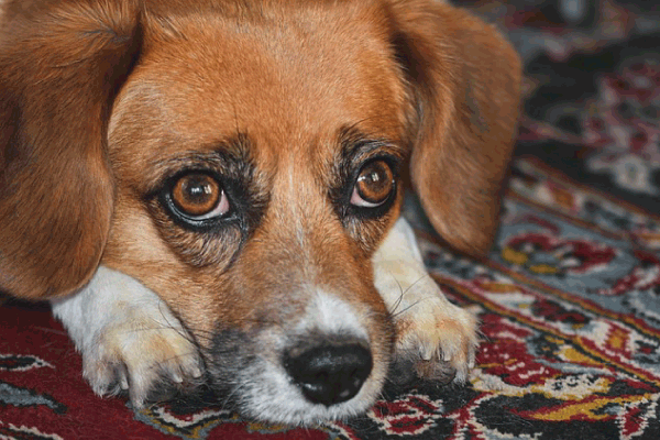
Mycetoma, also known as maduromycosis or fungal granuloma, is a chronic, progressive, and destructive infectious disease affecting the subcutaneous tissues, dermis, and sometimes underlying bone. While it can be caused by bacteria, the term mycetoma most commonly refers to fungal infections, particularly those caused by dematiaceous (darkly pigmented) fungi. These fungi are ubiquitous in the environment, found in soil, decaying vegetation, and on plant materials. In dogs, mycetoma is relatively rare but can lead to severe morbidity if not diagnosed and treated promptly. This guide will delve into the intricacies of mycetoma in dogs, providing a detailed understanding of its causes, manifestations, risk factors, diagnostic approaches, treatment strategies, prognosis, and preventive measures.
Causes of Mycetoma in Dogs
Mycetoma is caused by specific genera of fungi that gain entry into the body, typically through a breach in the skin. These fungi are saprophytic, meaning they live on dead organic matter. The most common culprits in dogs, as in humans, are dematiaceous fungi. While the exact species can vary geographically, some frequently implicated genera include:
- Madurella spp.: Often considered the most common cause of human mycetoma, and suspected in canine cases.
- Phialophora spp.: Another frequent cause, especially in tropical and subtropical regions.
- Exophiala spp.: Can also lead to mycetoma.
- Acremonium spp.: Formerly known as Cephalosporium, this genus has also been identified.
- Aspergillus spp.: While more commonly associated with respiratory infections, some species can cause cutaneous mycetoma.
The typical route of infection is traumatic inoculation. This occurs when the fungus, present in the environment (e.g., soil contaminated with animal feces, thorns, splinters), penetrates the skin. Minor wounds, abrasions, or even insect bites can serve as entry points. Once inside, the fungus establishes itself in the subcutaneous tissue, leading to a localized inflammatory response. The infection tends to be slow-growing and chronic, establishing granulomas – localized inflammatory nodules – filled with fungal elements.
It’s crucial to understand that mycetoma is not contagious between dogs or from dogs to humans through casual contact. The infection arises from environmental exposure.
Signs and Symptoms of Mycetoma in Dogs
The clinical presentation of mycetoma in dogs can be varied and often progresses slowly, making early diagnosis challenging. The typical lesions are found on the limbs, particularly the distal extremities (paws, legs), as these areas are most exposed to soil and plant material. However, mycetoma can occur anywhere on the body where the skin has been compromised.
Key signs and symptoms include:
- Subcutaneous Nodules and Swelling: The hallmark of mycetoma is the development of firm, painless to mildly painful nodules or swellings in the subcutaneous tissues. These can range in size from a few millimeters to several centimeters.
- Draining Fistulous Tracts: As the infection progresses, the nodules can ulcerate and form draining tracts or sinuses. These tracts ooze a serosanguineous (serum and blood-tinged) or purulent discharge. The discharge often contains characteristic “grains” – small, macroscopically visible aggregates of fungal hyphae and host cells. The color and consistency of these grains can vary depending on the causative fungus.
- Deformity: Chronic inflammation and tissue destruction can lead to significant swelling and deformities, particularly affecting the limbs.
- Pain and Lameness: While early lesions may be painless, advanced stages can cause pain due to inflammation, pressure on nerves, or involvement of underlying bone. This can manifest as lameness, reluctance to bear weight on the affected limb, or general discomfort.
- Fever and Lethargy: In severe or disseminated cases, systemic signs like fever, lethargy, and loss of appetite may be observed. However, this is less common in typical localized mycetoma.
- Skin Discoloration: The skin over the affected area may become hyperpigmented or hyperkeratotic (thickened).
- Bone Involvement (Osteomyelitis): In advanced cases, the infection can penetrate into the underlying bone, leading to a secondary fungal osteomyelitis. This significantly worsens the prognosis and can cause severe lameness and pain.
The progression of mycetoma is typically insidious. A dog might have a small, unnoticed lesion for months or even years before owners seek veterinary attention due to the noticeable swelling, draining tracts, or lameness.
Dog Breeds at Risk for Mycetoma
While mycetoma can theoretically affect any dog, certain breeds may exhibit a predisposition due to a combination of genetic factors, lifestyle, and anatomical features. It is important to note that direct breed-specific predisposition for mycetoma is not as well-documented as for some other diseases. However, breeds with specific traits that increase their exposure to soil and potential for skin trauma are more likely to encounter the fungi.
Breeds that spend a significant amount of time outdoors, particularly in environments with rich soil and abundant plant life, are at a higher risk of encountering the fungal spores. This includes working breeds, hunting dogs, and active companion breeds. For example, breeds like Retrievers (Labrador, Golden), Sheepdogs (Border Collie, Australian Shepherd), Herding breeds, and Terriers often have an active outdoor lifestyle. Their curiosity and tendency to dig or explore dense vegetation can lead to injuries that allow fungal entry.
Furthermore, breeds with pendulous ears or prominent skin folds might be more susceptible to infections in those areas if they become abraded or traumatized. However, mycetoma is predominantly seen on the extremities.
Another factor to consider is the thickness and integrity of the skin. While not breed-specific, dogs with thinner skin or those prone to allergies that cause scratching and subsequent skin lesions might be at a slightly increased risk.
Ultimately, the greatest risk factor is environmental exposure combined with trauma. Therefore, any dog breed that has access to environments where these fungi are prevalent and experiences minor skin injuries is at risk, regardless of specific breed predisposition. However, active outdoor breeds are statistically more likely to be exposed.
Susceptibility in Puppies, Adults, or Older Dogs
Mycetoma can affect dogs of any age, from puppies to senior dogs. However, there are some nuanced considerations for different age groups:
- Puppies: Puppies are generally more active and curious, often exploring their environment with less caution than adult dogs. They may be more prone to minor injuries from rough play, digging, or encountering foreign objects. Their immune systems are still developing, which could theoretically make them more susceptible to opportunistic infections, though this is not a primary factor in mycetoma. The slow, chronic nature of mycetoma means that a lesion acquired in puppyhood might not become clinically apparent until later in life due to the gradual growth of the fungal granuloma.
- Adult Dogs: Adult dogs, especially those that are highly active outdoors (e.g., working dogs, sporting dogs, avid hikers), are at the highest risk for acquiring the initial fungal inoculation. Their outdoor activities expose them to soil and decaying vegetation where the fungi reside. The chronic nature of the disease means that symptoms might develop over months or years, and by the time a dog is presented for diagnosis, it is usually in its adult life.
- Older Dogs: Older dogs may be at a slightly increased risk due to a potentially less robust immune system compared to younger adults. While not a direct cause, any decline in immune function can make it harder for the body to effectively fight off infections. Furthermore, older dogs might have pre-existing conditions or mobility issues that could lead to minor skin abrasions or slower wound healing, indirectly increasing the chance of infection. However, the disease is primarily acquired through exposure and trauma, which can happen at any age.
In summary, while puppies are susceptible to acquiring the initial infection due to their exploratory nature, and older dogs might have slightly diminished immune defenses, the adult dog engaged in outdoor activities remains the most commonly affected demographic due to higher exposure rates and the chronic, slow-progressing nature of the disease.
Diagnosis of Mycetoma in Dogs
Diagnosing mycetoma in dogs requires a multi-faceted approach, combining clinical signs with diagnostic tests to confirm the presence of the specific fungal pathogen and assess the extent of the disease.
- History and Physical Examination: A thorough history from the owner is crucial, noting the onset of swelling, any discharge, changes in activity or lameness, and the dog’s outdoor activities. The physical examination will focus on the characteristic lesions: nodules, swelling, draining tracts, and the presence of grains in the discharge. The location and size of the lesions are important.
- Cytology: A quick and relatively easy diagnostic step involves collecting a sample of the discharge from the draining tracts. Direct microscopy can reveal neutrophils, macrophages, and sometimes yeast-like cells or hyphal fragments. However, definitive identification of the causative fungus is usually not possible with cytology alone.
- Fungal Culture: This is a gold standard for diagnosis. Samples of the discharge, tissue from a biopsy, or even the excised lesion are submitted to a veterinary laboratory for fungal culture. The laboratory will attempt to grow the fungus on specialized media. Identification of the specific fungal species can be achieved through macroscopic and microscopic examination of the fungal colonies, and sometimes molecular techniques (PCR). It’s important to note that fungal cultures can take several weeks to yield results, as the fungi causing mycetoma are often slow-growing.
- Histopathology: A biopsy of the affected tissue is essential. The tissue sample is fixed, processed, and examined under a microscope by a veterinary pathologist. Histopathology can reveal the characteristic inflammatory response (granulomas), the presence of fungal hyphae within the tissue, and can help differentiate mycetoma from other conditions like bacterial abscesses or neoplasms. Special stains (e.g., Grocott’s methenamine silver, Periodic acid-Schiff) are often used to highlight the fungal elements.
- Imaging (Radiography/X-rays): If bone involvement is suspected (due to deep lesions or significant lameness), radiographs of the affected limb are vital. X-rays can reveal signs of osteomyelitis, such as bone lysis (destruction), periosteal reaction (new bone formation), or changes in the bone density. This helps determine the extent of the disease and guide treatment.
- Advanced Imaging (CT or MRI): In very complex or extensive cases, or when trying to assess the extent of involvement of deeper structures or bone, Computed Tomography (CT) or Magnetic Resonance Imaging (MRI) might be employed. These provide more detailed cross-sectional views of the affected tissues.
- Serological Tests: While not widely available or definitive for mycetoma in dogs, some serological tests that detect antibodies against specific fungi might exist for certain pathogens. However, their utility in routine diagnosis is limited.
The diagnosis is confirmed when characteristic clinical signs are coupled with direct visualization of fungal elements (hyphae or grains) in discharge or tissue, and ideally, confirmed by fungal culture and/or histopathology.
Treatment of Mycetoma in Dogs
Mycetoma is notoriously difficult to treat due to the chronic nature of the infection, the often-deep location of the fungal granulomas, and the slow growth of the causative fungi. Treatment strategies aim to eradicate the fungus, control the inflammation, and manage symptoms. A combination of approaches is often necessary.
- Surgical Excision: Complete surgical removal of the infected tissue is the most effective treatment, especially for localized and accessible lesions. However, this is often challenging for several reasons:
- Extent of Disease: The infection can be widespread within the subcutaneous tissue and may have spread to bone, making complete excision impossible without causing severe functional impairment or requiring amputation.
- Deep Lesions: Fungal granulomas can be deeply embedded, making it difficult to ensure all infected tissue is removed.
- Recurrence: Even with seemingly complete excision, microscopic fungal elements remaining in the surrounding tissue can lead to recurrence.
Surgery is often followed by long-term antifungal therapy. In cases of severe bone involvement or extensive limb deformity, amputation might be the most appropriate and humane option to eliminate the source of infection and intractable pain.
- Antifungal Medications: Systemic antifungal therapy is almost always employed, often in conjunction with surgery or as the sole treatment for non-surgical cases. The choice of antifungal depends on the suspected or identified fungal species.
- Itraconazole: This is a broad-spectrum triazole antifungal and is often the drug of choice due to its efficacy against a wide range of fungi that cause mycetoma. It is generally well-tolerated by dogs, although liver function should be monitored.
- Fluconazole: Another triazole, it can be used, but itraconazole is often preferred for mycetoma.
- Terbinafine: Effective against some fungi, it can be used in combination or as an alternative.
- Amphotericin B: This is a potent antifungal agent, but it is typically administered intravenously and can be nephrotoxic (kidney-damaging). It is usually reserved for severe, life-threatening infections or cases unresponsive to other treatments. Liposomal formulations of amphotericin B have reduced toxicity.
Antifungal therapy for mycetoma is long-term, often lasting for several months to a year or even longer, and requires consistent administration. Treatment is usually continued until resolution of clinical signs and confirmation of fungal clearance (e.g., negative cultures).
- Supportive Care:
- Wound Management: If there are draining tracts or ulcers, appropriate wound cleaning, debridement, and topical antimicrobial or antifungal agents may be used to manage secondary bacterial infections and promote healing.
- Pain Management: Non-steroidal anti-inflammatory drugs (NSAIDs) or other analgesics may be prescribed to manage pain and inflammation, especially in cases of bone involvement or before surgery.
- Nutritional Support: Ensuring adequate nutrition is vital for wound healing and overall recovery.
- Monitoring: Regular veterinary rechecks are crucial to monitor the response to treatment, assess for side effects of medications, and detect any signs of recurrence. This may involve repeat physical examinations, cytology, and imaging.
The success of treatment depends heavily on early diagnosis, the extent of fungal invasion, the dog’s immune status, and the owner’s commitment to long-term therapy.
Prognosis and Complications
The prognosis for mycetoma in dogs is variable and depends heavily on several factors:
- Extent of Disease: If the mycetoma is localized to the skin and subcutaneous tissues and can be completely surgically excised, the prognosis can be good to excellent. However, if there is significant bone involvement (osteomyelitis), extensive tissue destruction, or if the infection has spread systemically, the prognosis becomes guarded to poor.
- Timeliness of Diagnosis and Treatment: Early diagnosis and aggressive treatment offer the best chance of a favorable outcome. Delayed diagnosis often leads to more advanced disease, making it harder to treat effectively.
- Response to Therapy: The dog’s response to antifungal medications and the ability to achieve complete surgical remission are critical determinants of prognosis.
- Complications:
- Recurrence: Mycetoma can recur even after seemingly successful treatment, especially if complete eradication of the fungus is not achieved.
- Secondary Bacterial Infections: The open draining tracts are prone to secondary bacterial infections, which can complicate treatment and worsen the condition.
- Chronic Pain and Lameness: If the infection is not fully resolved or if there is permanent tissue damage or bone destruction, chronic pain and lameness can persist, impacting the dog’s quality of life.
- Amputation: In severe, intractable cases, amputation of the affected limb may be necessary, which significantly alters the dog’s mobility and lifestyle.
- Systemic Spread: While rare, in immunocompromised individuals, the fungus can potentially spread to other organs, leading to systemic mycosis.
In general, localized cutaneous mycetoma has a more favorable prognosis than deep-seated or bone-invasive forms. Owners must be prepared for potentially long-term treatment, frequent veterinary visits, and the possibility of recurrence or complications.
Prevention of Mycetoma in Dogs
Preventing mycetoma in dogs is challenging because the causative fungi are ubiquitous in the environment. However, certain measures can help reduce the risk of infection:
- Minimize Skin Trauma:
- Supervise Outdoor Activities: While it’s impossible to prevent all injuries, supervise your dog during activities where they are likely to encounter sharp objects, thorns, or rough terrain.
- Prompt Wound Care: Any cuts, abrasions, or puncture wounds should be promptly cleaned and disinfected. Monitor wounds for signs of infection and seek veterinary attention if they appear inflamed, swollen, or are not healing properly.
- Avoid High-Risk Areas: If you live in an area known for high incidence of mycetoma or if your dog has had previous issues, consider avoiding areas with particularly damp soil or dense, decaying vegetation where fungal spore concentration might be higher.
- Maintain Good Hygiene:
- Paw Care: Regularly inspect and clean your dog’s paws after outdoor excursions, especially if they have been walking or digging in soil.
- Environmental Management: While difficult to control on a large scale, maintaining good sanitation around your property can help reduce the presence of fungal spores. For example, clearing away decaying organic matter.
- Boost Immune System: While not solely for mycetoma prevention, a healthy immune system can better combat infections.
- Balanced Diet: Provide a high-quality, balanced diet appropriate for your dog’s age and activity level.
- Regular Veterinary Care: Ensure your dog receives regular veterinary check-ups, vaccinations, and parasite control. This helps maintain overall health and detect potential issues early.
- Awareness and Early Detection:
- Know Your Dog: Be familiar with your dog’s normal appearance and behavior. Regularly groom your dog and palpate their skin and limbs to detect any unusual lumps, bumps, or skin changes early on.
- Seek Veterinary Advice: If you notice any persistent swelling, draining sores, or lameness, consult your veterinarian immediately. Early diagnosis is key to successful treatment.
Given the environmental nature of the causative agents, absolute prevention is unlikely. The focus should be on minimizing opportunities for inoculation and ensuring prompt, appropriate care if an injury occurs.
Diet and Nutrition for Dogs with Mycetoma
While there is no specific diet that cures mycetoma, proper nutrition plays a crucial role in supporting the dog’s immune system, aiding in tissue repair, and improving their overall ability to fight infection and recover from treatment.
Key dietary considerations:
- High-Quality Protein: Adequate protein intake is essential for tissue repair and immune function. Look for high-quality protein sources in your dog’s food, such as meat, poultry, or fish. For dogs undergoing treatment, ensuring sufficient protein can help rebuild damaged tissues.
- Essential Fatty Acids (Omega-3 and Omega-6): These play a vital role in reducing inflammation and supporting skin health. A diet rich in omega-3 fatty acids (found in fish oil) can help modulate the inflammatory response associated with mycetoma. Omega-6 fatty acids are also important but should be balanced with omega-3s.
- Vitamins and Minerals:
- Vitamin E: An antioxidant that can support immune function and skin health.
- Zinc: Crucial for wound healing and immune response.
- Selenium: Another important antioxidant that works with Vitamin E.
- B Vitamins: Involved in energy metabolism and cell function, important for recovery.
- Antioxidants: Foods rich in antioxidants (e.g., blueberries, cranberries, certain vegetables) can help combat oxidative stress that may occur during chronic inflammation and healing.
- Palatability and Digestibility: If the dog is experiencing pain, lethargy, or side effects from medication, they may have a reduced appetite. Choosing a highly palatable and easily digestible food is important to ensure they are receiving adequate nutrition.
- Hydration: Adequate water intake is critical for all bodily functions, including flushing out toxins and supporting cellular processes involved in healing.
Recommendations:
- Veterinary Consultation: It is always best to discuss your dog’s nutritional needs with your veterinarian, especially when they are dealing with a chronic condition like mycetoma. They can recommend a specific diet or recommend supplements if necessary.
- High-Quality Commercial Food: Feeding a premium-quality commercial dog food formulated for adult maintenance or a specific therapeutic diet recommended by your vet is generally the easiest way to ensure a balanced intake of nutrients.
- Avoid Unnecessary Supplements: Unless specifically recommended by your veterinarian, avoid over-supplementing. Excessive intake of certain vitamins or minerals can be harmful.
- Consider Therapeutic Diets: In some cases, your vet might recommend a therapeutic diet designed to support immune function or reduce inflammation.
Ultimately, a well-balanced, nutrient-dense diet, combined with medical treatment, will provide the best support for a dog recovering from mycetoma.
Zoonotic Risk of Mycetoma in Dogs
The zoonotic risk of mycetoma from dogs to humans is considered extremely low to negligible.
Mycetoma is caused by fungi that are ubiquitous in the environment, primarily found in soil, decaying vegetation, and on certain plant materials. Humans and dogs are exposed to these fungi through environmental contact, most commonly via inoculation through skin wounds.
The crucial point is that dogs do not typically act as reservoirs or vectors for the transmission of mycetoma-causing fungi to humans in a contagious manner. You won’t “catch” mycetoma from petting your dog or from their saliva or bodily fluids through casual contact.
The risk, albeit very small, would theoretically arise from:
- Direct contact with infected discharge or grains and an open wound on the human: If a person were to directly handle the discharge from a dog’s mycetoma lesion and then have an open wound on their own skin (especially a puncture wound), there’s a theoretical, though highly unlikely, chance of fungal inoculation.
- Environmental contamination: The fungi themselves are present in the environment where both dogs and humans might be exposed.
In summary:
- No direct transmission: Dogs do not transmit mycetoma to humans through normal interaction.
- Environmental exposure: The risk is from the environment for both species, not from animal-to-human transmission.
- Extreme rarity: Even in veterinary settings where dogs with mycetoma are routinely handled, human infections are virtually unheard of.
Therefore, while general hygiene practices like washing hands after handling a dog’s wound are always recommended, there is typically no need for special precautions specifically for mycetoma beyond standard veterinary care protocols.
#MycetomaInDogs, #FungalInfectionDog, #DogHealth, #CanineMycetoma, #VeterinaryMedicine, #DogCare, #DermatologyDiva, #RareDogDiseases, #FungalGranuloma, #MaduraMycosis, #DogLimping, #PetHealth, #AntifungalTreatment, #DogInfection, #HealthyPaws, #DogWounds, #VeterinaryCare, #SoilFungi, #Mycology, #PetWellness, #DogEducation, #AskAVet, #CanineDermatology, #FungalDisease, #DogLife, #PetOwnerTips.


Add comment