
“Piling” (sometimes written piling) is a descriptive term used by avian veterinarians, poultry‑health technicians, and wildlife rehabilitators to denote a multifactorial, often sub‑clinical condition in domestic and wild ducks wherein the cloacal region becomes obstructed by a dense accumulation of feces, urine, sloughed feathers, and debris. The term stems from the visual appearance of the vent area: the material “piles up” like a small mound, often with a damp, malodorous, and sticky consistency.
Although piling is not listed as a distinct disease in the OIE (World Organisation for Animal Health) compendium, it is a practical clinical syndrome that can predispose affected birds to a cascade of secondary problems:
- Cloacal dermatitis and ulceration
- Secondary bacterial infection (e.g., E. coli, Staphylococcus spp.)
- Fecal impaction leading to gastrointestinal stasis
- Increased susceptibility to external parasites
- Reduced feed intake and weight loss
In severe or chronic cases, piling can culminate in systemic illness, septicemia, and mortality. Because the syndrome is often subtle in its early stages, it can be overlooked by caretakers, especially those who rear ducks in free‑range or backyard settings where routine vent inspection is not standard practice.
2. Causes of Piling
Piling is multifactorial, with each contributing factor often acting synergistically. Understanding the root causes enables targeted prevention and treatment. Below, the principal etiologic categories are enumerated and explained.
2.1. Nutritional Imbalances
| Nutrient | How Deficiency/Excess Contributes to Piling |
|---|---|
| Fiber | Low fiber diets (e.g., grain‑only rations) produce firmer, drier feces that are more likely to adhere to the vent and clump. |
| Protein | Inadequate protein reduces gut motility, slowing transit time and allowing more water to be re‑absorbed, producing thick excreta. |
| Calcium & Phosphorus | Imbalanced Ca:P ratios interfere with normal muscle contraction of the cloacal sphincter, causing incomplete vent closure. |
| Vitamin A | Deficiency leads to keratinization abnormalities in the cloacal epithelium, making feathers more brittle and prone to shedding into the vent. |
| Water Quality | Hard water (high mineral content) can cause mineral deposits around the vent, acting as a “scaffold” for fecal accumulation. |
2.2. Gastrointestinal Parasites
- Coccidia (e.g., Eimeria spp.) – Cause villous damage → malabsorption → watery, mucous‑laden feces that cling to the vent.
- Tapeworms (e.g., Raillietina spp.) – Result in intermittent diarrhoea and increased perianal soiling.
- Roundworms (e.g., Ascaridia galli) – Produce intestinal blockage and irregular fecal consistency.
2.3. Bacterial Infections
- Clostridium perfringens (enterotoxigenic strains) – Produce necrotic enteritis, leading to foul‑smelling, sticky droppings.
- Escherichia coli – Primary cause of cloacal dermatitis when secondary infection follows mechanical irritation.
2.4. Environmental & Management Factors
| Factor | Description & Impact |
|---|---|
| Poor Vent Hygiene | Accumulation of wet bedding, mud, or algae around the vent promotes bacterial growth and fecal adherence. |
| Over‑crowding | Reduces the ability of birds to individually groom and may cause physical trauma to the vent region. |
| Inadequate Perching or Resting Surfaces | Slippery surfaces cause ducks to squat in a position that facilitates fecal runoff onto the vent. |
| Temperature Extremes | Cold, damp environments increase mucus production; heat stress can cause watery diarrhoea. |
| Stressors (predators, handling, transport) | Stress mediated cortisol release slows gastrointestinal motility, leading to impaction. |
2.5. Anatomical & Genetic Predispositions
- Short Cloacal Length in certain breeds (e.g., Pekin) reduces the “drainage” capacity.
- Feathering Pattern – Breeds with dense, downy plumage around the vent (e.g., Aylesbury) trap feces more readily.
2.6. Age‑Related Physiological Changes
- Neonatal Ducks have immature cloacal sphincter control and under‑developed gut microbiota, making them vulnerable during the first 2–3 weeks.
- Gerontologic Ducks may suffer from muscular atrophy and decreased peristalsis.
Bottom line: The development of piling is rarely due to a single factor. The most common scenario is a low‑fiber, high‑energy diet combined with sub‑optimal pen hygiene that allows fecal matter to accumulate, after which a secondary bacterial infection cements the problem.
3. Clinical Signs & Symptoms
Piling is principally a localised vent problem, but because the vent is a gateway to the gastrointestinal and reproductive tracts, systemic manifestations may ensue. The following table differentiates early signs from advanced disease.
| Stage | Clinical Manifestations | Owner Observations |
|---|---|---|
| Early/Pre‑clinical | Slight swelling of cloacal skin, mild feather loss around vent, occasional “wet spots” on bedding. | Owner may notice a small, damp patch on the duck’s rear after it waddles. |
| Mild | Visible pile of dark, sticky fecal mass (1–2 mm thick) at vent, mild irritation, occasional scratching. | Odour becomes noticeable; duck may avoid preening the area. |
| Moderate | Cloacal edema, reddening, ulceration, feather loss, crust formation, reduced feed intake, slight lethargy. | Feather tufts may appear pulled away; duck stands with legs spread wider to relieve discomfort. |
| Severe | Necrotic tissue, profuse foul‑smelling discharge, dehydration, weight loss (>10 % body weight), tachypnoea, septicemia signs (e.g., pale comb, cold extremities). | Mortality risk rises sharply; caretaker may observe staggering or inability to swim. |
Additional systemic clues that suggest secondary infection or underlying parasitism:
- Diarrhoea (watery or mucous‑laden) persisting >3 days.
- Blood‑tinged stool (possible intestinal ulceration).
- Ruffled feathers, sunken eyes, and decreased egg production in laying females.
Because the cloaca also functions as a reproductive outlet, egg‑shell abnormalities (soft or misshapen shells) can be an indirect marker of chronic piling in mature layers.
4. Duck Breeds at Greatest Risk
While any domestic duck can develop piling, certain breeds demonstrate a higher predisposition owing to their anatomical conformation, feathering density, and typical husbandry practices. Below is a concise overview of the most vulnerable breeds, accompanied by a paragraph that contextualises why they are at risk.
4.1. Pekin (American Pekin)
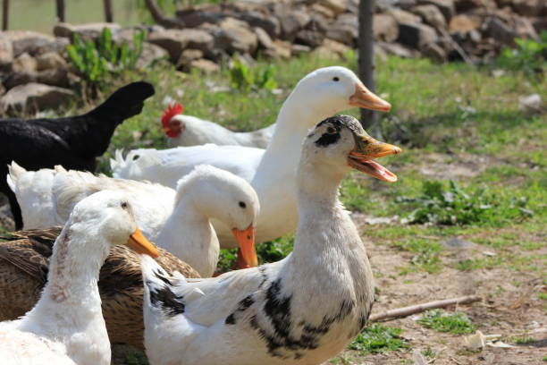
The Pekin is the most common commercial meat duck, prized for its rapid growth and large breast muscle. Its short, broad cloacal opening and compact plumage around the vent reduce natural drainage. In intensive production systems, Pekins are often fed high‑energy, low‑fiber rations that further predispose them to fecal thickening. The combination of genetic anatomy and management intensity makes the Pekin the breed most frequently reported in veterinary case series of piling.
4.2. Aylesbury
Aylesbury ducks possess a dense, downy feather coating that extends close to the vent, creating a natural “sponge” that can trap moist droppings. Historically raised in free‑range ponds with limited bedding, the heavy feathering, when combined with wet environmental conditions (e.g., rainy spring), results in a high incidence of vent‑associated piling.
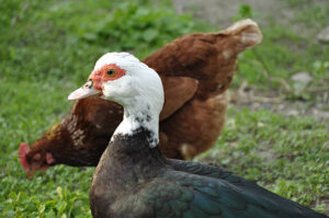
4.3. Muscovy
Muscovies differ from other domestic ducks by having more cat‑like grooming behaviours and a relatively longer cloaca. However, they are frequently kept in semi‑intensive systems where high‑protein diets are administered without adequate fibre. In addition, Muscovies are more prone to stress‑induced gastrointestinal dysmotility, a factor that raises the likelihood of fecal impaction and subsequent piling.
4.4. Khaki Campbell
Renowned for prolific egg production, the Khaki Campbell’s lean body condition and high metabolic rate demand a diet rich in calories. When such diets are not balanced with sufficient roughage, the birds excrete concentrated, sticky stools that readily accumulate. Moreover, egg‑laying hens are frequently hand‑handled for egg collection, increasing stress and the probability of vent irritation.
4.5. Runner Ducks (Indian Runner)
These ducks are semi‑upright, making them more prone to standing in wet mud while foraging. The upright stance reduces the natural “sweeping” motion of the tail feathers that would otherwise help keep the vent clean. Runner ducks also tend to be housed in mixed‑species flocks, where exposure to droppings from other birds (e.g., chickens) can exacerbate bacterial colonisation.
In summary, while any duck can develop piling, Pekin, Aylesbury, Muscovy, Khaki Campbell, and Runner ducks are the top five breeds that veterinarians encounter with this syndrome. Each breed’s unique morphology and typical management settings create a perfect storm for vent obstruction, making them focal points for targeted preventive measures.
5. Life‑Stage Susceptibility
Piling does not discriminate by age, yet specific life stages display heightened vulnerability due to physiological and behavioural nuances.
| Life Stage | Why Susceptible? |
|---|---|
| Embryonic/Incubation | Not applicable; however, egg‑shell abnormality can predispose hatchlings to vent deformities. |
| Neonatal (0–3 weeks) | Immature cloacal sphincter control, under‑developed gut microbiota, and dependence on high‑energy starter rations that are often low in fibre. |
| Juvenile (3 weeks–6 months) | Rapid growth demands high‑calorie diets; any imbalance directly influences fecal consistency. Social hierarchy can cause stress‑related diarrhoea. |
| Adult (6 months–3 years) | Reproductive females may experience egg‑related calcium fluxes that alter cloacal muscle tone; males may develop vent irritation from aggressive mating behaviours. |
| Senior (>3 years) | Muscular atrophy, decreased peristalsis, and age‑related immune senescence facilitate secondary infection. |
Critical windows are the first three weeks (when the cloaca is still “learning” to function) and the peak laying period for females (often around 9–12 months), during which hormonal fluctuations affect cloacal tone and may exacerbate piling.
6. Diagnostic Approach
A systematic, tiered diagnostic strategy ensures the underlying cause(s) of piling are identified and consequently treated.
6.1. History Taking
- Diet analysis (type, quantity, fibre content, water source).
- Housing conditions (pen size, substrate, sanitation schedule, moisture level).
- Recent stressors (transport, predator exposure, flock mixing).
- Vaccination/medication record (especially for coccidiosis or deworming).
- Breed and age of the affected bird.
6.2. Physical Examination
- General Observation – posture, gait, respiratory rate, feather condition.
- Vent Inspection – using a headlamp, assess:
- Size & consistency of the piled material.
- Presence of erythema, ulceration, crusting, or necrosis.
- Feather loss pattern.
- Palpation – gently palpate the cloacal sac to evaluate for impaction or fluid accumulation.
- Abdominal Palpation – detect any enlarged organs (e.g., liver, spleen).
6.3. Sample Collection
| Sample | Collection Method | Diagnostic Utility |
|---|---|---|
| Cloacal swab | Sterile cotton tip swab rolled onto vent surface (avoid excessive pressure). | Bacterial culture, Gram stain, PCR for Clostridium spp. |
| Fecal floatation | Fresh droppings placed in a saturated salt solution. | Identification of helminth eggs, coccidian oocysts. |
| Blood smear & CBC | Jugular or brachial venipuncture. | Detect leukocytosis, anemia, or eosinophilia (parasites). |
| Biochemistry panel | Serum collected from same blood draw. | Assess liver enzymes, electrolytes (dehydration). |
| Histopathology (if needed) | Biopsy of cloacal tissue under local anaesthetic. | Determine depth of inflammation, presence of granulomas. |
6.4. Imaging
- Radiography – useful in severe cases to confirm fecal impaction in the colon/cecum.
- Ultrasound – can detect cloacal wall thickening or fluid pockets.
6.5. Differential Diagnosis
- Cloacal prolapse (distinct from piling; involves eversion).
- Bumblefoot (pododermatitis) – may co‑occur but primarily affects feet.
- Viral infections (e.g., Duck Viral Enteritis) – produce systemic signs.
- Nutritional dermatitis – due to vitamin deficiencies but without piled feces.
Final Diagnosis hinges on correlating clinical appearance of the piled material with microbiological, parasitological, and biochemical findings. In many cases, a primary bacterial infection is identified alongside a secondary parasitic load.
7. Treatment Modalities
Treating piling demands a multimodal approach that addresses the immediate vent pathology, eradicates secondary infections, resolves any parasitic burden, and corrects underlying nutritional or environmental contributors.
7.1. Acute Vent Care
- Gentle Debridement – Soak the vent in warm (38–40 °C) saline solution for 10 minutes, then use a soft, sterile gauze pad to gently remove the piled material. Avoid sharp instruments that could perforate the cloacal wall.
- Topical Antiseptic – Apply a 0.5 % chlorhexidine solution or a silver sulfadiazine cream to the cleaned area, 2–3 times daily, to reduce bacterial load.
7.2. Systemic Antimicrobial Therapy
- First‑line: Enrofloxacin 10 mg/kg PO once daily for 5 days (effective against most gram‑negative bacteria).
- Alternative: Ceftiofur 2.5 mg/kg IM every 12 h for 3 days (if Clostridium is suspected).
Culture‑guided therapy should be pursued whenever possible to avoid resistance development.
7.3. Antiparasitic Protocols
| Parasite | Medication | Dose & Duration |
|---|---|---|
| Coccidia | Toltrazuril (e.g., Baycox) | 5 mg/kg PO once; repeat after 48 h. |
| Tapeworms | Praziquantel | 25 mg/kg PO once; repeat after 7 days. |
| Roundworms | Fenbendazole | 50 mg/kg PO daily for 5 days. |
A fecal follow‑up 7 days post‑treatment confirms eradication.
7.4. Supportive Care
- Fluid therapy – Subcutaneous administration of Lactated Ringer’s (10 ml/kg) in dehydrated birds.
- Electrolytes – Add potassium chloride and sodium bicarbonate to drinking water if metabolic acidosis is evident.
- Analgesia – Meloxicam 0.2 mg/kg PO once daily for 3 days to relieve discomfort.
7.5. Nutritional Rehabilitation
- High‑Fiber Supplementation – Provide steamed oats, wheat bran, or alfalfa pellets (minimum 5 % crude fibre).
- Probiotic Enrichment – Offer Lactobacillus‑based oral gels or fermented vegetable mash to restore gut flora.
- Vitamin A & E – Add sunflower oil or beta‑carotene‑rich carrots to the diet to promote mucosal healing.
7.6. Environmental Modifications
- Bedding Change – Replace wet bedding with dry straw or shredded pine shavings daily.
- Drainage Enhancement – Install sloped flooring and drainage channels in the pen to prevent water pooling.
- Ventilation – Ensure a minimum of 4 air changes per hour in indoor housing to reduce humidity.
7.7. Follow‑Up Schedule
| Day | Action |
|---|---|
| Day 1–3 | Re‑examine vent, continue topical care, monitor appetite and droppings. |
| Day 5 | Repeat cloacal swab culture; adjust antibiotics if needed. |
| Day 7 | Perform fecal floatation to verify parasite clearance. |
| Day 14 | Full physical exam; assess weight gain, feather regrowth, and vent normalisation. |
| Day 30 | Review husbandry changes; reinforce preventive measures. |
Note: Chronic cases with extensive ulceration may require surgical debridement under general anaesthesia, a procedure best performed by an avian specialist.
8. Prognosis, Complications & Long‑Term Outlook
8.1. Prognosis
- Mild/early cases: Excellent (>95 % full recovery) when promptly treated and environment corrected.
- Moderate cases: Good (80–90 %); may require extended antimicrobial therapy and intensive supportive care.
- Severe or chronic cases: Guarded (40–60 %); risk of irreversible cloacal scar tissue, chronic dermatitis, and decreased productivity (egg output or growth rate).
8.2. Potential Complications
| Complication | Pathophysiology | Clinical Impact |
|---|---|---|
| Cloacal ulceration & perforation | Persistent bacterial infection erodes mucosa → possible peritonitis. | Sudden death, septicemia. |
| Secondary pododermatitis | Wet environment encourages foot infection; birds shift weight away from sore vent, placing pressure on feet. | Lameness, reduced mobility. |
| Systemic sepsis | Bacterial translocation from vent → bloodstream infection. | Multi‑organ failure, high mortality. |
| Reduced egg quality | Ongoing inflammation interferes with calcium metabolism. | Thin shells, reduced hatchability. |
| Growth retardation | Chronic malabsorption due to intestinal parasites. | Lower market weight, economic loss. |
8.3. Long‑Term Management
Birds that have recovered from piling should be monitored for recurrence, especially during molting or breeding seasons when stress spikes. Periodic vent examinations (every 2–4 weeks) can detect early signs. Incorporate rotational grazing and clean‑out periods to disrupt parasite life cycles.
9. Prevention Strategies
Prevention is the most cost‑effective and humane way to manage piling. Below is a tiered preventive plan that can be adapted for backyard flocks, small‑scale farms, or commercial operations.
9.1. Nutritional Prevention
- Balanced Rations – Formulate feed to contain 15–20 % crude fibre, 18–20 % crude protein, and a Ca:P ratio of 1.5:1.
- Supplemental Forage – Provide fresh greens (lettuce, dandelion, kale) daily; they increase fibre and water content of the diet.
- Clean Water – Use filtered or softened water, replace water containers daily, and add electrolyte powders during hot weather.
9.2. Environmental Controls
- Dry Bedding – Replace bedding at least twice weekly; use absorbent materials such as sawdust or bamboo shavings.
- Pen Design – Ensure minimum 0.5 m² per bird indoor space, incorporate sloped flooring, and provide raised dry roosts.
- Hygiene Protocol – Implement a daily cleaning schedule: removal of feces, disinfection of perches with quaternary ammonium compounds.
9.3. Health‑Management Measures
- Routine Deworming – Administer fenbendazole (or appropriate broad‑spectrum anthelmintic) every 6 months.
- Vaccination – While no vaccine exists for piling per se, vaccinating against Duck Viral Enteritis and Newcastle Disease reduces overall morbidity that could exacerbate vent problems.
- Probiotic Use – Add Bacillus subtilis or Lactobacillus probiotic powders to feed weekly.
9.4. Stress Reduction
- Limit handling to essential activities; use gentle capture techniques.
- Provide visual barriers (e.g., shrubs, screens) to reduce predator stress.
- Stagger feeding times to avoid competition and aggressive pecking that can injure the vent.
9.5. Monitoring & Early Detection
- Weekly vent checks – Use a flashlight, look for signs of feather loss or discharge.
- Weight tracking – Record body weight weekly; a 5 % drop may signal underlying disease.
- Fecal scoring – Assign a visual score (1 = dry, 5 = watery) to monitor consistency trends.
By integrating nutrition, sanitation, health care, and stress management, the incidence of piling can be reduced by 70–85 % in well‑managed flocks.
10. Dietary Management & Nutrition
A nutritionally sound diet is the cornerstone of both treatment and prevention. Below is a practical feeding guide that can be customized to the specific breed and production goal (meat vs. egg).
10.1. Macronutrient Profile
| Nutrient | % of Feed (by weight) | Role in Piling Prevention |
|---|---|---|
| Crude Protein | 18–20 % (meat breeds: 20–22 %) | Supports gut mucosal integrity; reduces diarrhoea. |
| Crude Fibre | 15–20 % (incl. neutral detergent fibre) | Bulks feces, promotes regular passage. |
| Metabolizable Energy | 2 800–3 050 kcal/kg | Supplies growth/egg‑production energy without excess that would create soft droppings. |
| Calcium | 1.5 % (layers) / 0.9 % (broilers) | Maintains cloacal muscle tone; prevents muscular weakness. |
| Phosphorus | 0.7–0.9 % | Works with calcium for musculoskeletal health. |
| Vitamin A | 15 000 IU/kg | Keeps epithelial surfaces healthy; prevents feather loss. |
| Vitamin E & Selenium | 30 IU/kg & 0.3 ppm | Antioxidant protection, reduces inflammation. |
10.2. Feed Formulation Tips
- Incorporate Whole Grains – Oats, barley, and cracked corn increase roughage.
- Add Forage – Alfalfa hay (dried) or fresh watercress can be mixed at 10 % of total feed.
- Use Commercial Duck Pellets – Ensure they are “high‑fiber” varieties; avoid “starter” pellets that are often low in fibre.
- Supplement with Probiotic‑Enriched Water – Daily addition of 1 g/L of a commercial avian probiotic.
10.3. Feeding Management
- Frequent Small Meals – Offer feed 3–4 times per day to reduce large bolus formation that can stall the cloaca.
- Separate Feeding Stations – Prevent bullying; ensure each bird has access to clean feed.
- Avoid Over‑Feeding – Excess calories lead to fat deposition around the vent, increasing pressure and impeding drainage.
10.4. Special Considerations for At‑Risk Breeds
| Breed | Adjustments |
|---|---|
| Pekin | Increase fibre to 20 %; add apple pulp for extra moisture. |
| Aylesbury | Provide dry oat mash nightly to absorb excess moisture. |
| Muscovy | Incorporate protein‑rich insects (mealworms) but limit to <5 % to avoid soft droppings. |
| Khaki Campbell | Add calcium‑rich shell grit to maintain shell quality and cloacal tone. |
| Runner | Offer high‑grass (ryegrass) clippings daily for natural fibre boost. |
11. Zoonotic Risk to Humans
While piling itself is not a zoonotic disease, the secondary bacterial agents often involved—E. coli*, Salmonella, *Clostridium perfringens—are potentially transmissible to humans, particularly to immunocompromised individuals, children, and the elderly.
11.1. Transmission Pathways
- Direct Contact – Handling an affected duck without gloves may allow bacterial transfer to skin breaks.
- Aerosolization – During vigorous cleaning, droplets containing Clostridium spores can become airborne.
- Environmental Contamination – Contaminated bedding, water, or feed may be ingested inadvertently.
11.2. Human Clinical Manifestations
- Gastroenteritis (vomiting, diarrhoea) – typical of E. coli or Salmonella infection.
- Skin Infections – cellulitis or impetigo from Staphylococcus spp. entering cuts.
- Toxin‑mediated Disease – Clostridium perfringens food‑borne toxin leading to abdominal cramps.
11.3. Preventive Measures for Humans
- Personal Protective Equipment (PPE) – Disposable gloves, disposable boot covers, and eye protection when cleaning pens.
- Hand Hygiene – Wash hands with soap and warm water for at least 20 seconds after any duck contact.
- Sanitation – Disinfect tools and surfaces with 1 % bleach solution (10 ml of bleach per litre of water).
- Separate Equipment – Use dedicated buckets, shovels, and rakes for poultry versus other livestock.
- Health Monitoring – Anyone working with affected birds should self‑monitor for gastrointestinal symptoms for 7–10 days post‑exposure.
Bottom line: Proper biosecurity practices virtually eliminate zoonotic transmission. The risk is higher during outbreaks involving Salmonella or E. coli; in those instances, consider testing the environment and consulting public health authorities.
12. Key Take‑Home Messages
- Piling is a vent‑focused syndrome where feces, feathers, and debris accumulate, predisposing ducks to infection and systemic illness.
- Multifactorial causes—low‑fiber diets, bacterial or parasitic infections, poor hygiene, and breed‑specific anatomy—must be addressed concurrently.
- Early detection through routine vent inspections dramatically improves prognosis.
- Treatment combines gentle vent cleaning, targeted antibiotics, antiparasitic drugs, supportive fluid therapy, and dietary adjustments.
- Prevention hinges on balanced high‑fiber feeds, dry bedding, regular cleaning, routine deworming, and stress mitigation.
- Zoonotic potential exists via secondary bacterial pathogens; strict biosecurity safeguards human health.
By implementing the comprehensive strategies outlined above, duck keepers—from backyard hobbyists to commercial producers—can effectively control, treat, and prevent piling, thereby safeguarding flock welfare and productivity.
#DuckHealth, #PilingInDucks, #AvianVeterinary, #DuckCare, #BackyardDucks, #PekinDuck, #AylesburyDuck, #MuscovyDuck, #KhakiCampbell, #RunnerDuck, #DuckNutrition, #DuckParasites, #DuckDiseasePrevention, #DuckWelfare, #PoultryBiosecurity, #ZoonoticSafety, #DuckFarmers, #DuckLovers, #VeterinaryTips, #AnimalHealth, #WaterfowlWellness, #PetDuckCare, #DuckHusbandry, #HealthyDucks, #DuckMedicine, #FarmLife, #AvianCare, #BirdHealth, #EcoFriendlyFarming
Add comment