
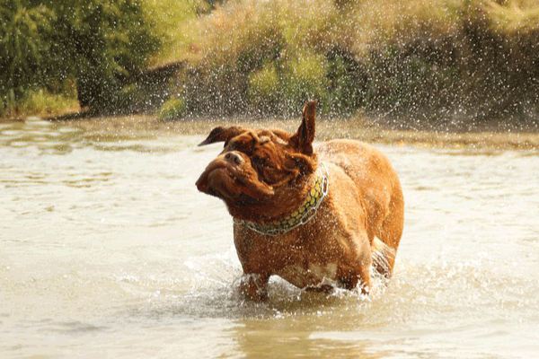
Sarcocystosis, often overlooked in routine veterinary practice, is an infection caused by protozoan parasites of the genus Sarcocystis. While it is generally regarded as a silent or subclinical disease in dogs, its importance lies in the dog’s role as the definitive host, completing the parasite’s life cycle and acting as a major source of infection for livestock worldwide. Understanding canine sarcocystosis is critical for veterinary medicine, public health control, and global livestock management, necessitating a deep dive into its complex biology, clinical presentation, and preventive strategies.
I. Etiology and Life Cycle (Causes of Sarcocystosis)
Sarcocystosis is caused by various species of the genus Sarcocystis, which are obligate heteroxenous coccidian parasites, meaning their life cycle requires two distinct host species: a definitive host (carnivore, usually the dog) and an intermediate host (herbivore, typically livestock).
A. Key Species Affecting Dogs
The specific clinical outcome depends heavily on the Sarcocystis species ingested, determined by the type of intermediate host consumed:
- Sarcocystis cruzi: The most common species, transmitted when dogs consume raw or undercooked infected beef (cattle are the intermediate hosts). This species is highly pathogenic in cattle, causing significant economic losses, but usually results in mild, self-limiting enteritis in the dog.
- Sarcocystis ovicanis (formerly S. tenella): Transmitted when dogs consume raw or undercooked infected mutton (sheep and goats are the intermediate hosts). Similar to S. cruzi, it primarily causes intestinal infection in the dog.
- Sarcocystis canis: Historically used, but the taxonomy is complex. Some classifications suggest dogs are intermediate hosts for certain rare, non-pathogenic species, though this is not the primary mode of transmission.
- Sarcocystis suihominis: Transmitted if dogs consume infected swine (pigs). While pigs are typically associated with S. suihominis and S. meischeriana, the involvement of dogs as definitive hosts for pig-related species is less common than with cattle and sheep but still possible in areas where dogs scavenge hog carcasses.
B. The Obligate Two-Host Life Cycle
The infection cycle is strictly dependent on the predator-prey relationship:
- Ingestion (Infection of the Definitive Host): The dog (definitive host) ingests bradyzoites—the term for the slow-growing, highly infectious stage encased within sarcocysts—found in the muscle tissue (meat or offal) of the intermediate host (e.g., cow, sheep).
- Intestinal Development (Asexual and Sexual Stages): Once ingested, stomach acids dissolve the cyst wall, releasing bradyzoites. These penetrate the cells lining the dog’s small intestine. They immediately undergo the sexual reproduction phase (gametogony).
- Sporogony and Shedding: Male and female gametes fuse, forming oocysts. These oocysts sporulate rapidly within the intestinal wall, generating two mature sporocysts, each containing four infective sporozoites. Unlike many other coccidia (like Cystoisospora), the oocysts often rupture within the intestinal mucosa, releasing the more fragile, immediately infective sporocysts into the feces.
- Environmental Contamination: The dog sheds these sporocysts into the environment via feces.
- Intermediate Host Infection: The intermediate host (livestock) ingests the sporocysts via contaminated feed, water, or pasture.
- Systemic and Muscle Invasion (Intermediate Host): Inside the livestock, sporozoites migrate through the bloodstream and undergo multiple rounds of asexual multiplication (schizogony) in endothelial cells. Eventually, they invade muscle tissue, where they develop into the characteristic, encapsulated sarcocysts (containing bradyzoites), completing the cycle.
The primary cause of canine sarcocystosis is the consumption of raw, uncooked, or improperly handled infected meat or offal from livestock.
II. Clinical Manifestations (Signs and Symptoms)
The pathogenicity of Sarcocystis in the definitive host (dog) is low compared to the disease it causes in the intermediate host (livestock). Most canine infections are subclinical or asymptomatic. However, clinical disease can occur, usually correlating directly with the size of the infectious dose ingested, the dog’s immune status, and the specific Sarcocystis species involved.
A. Gastrointestinal Syndromes (Most Common)
When symptoms do occur, they are typically limited to the small intestine where the parasites are undergoing schizogony and gametogony. Symptoms usually appear 5 to 10 days post-ingestion.
- Transient, Mild Diarrhea: This is the most prevalent sign. The diarrhea is often watery or semi-formed and may clear up spontaneously as the dog’s immune system clears the infection or the parasite shedding phase concludes.
- Anorexia and Lethargy: A general feeling of malaise often accompanies the intestinal inflammation (enteritis).
- Mild Vomiting: Less common than diarrhea, but may occur due to generalized GI irritation.
- Weight Loss: In cases of chronic, heavy infection, or if the dog is concurrently infected with other pathogens, repeated diarrhea and malabsorption can lead to noticeable weight loss.
- Tenesmus: Straining to defecate, resulting from the inflammatory process in the colon and rectum, though the infection primarily affects the small intestine.
B. Severe or Systemic Syndromes (Rare)
In rare instances, particularly in immunocompromised dogs or those exposed to extraordinarily high doses of certain Sarcocystis species (though this is debated and often linked to concurrent infection or misdiagnosis with Neospora or Toxoplasma), systemic disease or generalized toxemia can occur.
- Neurological Signs: Very rarely reported, but theoretically, migrating schizonts could cause local damage, leading to ataxia (wobbliness), muscle tremors, or weakness.
- Myocarditis (Inflammation of the Heart Muscle): Extreme cases of systemic dissemination could theoretically affect heart tissue, though clinical evidence in dogs points toward other protozoa as the primary cause of such symptoms.
- Generalized Inflammatory Response: Fever, depression, and generalized muscle pain (myositis) resulting from massive schizont development in non-intestinal tissues.
III. Dog Breeds at Risk (Lifestyle vs. Genetics)
Unlike many hereditary diseases, susceptibility to Sarcocystis is not linked to the dog’s genetic makeup or specific breed predisposition. Instead, the risk is almost entirely determined by lifestyle, diet, and occupation. Breeds whose roles or environment put them in direct contact with uncooked livestock products or carrion face the highest risk.
| Category | Breeds at Risk (Examples) | Explanation of Risk |
|---|---|---|
| Hunting and Working Dogs | Beagles, Pointers, Retrievers, Hounds, Sled Dogs (Huskies, Malamutes) | These dogs are frequently exposed to raw offal, field-dressed carcasses, and sometimes consume parts of prey or game animals while in the field. Sled dogs and performance dogs are sometimes fed diets heavily reliant on raw, unprocessed meats and organs as a highly caloric fuel source, directly increasing the risk of ingesting sarcocysts. |
| Farm Dogs and Herding Breeds | Border Collies, Shepherds, Livestock Guardians | Dogs living on farms have constant exposure to environments shared with livestock (cattle, sheep). While consumption of meat is the main route, scavenging dead birth material or carcasses that died of unknown causes is a significant risk factor. |
| Scavenging and Shelter Dogs | Mixed Breeds, Strays, Dogs in High-Ecology Areas | Dogs that scavenge in uncontrolled environments (garbage, dumps, uncleaned slaughter areas) frequently consume raw or spoiled meat, offal, and rodent/carrion flesh, significantly raising their probability of exposure. |
| Dogs on Raw Food Diets | All Breeds | Dogs consuming commercially available or home-prepared raw meat diets are at higher risk if the meat is sourced from animals infected with Sarcocystis and has not been properly handled (e.g., flash-frozen at specific temperatures or handled under strict veterinary guidelines). |
IV. Age and Susceptibility
The impact of Sarcocystis infection varies based on the age and immunological maturity of the dog.
A. Puppies (High Susceptibility to Clinical Disease)
Puppies possess naïve immune systems that are less efficient at mounting a rapid and effective response against protozoan invaders.
- Increased Severity: If a puppy ingests a sufficient dose of infected meat, the resulting enteritis (intestinal inflammation) is likely to be more severe, leading to pronounced dehydration, weight loss, and more persistent diarrhea.
- Higher Risk of Complications: Rapid dehydration from severe diarrhea poses a lethal threat to neonates.
B. Adult and Older Dogs (Mostly Asymptomatic)
Adult dogs with competent immune systems usually tolerate the infection well.
- Subclinical Infection: Most adult dogs show no visible signs of illness, merely shedding sporocysts intermittently in their feces.
- Immunocompromised Risk: Older dogs or those suffering from chronic illnesses (e.g., Cushing’s disease, diabetes), or dogs on immunosuppressive medications (e.g., chemotherapy, high-dose steroids), are at risk for more severe, prolonged, or even systemic forms of the disease, though this is rare. The immunosuppression allows the parasite to multiply unchecked in the gut lining.
V. Diagnosis
Diagnosing clinical sarcocystosis in dogs can be challenging because symptoms are non-specific (mimicking other GI diseases) and the fecal shedding of sporocysts is often intermittent.
A. Fecal Examination (Detection of Shedding)
The primary antemortem diagnostic method involves identifying the parasite stages in the feces.
- Fecal Flotation: Standard zinc sulfate or sugar flotation techniques are used to concentrate the sporocysts.
- Identification: Sarcocystis sporocysts are small (typically 9-16 µm long), oval, and contain four characteristic sporozoites.
- Crucial Differential Diagnosis: Microscopic identification must carefully distinguish Sarcocystis sporocysts from the similar-looking oocysts of other common coccidians, especially Cystoisospora (formerly Isospora), which are generally larger and do not usually sporulate until after they are passed in the environment. Sarcocystis sporocysts are already fully sporulated when passed in the feces.
- Intermittency: Because the shedding phase is tied to the parasite’s intestinal life cycle (which is relatively short post-ingestion), a single negative fecal test does not rule out exposure. Repeated fecal examinations over several weeks may be necessary if clinical signs persist.
B. Other Diagnostic Techniques
- History and Diet Review: A definitive diagnosis often relies heavily on the veterinary history, noting the recent consumption (within the last 1-3 weeks) of raw beef, mutton, or offal.
- PCR (Polymerase Chain Reaction): Used primarily in research or complex diagnostic settings, PCR can identify the specific Sarcocystis DNA in fecal samples, differentiating it from other coccidians and even classifying the species (cruzi, ovicanis, etc.).
- Histopathology (Postmortem): In extremely rare cases of suspected systemic involvement (neurological or muscular), tissue samples collected during necropsy can reveal schizonts or bradyzoites outside the intestinal tract.
VI. Treatment
The goal of treatment is twofold: to eliminate the intestinal stage of the parasite to stop the shedding of infectious sporocysts, and to provide supportive care for clinical symptoms.
A. Antiprotozoal Medications
There is no single drug definitively licensed or proven to be 100% effective in completely clearing Sarcocystis infection in all dogs, but several antiprotozoals have demonstrated efficacy in reducing shedding.
- Sulfonamides and Trimethoprim-Sulfadiazine (TMS): These drugs, commonly used for coccidiosis, are often prescribed. They act by interfering with the protozoa’s folic acid synthesis. Treatment typically lasts 5–10 days.
- Amprolium: While more commonly used in livestock for coccidiosis, Amprolium (a thiamine analog) can be used off-label in dogs, though its efficacy against Sarcocystis specifically is debated.
- Clindamycin: Although primarily used for toxoplasmosis and some bacterial infections, Clindamycin has been used experimentally against some tissue-dwelling protozoa, but is not a first-line treatment for intestinal Sarcocystis.
- Toltrazuril/Ponazuril: These triazine derivatives have high efficacy against related coccidia (Cystoisospora and Neospora) and are increasingly used in veterinary medicine; they often achieve better clearance of the intestinal stages than older drugs, though specific data for Sarcocystis in dogs is limited.
B. Supportive Care
For dogs showing clinical signs (diarrhea, vomiting).
- Fluid Therapy: Essential for correcting dehydration and electrolyte imbalances, especially in young puppies with profuse diarrhea.
- Dietary Management: Transitioning the dog to a bland, easily digestible diet (e.g., boiled chicken and rice) helps soothe the inflamed intestinal lining and reduces the burden on the GI tract during recovery.
- Gastroprotectants: Medications to reduce nausea and vomiting (antiemetics) or intestinal hypermotility may be used symptomatically.
VII. Prognosis and Complications
A. Prognosis
The prognosis for canine sarcocystosis is overwhelmingly excellent.
- Asymptomatic Cases: Dogs that are asymptomatic or have mild, transient diarrhea recover completely without intervention, often within a week or two, as the intestinal phase of the parasite is self-limiting.
- Treated Cases: Dogs receiving appropriate antiprotozoal and supportive care typically recover fully and cease shedding the parasite.
B. Complications
Complications, while rare, are generally associated with severe intestinal inflammation or immunosuppression.
- Dehydration and Metabolic Acidosis: Severe, unmanaged diarrhea, especially in small puppies, can rapidly lead to life-threatening dehydration and metabolic disturbances.
- Chronic Enteritis/Malabsorption: A heavy parasitic load can cause severe damage to the intestinal villi, potentially leading to chronic inflammation, malabsorption syndrome, and persistent weight loss, even after the parasite has been cleared.
- Relapse: If the dog is returned to a diet that includes raw, infected meat, reinfection (or patent shedding) is guaranteed to occur.
VIII. Prevention Protocols
Prevention focuses on breaking the cycle by stopping the dog from consuming infected intermediate host tissue and preventing the intermediate host (livestock) from consuming dog feces.
A. Dietary Control (Primary Prevention)
This is the most critical and effective preventative measure.
- Cook All Meat: All meat products, offal, and homemade raw diets fed to dogs must be thoroughly cooked (reaching an internal temperature sufficient to kill protozoa, typically 145–165°F or 63–74°C). Cooking destroys the heat-sensitive sarcocysts and the bradyzoites within.
- Freezing: Freezing can also be effective, though less reliable than cooking, as the requirements are precise. Meat must be frozen at extremely low temperatures (e.g., below -20°C or -4°F) for several days to ensure destruction of all life stages.
- Commercial Processed Feed: Using commercially prepared kibble or canned food eliminates the risk, as these products are rendered and cooked.
- Restrict Scavenging: Prevent dogs, particularly farm or hunting dogs, from scavenging carcasses, placenta, aborted materials, or raw trimmings in the field or slaughter areas.
B. Environmental Hygiene
- Fecal Management: Prompt and sanitary disposal of dog feces is essential, especially in areas where livestock graze or are housed. Feces should not be deposited in or near feed troughs, hay storage areas, or waterways used by cattle or sheep.
- Rodent Control: While Sarcocystis primarily involves livestock, strict pest control prevents dogs from preying on potentially infected rodents or other small intermediate hosts.
IX. Diet and Nutritional Support
Appropriate nutritional management plays a dual role: managing acute symptoms and restoring gut health after treatment.
A. Acute Phase Management
During active enteritis and treatment, the diet should support intestinal recovery.
- Bland and Highly Digestible: Feed easily digestible, low-fat diets to minimize stress on the inflamed intestine. Fiber (such as that from pumpkin or psyllium) may be added gently to help regulate fecal consistency.
- Small, Frequent Meals: Reduces the volume burden on the gut, aiding nutrient absorption.
B. Recovery and Long-Term Gut Health
- Probiotics and Prebiotics: The intestinal life cycle of Sarcocystis and the subsequent use of antiprotozoal drugs can disrupt the delicate balance of the gut microbiome. Supplementation with veterinary-approved probiotics (beneficial bacteria) and prebiotics (fiber that feeds the good bacteria) helps restore normal flora and repair the GI mucosal barrier.
- High-Quality Protein and Essential Fatty Acids: Ensuring the dog receives a diet rich in high-quality, biologically available protein supports tissue repair. Omega-3 fatty acids (EPA/DHA) are beneficial for their anti-inflammatory properties, helping to reduce chronic inflammation caused by the intestinal damage.
X. Zoonotic Risk Assessment (Human Health Implications)
The term “zoonotic risk” refers to the danger of disease transmission from animals (dogs) to humans. For Sarcocystis, this risk is complex and often misunderstood.
A. The Dog’s Role in Human Infection
Dogs are the definitive hosts for species that primarily infect livestock (S. cruzi, S. ovicanis). These specific species that dogs shed are generally not considered a cause of symptomatic muscular sarcocystosis in humans.
- Fecal-Oral Route Risk: While sporocysts shed in dog feces are infective for intermediate hosts, the risk of humans contracting Sarcocystosis directly from dog feces (via poor hygiene) is low for the debilitating muscular disease. However, strict hygiene (hand washing after handling feces) must always be practiced to prevent general fecal-oral transmission of any parasite or bacteria.
B. Human Infection—The Real Threat (Foodborne)
Humans typically become infected with Sarcocystis through the same means as the dog: by consuming infected intermediate host tissue.
- Intestinal Sarcocystosis (Symptomatic): Humans are definitive hosts for two specific species: Sarcocystis hominis (from cattle) and Sarcocystis suihominis (from pigs). Humans contract gastrointestinal symptoms by eating undercooked beef or pork infected with the cysts of these species. Symptoms may include nausea, abdominal pain, and diarrhea.
- Muscular Sarcocystosis (Rare and Severe): Other, less common Sarcocystis species (some of which involve non-domestic intermediate hosts) can infect the human muscle tissue, leading to myalgia, fever, and fatigue, but these are not lifecycle-dependent on the dog.
Summary of Zoonotic Risk: While dogs are critical reservoirs for livestock contamination, the primary risk to human health from Sarcocystis comes directly from consuming improperly cooked beef or pork (S. hominis or S. suihominis), not usually from contact with dog feces. Comprehensive food safety practices are the best defense for human health.
#Sarcocystosis #DogParasite #CanineHealth #VetMed #Protozoan #RawDogFoodSafety #Sarcocystis #DogDisease #Worming #VeterinaryMedicine #ZoonoticRisk #PetHealthGuide #DogNutrition #Parasitology #DogSafety #PreventativeCare #FarmDogs #Coccidia


Add comment