
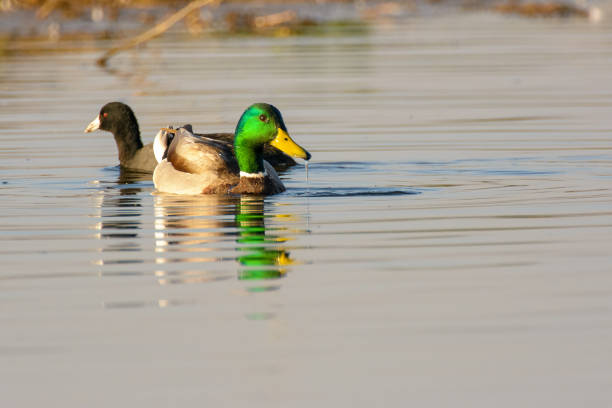
Relapsing fever, commonly referred to as spirochetosis, is a systemic bacterial infection caused by motile, helical microorganisms of the genus Borrelia and related spirochetes. While the disease is best known for affecting mammals—particularly humans, rodents, and domestic ruminants—it also afflicts avian species, especially waterfowl such as domestic ducks (Anas spp.). In ducks, spirochetosis manifests as intermittent bouts of fever, anemia, and severe weight loss, often leading to high mortality in poorly managed flocks. Understanding the disease’s etiology, epidemiology, clinical picture, and control measures is essential for veterinarians, duck producers, and hobbyists who aim to maintain healthy, productive flocks.
2. Causative Agents (Etiology)
| Taxonomic Group | Representative Species | Key Features | Primary Reservoir in Ducks |
|---|---|---|---|
| Borrelia | Borrelia anserina (formerly Spirillum anserinum) | Thin, flexible, Gram‑negative spirochete; 10–30 µm length; periplasmic flagella give corkscrew motility | Wild waterfowl, especially migratory ducks; occasionally domestic ducks |
| Spirochaeta | Spirochaeta spp. (rare) | Larger diameters; often environmental | Soil and stagnant water |
| Leptospira (occasionally confused) | Leptospira interrogans | Spiral, obligate aerobe; not true cause of “relapsing fever” in ducks but can produce similar hemolysis | Rodents, contaminated water |
The principal pathogen responsible for classic relapsing fever in ducks is Borrelia anserina. It is transmitted primarily by the hard tick Argas spp. (e.g., Argas persicus, Argas walkerae) that feed on waterfowl. In some geographical regions, soft ticks (Ornithodoros spp.) can also act as vectors.
3. Epidemiology & Transmission
- Geographic Distribution – Endemic in temperate and subtropical zones where both suitable tick vectors and dense duck populations coexist. Notable hotspots include:
- Europe (especially the United Kingdom, Germany, and France)
- North America (mid‑Atlantic and Pacific coastal states)
- Asia (Japan, China, and parts of Southeast Asia)
- Australia and New Zealand
- Transmission Routes
- Vector‑borne – The most efficient route. Infected ticks become infectious after a blood meal; spirochetes replicate in the tick’s salivary glands and are inoculated during subsequent feedings.
- Sexual & Vertical – Rare but documented; infected embryos may hatch with low‑level bacteremia.
- Fecal–Oral – Contaminated water or feed contaminated with tick feces can lead to ingestion of spirochetes.
- Seasonality – Peaks during warm, humid months when tick activity is maximal (late spring through early autumn). In colder climates, infections may persist in indoor or heated housing where ticks survive in cracks and crevices.
- Risk Factors – Overcrowding, poor sanitation, inadequate litter management, and lack of routine tick control dramatically increase infection pressure. Water sources that are stagnant or shared with wild migratory birds also serve as reservoirs for both the pathogen and its arthropod vector.
4. Pathogenesis
- Entry & Dissemination – Following inoculation, spirochetes rapidly penetrate the dermal capillary bed and enter the bloodstream, producing an acute bacteremia that may reach 10⁸–10⁹ cfu/mL.
- Immune Evasion – B. anserina expresses variable surface proteins (Vsp) that undergo antigenic variation, allowing the bacteria to escape host antibodies. This mechanism underlies the characteristic “relapsing” pattern: fever subsides as the immune system clears the dominant serotype, only to re‑emerge when a new Vsp variant proliferates.
- Organ Involvement
- Spleen & Liver – Spirochetes localize in the reticuloendothelial system, causing splenomegaly and hepatic congestion.
- Bone Marrow – Hematopoietic suppression leads to anemia and leukopenia.
- Central Nervous System – Occasionally, spirochetes cross the blood–brain barrier, producing neurologic signs such as ataxia and tremors.
- Inflammatory Response – Cytokine release (TNF‑α, IL‑1β) triggers fever, while complement activation contributes to hemolysis and endothelial damage, manifesting clinically as petechial hemorrhages and ecchymoses.
5. Clinical Signs & Symptoms
| Phase | Typical Signs | Comments |
|---|---|---|
| Acute (Day 0‑5) | Sudden high fever (41–42 °C), lethargy, loss of appetite, rapid breathing (tachypnea), pale combs and wattles, dark‑red or black‑filled droppings (due to hemoglobinuria) | Often misdiagnosed as viral gastroenteritis. |
| Relapse (Day 7‑14) | Re‑appearance of fever, icterus (jaundice), increased mortality, intermittent seizures, tremors, ataxia | May coincide with tick feeding cycles. |
| Chronic/Convalescent | Mild anemia, emaciation, poor feather quality, reduced egg production (in layers), weight loss up to 30 % of body condition | Birds that survive become carriers. |
Additional observations
- Mucosal petechiae on the oral cavity, eyelids, and vent.
- Swollen, boggy abdomen due to ascites or peritoneal effusion in severe cases.
- Plantar dermatitis (foot pad lesions) secondary to reduced grooming and wet litter.
The mortality rate in untreated flocks can reach 40‑70 %, especially in young ducklings and in flocks where tick infestations are heavy.
6. Duck Breeds at Risk
While spirochetosis can infect any domestic duck, certain breeds exhibit higher susceptibility due to genetic, behavioral, or management characteristics.
6.1 Mallard‑Derived Commercial Breeds (e.g., Pekin, Aylesbury, Rouen)
These breeds are the backbone of intensive duck production. Their fast growth rate and high stocking densities create an environment where tick vectors thrive. Moreover, commercial flocks often rely on indoor housing, where ticks can establish hidden reservoirs in cracks, bedding, and ventilation ducts. Consequently, Pekin and Aylesbury ducks frequently suffer severe outbreaks, especially during the first 6 weeks of life.
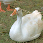
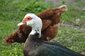
6.2 Heritage & Free‑Range Breeds (e.g., Muscovy, Khaki Campbell, Indian Runner)
Heritage breeds are often raised in extensive systems with access to ponds and natural vegetation. While outdoor access reduces crowding, it simultaneously increases exposure to wild birds that carry infected ticks. Muscovy ducks (Cairina moschata) are particularly vulnerable because they spend considerable time foraging on the ground, where questing ticks wait. Their relatively longer lifespan also provides a larger window for infection.
6.3 Bantam and Ornamental Ducks (e.g., Call, Crested, Silver Appleyard)
These smaller breeds are commonly kept in backyard settings with limited biosecurity. Owners may underestimate the need for tick control, allowing infestations of pet or farm cats—which can carry larval ticks—to infiltrate duck coops. The compact size of bantams means even a modest bacteremia can cause dramatic clinical signs, leading to rapid loss of the bird.
6.4 Ducks Used for Research or Exhibition
Laboratory and show ducks are often immunologically naïve because they are bred for specific traits rather than disease resistance. Their high‑value status leads to intense handling, which may inadvertently spread ticks between enclosures. The stress of transport and exhibition can precipitate relapses, making vigilant monitoring essential.
Take‑away: Any duck breed can become infected, but high‑density commercial breeds, ground‑foraging heritage ducks, and poorly bio‑secured ornamental or research birds are the most at risk. Tailoring management practices to breed‑specific behaviors dramatically reduces the likelihood of severe disease.
7. Affected Life‑Stage
| Life‑Stage | Susceptibility | Typical Clinical Course |
|---|---|---|
| Embryo | Very low (vertical transmission rare) | May hatch with sub‑clinical bacteremia; usually asymptomatic until 2‑3 weeks of age. |
| Ducklings (0‑6 weeks) | Highest – immature immune system, thin skin, rapid growth demands | Sudden onset of fever, severe anemia, high mortality (up to 80 % in naïve flocks). |
| Juveniles (6 weeks‑4 months) | Moderate – immune system more mature but still developing | Relapsing fevers, reduced feed conversion, occasional neurologic signs. |
| Adult Breeders & Layers | Low‑moderate – developed immunity but can become carriers | Subclinical bacteremia, intermittent drops in egg production, occasional relapse during stress (molting, breeding). |
| Seniors (>2 years) | Variable – chronic carriers may develop secondary infections | Weight loss, poor feather condition, increased susceptibility to secondary pathogens (e.g., E. coli septicemia). |
Key point: The first six weeks of life represent a critical window. Early tick control, prophylactic antibiotic administration (under veterinary guidance), and strict biosecurity can dramatically improve survival rates in ducklings.
8. Diagnosis
8.1 Clinical Assessment
- History – Recent tick exposure, recent introduction of new birds, seasonal timing.
- Physical Exam – Fever, pallor, icterus, splenomegaly, petechiae, neurological deficits.
8.2 Laboratory Techniques
- Microscopic Examination of Blood Smears
- Fresh peripheral blood smear stained with Giemsa or Wright.
- Look for slender, helical organisms (10‑30 µm) moving in a characteristic “corkscrew” motion.
- Sensitivity increases during peak bacteremia (first 3‑5 days of fever).
- Dark‑field or Phase‑contrast Microscopy
- More sensitive for low‑level bacteremia; visualizes spirochetes without staining.
- Polymerase Chain Reaction (PCR)
- Targeted primers for Borrelia anserina 16S rRNA or flagellin genes.
- Provides definitive diagnosis even when blood smears are negative.
- Serology (ELISA, IFA)
- Detects antibodies against variable surface proteins. Useful for herd screening and identifying carriers.
- Culture
- Highly specialized; requires Barbour–Stoermer–Kelly (BSK) medium under microaerophilic conditions. Practically limited to reference laboratories.
8.3 Necropsy Findings (if mortality is high)
- Gross: Enlarged, dark spleen; friable liver with petechial hemorrhages; serous effusions in body cavities.
- Histopathology: Perivascular infiltrates of macrophages and lymphocytes; spirochetes seen in the lumen of small vessels using silver stains (Warthin‑Starry).
8.4 Differential Diagnosis
- Avian influenza – respiratory signs, high mortality.
- Duck viral hepatitis (DVH) – liver necrosis, jaundice.
- Pasteurellosis – sudden death, septicemia.
- Clostridial enteritis – watery diarrhea, foul odor.
A combined diagnostic approach (clinical + PCR + microscopy) yields the most reliable confirmation.
9. Treatment
9.1 Antibiotic Therapy
| Antibiotic | Dose (per kg bw) | Route | Duration | Remarks |
|---|---|---|---|---|
| Tetracycline (or oxytetracycline) | 10–15 mg/kg | IM or oral (via water) | 5–7 days | First‑line; bacteriostatic, penetrates intracellularly |
| Doxycycline | 5 mg/kg | Oral (water) | 5 days | More stable in water; works against resistant strains |
| Enrofloxacin | 5 mg/kg | IM | 3 days | Fluoroquinolone – reserved for severe cases or tetracycline‑failure |
| Ceftriaxone (parenteral) | 30 mg/kg | IM/SC | 3 days | Effective for CNS involvement; expensive |
Therapeutic guidelines
- Treat all birds in the affected flock, not only clinically ill individuals, to eradicate the carrier state.
- Add antibiotics to drinking water with a stabilizer (e.g., ascorbic acid) to maintain potency.
- Monitor water intake to ensure therapeutic dosing; adjust concentrations for warm weather (higher consumption).
9.2 Supportive Care
- Fluid therapy – Isotonic saline (20 mL/kg) subcutaneously or via oral electrolytes to correct dehydration from fever and hemoglobinuria.
- Iron supplementation – Oral ferric glycinate (0.5 mg/kg) for recovering anemic birds.
- Vitamin A & E – Antioxidant support to protect hepatic cells (e.g., 200 IU/kg vitamin A, 10 IU/kg vitamin E).
- Heat support – Maintain ambient temperature 30‑32 °C for ducklings during acute phase.
9.3 Anti‑Tick Treatment
Simultaneous control of the vector is indispensable:
- Acaricide sprays – Permethrin 0.5 % (spray coat all surfaces, especially crevices).
- Dusts – Deltamethrin 0.05 % applied to litter.
- Environmental – Steam cleaning of housing; replace old bedding.
Note: Re‑treat every 14 days for at least two cycles to cover the tick life cycle.
9.4 Monitoring & Follow‑up
- Re‑evaluate blood smears 48 h after starting therapy.
- Perform PCR on a random sample of 10 % of the flock 7 days post‑treatment to confirm eradication.
- Continue prophylactic tick control for an additional 4 weeks.
10. Prognosis & Complications
| Condition | Likelihood | Clinical Impact |
|---|---|---|
| Full Recovery | High (80‑90 % in treated ducks) | Return to normal weight and egg production within 2‑3 weeks. |
| Chronic Carrier State | Moderate (30‑40 % of survivors) | Persistent low‑level bacteremia; risk of future relapses, especially during stress. |
| Neurologic Sequelae | Low (≤5 %) | Ataxia, tremors may persist for weeks, occasionally permanent. |
| Secondary Bacterial Sepsis | Moderate (10‑20 % in severe cases) | E. coli or Clostridium overgrowth → sudden death. |
| Reproductive Decline | Moderate (10‑15 % of layers) | Egg yolk discoloration, reduced clutch size for 2‑4 weeks. |
| Mortality | Variable (30‑70 % untreated, <10 % treated) | Dependent on age, breed, and timeliness of treatment. |
Prognostic factors
- Age – Ducklings < 2 weeks have the poorest outlook.
- Tick burden – Heavy infestations correlate with higher mortality.
- Promptness of therapy – Beginning antibiotics within 24 h of fever onset improves survival > 90 %.
11. Prevention & Control
11.1 Integrated Tick‑Management (ITM)
- Environmental Hygiene
- Remove debris, replace litter weekly, and keep water sources clean.
- Seal cracks in walls, floors, and ventilation ducts where ticks hide.
- Chemical Acaricides
- Rotate classes (pyrethroids → organophosphates → amidines) to prevent resistance.
- Apply permethrin (0.5 %) as a spray every 30 days during peak tick season.
- Biological Controls
- Introduce Entomopathogenic fungi (Beauveria bassiana) as a dust in nesting boxes.
- Use predatory mites (Hypoaspis miles) that prey on tick eggs and larvae.
- Physical Barriers
- Install fine mesh (≤ 0.5 mm) on ventilation openings.
- Use raised platforms for nest boxes to reduce ground contact.
11.2 Herd Health Practices
- Quarantine all new arrivals for a minimum of 21 days, with tick inspections and PCR testing before integration.
- Vaccination – No commercial vaccine exists for avian spirochetosis; experimental recombinant Vsp vaccines show promise but are not yet field‑available.
- Regular Screening – Perform monthly blood smear examinations during high‑risk months.
11.3 Nutritional Immunomodulation
- Vitamin C (250 mg/L water) boosts macrophage activity.
- Beta‑glucans (derived from yeast) added to feed (0.2 % of diet) enhance innate immunity.
11.4 Biosecurity Checklist (Weekly)
| Task | Frequency | Responsible |
|---|---|---|
| Inspect all bird areas for live ticks | Daily | Farm staff |
| Replace water troughs and clean disinfect | Every 3 days | Keeper |
| Rotate bedding material | Weekly | Manager |
| Apply acaricide dust in nesting boxes | Bi‑weekly | Veterinarian |
| Conduct random blood smear checks | Weekly (high‑risk season) | Vet tech |
| Record mortality and clinical signs | Ongoing | Keeper |
12. Diet & Nutrition
A balanced, high‑quality diet fortifies the immune system and accelerates recovery from spirochetosis.
| Nutrient | Recommended Level (per kg feed) | Source | Rationale |
|---|---|---|---|
| Protein | 20‑24 % (starter), 18‑20 % (grower) | Soybean meal, fish meal, dried insects | Provides amino acids for hemoglobin synthesis & tissue repair. |
| Energy | 2 800‑3 000 kcal ME/kg | Corn, wheat, barley | Supports thermoregulation during fever. |
| Methionine + Cysteine | 0.6 % total | DL‑methionine, powdered egg yolk | Sulphur amino acids aid in glutathione production, an antioxidant critical during oxidative stress from hemolysis. |
| Vitamin A | 12 000 IU/kg | Carrots, synthetic retinyl acetate | Supports mucosal integrity and ocular health (prevent icteric lesions). |
| Vitamin E | 200 IU/kg | Wheat germ oil, tocopherol | Reduces lipid peroxidation in membranes damaged by spirochete toxins. |
| Vitamin C | 500 mg/L water (supplement) | Ascorbic acid powder | Enhances leukocyte function. |
| Minerals | Calcium 0.9 %, Phosphorus 0.7 % ; trace Zn, Se, Mn | Limestone, dicalcium phosphate, mineral premix | Zinc and selenium are essential for superoxide dismutase activity. |
| Probiotics | 10⁸ CFU/g feed | Lactobacillus spp., Bifidobacterium spp. | Maintain gut flora balance, reducing secondary bacterial overgrowth. |
Feeding Recommendations During Outbreak
- Offer easily digestible feed (e.g., boiled rice, soft mash) for ducklings with anorexia.
- Provide supplemental electrolytes (sodium chloride 0.5 %) in drinking water to replace losses from fever and urination.
- Increase feed frequency – offer small meals every 2‑3 hours to encourage intake.
13. Zoonotic Risk
13.1 Human Infection
- Borrelia anserina is not considered a common human pathogen, but related Borrelia spp. (e.g., B. hermsii, B. recurrentis) cause relapsing fever in humans.
- Occupational exposure (farm workers, veterinarians) to infected ticks or contaminated duck droppings can theoretically result in transient bacteremia, though documented cases are exceedingly rare.
13.2 Public Health Considerations
- Personal Protective Equipment (PPE) – Gloves, long sleeves, and boots when handling sick ducks or cleaning enclosures.
- Tick Bite Prevention – Use of permethrin‑treated clothing and regular tick checks for personnel.
- Hand Hygiene – Wash hands with soap and water after contact with birds or their environment.
13.3 Reporting & Regulations
- In many countries, relapsing fever caused by any Borrelia species is a notifiable disease. If a veterinarian suspects zoonotic transmission, they should inform local public‑health authorities.
14. Summary of Key Points
- Spirochetosis in ducks is primarily caused by Borrelia anserina, transmitted by Argas ticks.
- Clinical picture includes cyclic fever, anemia, icterus, and possible neurologic signs; mortality can exceed 70 % without treatment.
- Young ducklings and high‑density commercial breeds are most vulnerable; however, free‑range heritage ducks and ornamental breeds are also at risk due to environmental exposure.
- Diagnosis is best achieved with a combination of blood smear microscopy, PCR, and serology.
- Treatment relies on tetracycline‑class antibiotics plus aggressive tick control; supportive fluid therapy and nutritional supplementation improve outcomes.
- Prevention hinges on Integrated Tick‑Management, strict biosecurity, routine screening, and optimal nutrition.
- Zoonotic potential is low but not negligible; proper PPE and tick‑bite precautions protect farm workers.
By adhering to the preventive and therapeutic strategies outlined above, producers can markedly reduce the incidence of relapsing fever in ducks, safeguard animal welfare, and protect public health.
#DuckHealth, #RelapsingFever, #Spirochetosis, #BorreliaAnserina, #PoultryMedicine, #AvianVeterinary, #DuckFever, #TickControl, #WaterfowlCare, #DuckBreeds, #PoultryBiosecurity, #AvianZoonoses, #DuckNutrition, #FarmSafety, #VeterinaryGuide, #DuckLovers, #BackyardPoultry, #OrganicFarming, #AnimalWelfare, #OneHealth


Add comment